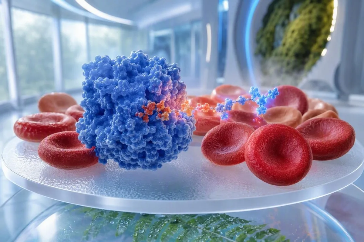
Una bassa attività enzimatica della G6PD significa che i globuli rossi potrebbero essere meno in grado di gestire lo stress ossidativo, ma un risultato normale durante o subito dopo l’emolisi può dare una falsa rassicurazione. Il passo successivo più sicuro è di solito un test quantitativo di ripetizione dopo la ripresa, con le decisioni sui farmaci guidate dal clinico prescrittore.


- Bassa attività enzimatica della G6PD può aumentare il rischio di rottura dei globuli rossi dopo alcuni farmaci, infezioni, fave o esposizioni chimiche.
- Tempistica di falso-normale è comune durante un’emolisi acuta perché i globuli rossi più anziani, più carenti di enzima, vengono rimossi per primi, lasciando cellule più giovani con un’attività relativamente più alta.
- Ripetere il test è comunemente considerata circa 2-3 mesi dopo un episodio emolitico acuto o una trasfusione, a meno che uno specialista non consigli una tempistica diversa.
- Test quantitativo riporta l’attività in unità per grammo di emoglobina o come percentuale di una mediana maschile corretta; conta l’intervallo di riferimento proprio del laboratorio.
- Risultati intermedi nell’intervallo di attività approssimativamente 30%-70% meritano particolare attenzione nelle donne perché l’inattivazione del cromosoma X può creare popolazioni miste di globuli rossi.
- Sintomi urgenti includono urine di colore bruno-nero tipo tè, ittero, fiato corto, sensazione di svenimento, grave debolezza o pallore che peggiora rapidamente dopo un nuovo farmaco o un’illness.
- Rasburicase e blu di metilene richiede particolare cautela nella confermata carenza di G6PD; i prescrittori dovrebbero valutare alternative prima dell’uso.
- Fave e palline antitarme alla naftalina sono classici fattori scatenanti senza prescrizione, ma gli elenchi dei farmaci differiscono in base alla dose, alla gravità della malattia e alla variante coinvolta.
Cosa significa una bassa attività enzimatica della G6PD nei risultati degli esami di laboratorio
A risultato basso di G6PD significa che i tuoi globuli rossi hanno una protezione ridotta contro lo stress ossidativo e possono rompersi più rapidamente quando vengono sfidati da un fattore scatenante. Alla data del 7 agosto 2026, il risultato dovrebbe essere trattato come un riscontro di sicurezza del farmaco piuttosto che come una diagnosi basata su un singolo numero.
G6PD, o glucosio-6-fosfato deidrogenasi, aiuta i globuli rossi a produrre NADPH, che mantiene la glutatione nella sua forma protettiva ridotta. I globuli rossi non possono produrre nuove proteine enzimatiche una volta maturi, quindi l’attività diminuisce naturalmente mentre una cellula si avvicina alla sua normale durata di vita di 120 giorni.
Un risultato al di sotto del limite inferiore del laboratorio di refertazione supporta la carenza di G6PD, ma il valore non è universalmente confrontabile tra laboratori. Molti saggi quantitativi riportano circa 5,5–20,5 U/g di emoglobina negli adulti, tuttavia il metodo analitico, la correzione per l’emoglobina e la popolazione locale determinano ciò che è davvero basso.
Kantesti è un Analizzatore di analisi del sangue AI che legge un risultato di G6PD insieme a emoglobina, bilirubina, LDH, reticolociti e la data di qualsiasi malattia acuta, invece di trattare un singolo indicatore come definitivo. Il nostro team clinico vede regolarmente preoccupazione causata da un risultato tecnicamente normale ma raccolto esattamente nel momento sbagliato.
Il dott. Thomas Klein, MD, ha riscontrato che i pazienti spesso assumono che una bassa attività significhi che stanno male ogni giorno. La maggior parte delle persone con varianti comuni si sente completamente bene tra un’esposizione e l’altra; il problema pratico è documentare chiaramente il risultato e sapere quali situazioni meritano una chiamata a un clinico. Per il contesto sugli indicatori di laboratorio, vedi significati dei risultati fuori intervallo.
Come vengono misurati e classificati i risultati dei test della G6PD
A saggio quantitativo di G6PD misura l’attività enzimatica in un campione di sangue intero e fornisce informazioni più utili di uno screening semplice quando un risultato può influenzare il trattamento. L’attività è comunemente espressa come U/g Hb o come percentuale di un valore mediano maschile corretto, non come una concentrazione universale.
Il campione diagnostico preferito è tipicamente sangue intero anticoagulato con EDTA, con l’attività enzimatica normalizzata alla concentrazione di emoglobina. Un campione con emoglobina molto bassa può produrre un rapporto U/g Hb fuorviante, apparentemente elevato, perché nel denominatore c’è meno emoglobina.
Molti servizi considerano carente un’attività inferiore a 30% del valore mediano maschile corretto , circa 30% a 70% come intermedia, e sopra 70% come normale; queste fasce operative non sono intercambiabili con l’intervallo di riferimento di ogni laboratorio. Il nome del saggio e il limite di riferimento inferiore del referto dovrebbero accompagnare il risultato.
Uno screening a spot fluorescente può identificare molti campioni marcatamente carenti, ma può perdere attività intermedia, soprattutto nelle femmine eterozigoti o durante la reticolocitosi. Un saggio quantitativo è generalmente la scelta migliore quando si sta considerando tafenoquina, primaquina, dapsone o un medicinale ossidante simile.
I reticolociti sono globuli rossi giovani e di solito contengono più attività di G6PD rispetto alle cellule più vecchie, motivo per cui qui sono così importanti. Il nostro guida su reticolociti ed ematologia spiega perché un conteggio superiore a circa 100 × 10⁹/L durante l’anemia cambia l’interpretazione dei test enzimatici.
Perché un test di carenza di G6PD può risultare normale dopo l’emolisi
Un test G6PD può risultare normale durante o subito dopo l’emolisi perché le più vecchie e più carenti di enzima vengono eliminate in via preferenziale. La circolazione residua si arricchisce di reticolociti e cellule più giovani che hanno un’attività misurata relativamente più alta.
Questo è un effetto di selezione, non una cura. Se una persona perde una grande frazione di cellule vulnerabili nell’arco di 24-72 ore, l’analisi può misurare principalmente cellule appena rilasciate il cui contenuto di G6PD non è ancora diminuito.
Un risultato falsamente rassicurante è più probabile quando, vicino alla data di prelievo, erano presenti anemia, ittero, urine scure, LDH elevata, aptoglobina bassa o un aumento del conteggio dei reticolociti. Anche una diminuzione dell’emoglobina da 145 g/L a 105 g/L può modificare in modo sostanziale la miscela di età cellulare circolante.
La trasfusione recente crea un secondo problema: i globuli rossi del donatore possono avere un’attività enzimatica normale e possono mascherare la carenza del ricevente per settimane. Salva la data e il numero di unità trasfuse; spesso questo conta più di un valore borderline nel PDF.
Questa lezione più ampia si applica a molti risultati di test di laboratorio dopo una malattia acuta. Confrontare il prelievo attuale con il basale precedente alla malattia in una revisione del sangue tra una visita e l’altra può mostrare se il risultato dell’enzima è stato ottenuto durante una finestra biologicamente instabile.
Come i clinici riconoscono un’emolisi attiva in prossimità di un risultato della G6PD
L’emolisi attiva è suggerita da emoglobina in calo più reticolociti aumentati, LDH elevata, aumento della bilirubina indiretta e aptoglobina bassa; nessun singolo marcatore la dimostra. Il pattern combinato determina se un risultato di G6PD debba essere rimandato o ripetuto.
Un risultato di aptoglobina al di sotto di circa 0,3 g/L supporta l’emolisi intravascolare nel contesto giusto, ma aptoglobina bassa può anche riflettere una ridotta produzione epatica. L’LDH può aumentare di alcune volte oltre il limite superiore di riferimento, anche se il danno epatico, il danno muscolare e la gestione del campione possono anch’essi aumentarla.
La bilirubina non coniugata spesso aumenta quando accelera la clearance dei globuli rossi, mentre la bilirubina nelle urine è di solito negativa perché la bilirubina non coniugata non è solubile in acqua. Un risultato positivo della bilirubina nelle urine invece indirizza i clinici verso la bilirubina coniugata e un diverso ramo diagnostico.
Quando rivedo un pannello con emoglobina a 96 g/L, reticolociti a 180 × 10⁹/L, LDH a 780 UI/L e aptoglobina non rilevabile, non accetto un risultato di attività enzimatica normale così com’è. Thomas Klein, MD, di solito chiederebbe se il campione sia stato prelevato dopo un trigger piuttosto che etichettare il paziente come non affetto da deficit.
Un vetrino con campione di cellule periferiche può mostrare cellule a morso o cellule vescicolari dopo stress ossidativo, sebbene la loro assenza non escluda l’emolisi correlata a G6PD. Leggi di più su quando la morfologia cellulare cambia e richiede una revisione tempestiva nella nostra guida a reperti urgenti di schistociti E interpretazione dell’aptoglobina.
Quando ripetere un test della G6PD dopo emolisi o trasfusione
La ripetizione della determinazione quantitativa della G6PD è spesso più affidabile 2-3 mesi dopo che l’emolisi si è completamente stabilizzata e almeno 3 mesi dopo una trasfusione, perché i reticolociti e le cellule del donatore possono mascherare il valore basale. Un ematologo può modificare tale tempistica quando il trattamento non può attendere.
Non usare solo il calendario. Prima di ripetere il test, i clinici cercano un’emoglobina stabile, una conta dei reticolociti che ritorni verso il suo intervallo personale, la bilirubina e l’LDH che si stabilizzano e l’assenza di esposizione in corso a un trigger sospettato.
Dopo un episodio non complicato, un intervallo di 12 settimane in genere consente alla maggior parte dell’arricchimento transitorio di cellule giovani di svanire. Dopo diverse unità di trasfusione di emazie, l’intervallo potrebbe dover riflettere la sopravvivenza delle cellule del donatore, l’anemia in corso e l’urgenza della decisione terapeutica.
Se un farmaco deve essere iniziato urgentemente, il prescrittore può usare una combinazione di documentazione pregressa, test molecolari, consulenza specialistica e una decisione basata sul rapporto rischio-beneficio, invece di attendere un singolo esame perfetto. Questo è particolarmente rilevante nel trattamento della malaria, in cui ritardare la terapia può essere pericoloso di per sé.
Kantesti AI può visualizzare la sequenza di emoglobina, bilirubina, LDH e reticolociti attraverso più referti, così che la data di ripetizione sia spiegabile clinicamente. Per una tempistica pratica di recupero dopo un ricovero, vedi esami ematici in evoluzione dopo la dimissione ospedaliera e il nostro guida all’analisi delle tendenze di laboratorio.
Medicinali che richiedono cautela guidata dal clinico in caso di carenza di G6PD
Un deficit di G6PD confermato o fortemente sospettato richiede una revisione da parte del prescrittore o del farmacista prima dell’uso di farmaci ad azione ossidante; il rischio varia in base a dose, variante, età, infezione e anemia basale. Non sospendere un farmaco prescritto solo da un elenco online di evitamento.
Rasburicase è controindicata in caso di deficit di G6PD noto, perché può generare un significativo stress ossidativo, e blu di metilene può essere inefficace o dannoso quando la generazione di NADPH è compromessa. Queste sono decisioni a livello ospedaliero, in cui un’alternativa dovrebbe essere selezionata prontamente.
Primaquina e tafenoquina richiedono una valutazione quantitativa della G6PD validata prima dell’uso, perché possono causare emolisi correlata alla dose in persone suscettibili. Anche dapsona, nitrofurantoina, farmaci contenenti sulfonamide e alcuni antimalarici meritano una revisione individualizzata, invece di assunzioni generiche.
Luzzatto e Seneca hanno descritto il deficit di G6PD come un problema farmacogenetico continuo proprio perché il rischio dei farmaci è condizionale, non binario (Luzzatto e Seneca, 2014). Una malattia virale, la febbre, un danno renale o un ciclo di trattamento di 2 settimane possono trasformare un’esposizione precedentemente tollerata in una situazione clinicamente rilevante.
Porta al prescrittore il referto di laboratorio—non solo una diagnosi ricordata. La preparazione di farmaci e integratori può anche alterare altri esami, quindi il nostro articolo su integratori prima degli esami del sangue è utile quando è prevista una nuova raccolta.
Alimenti ed esposizioni domestiche associati a emolisi correlata a G6PD
I fagioli di fava sono il trigger alimentare più consolidato per l’emolisi correlata al G6PD, mentre le palline antitarme contenenti naftalene sono un’esposizione domestica riconosciuta da evitare. La reazione può verificarsi entro poche ore fino a diversi giorni e non richiede una porzione enorme in una persona altamente suscettibile.
La favismo non è semplicemente un’allergia alimentare. I metaboliti della vicina e della convicina possono creare stress ossidativo in globuli rossi vulnerabili e la suscettibilità varia in modo sostanziale tra le diverse varianti del G6PD e tra individui.
Il naftalene può essere presente in alcune palline antitarme e nei prodotti deodoranti; l’esposizione accidentale è più preoccupante nei lattanti e nei bambini piccoli perché una dose relativamente piccola rappresenta un’esposizione maggiore per chilogrammo. Conservare i prodotti nella confezione originale e richiedere un parere del servizio di tossicologia dopo un’ingestione sospetta o un’esposizione inalatoria intensa.
Le diete ordinarie non devono diventare restrittive di default. Aglio, legumi diversi dai fagioli di fava, mirtilli, alimenti ricchi di vitamina C e la maggior parte delle erbe aromatiche da cucina sono spesso inclusi in liste poco affidabili senza prove cliniche coerenti di danno alle quantità alimentari.
Kantesti è un Strumento di analisi degli esami del sangue basato su AI che mantiene un risultato enzimatico documentato con la data, i sintomi e il potenziale trigger, il che può rendere meno caotica una futura conversazione sui farmaci. Le evidenze sugli integratori sono disomogenee; la nostra revisione di integratori di glutatione e valori di laboratorio spiega perché un’etichetta di prodotto non è la stessa cosa della protezione clinica dimostrata.
Perché i risultati della G6PD differiscono in donne, neonati e gravidanza
L’interpretazione del test del G6PD è meno lineare in molte donne perché l’inattivazione casuale del cromosoma X può lasciare una miscela di globuli rossi normali e carenti. Anche i neonati hanno proporzioni più elevate di reticolociti, quindi non dovrebbero essere applicati cut-off per adulti senza un intervallo di riferimento pediatrico.
Una donna può portare un allele alterato del G6PD e avere comunque un’attività intermedia clinicamente significativa, anche se uno screening qualitativo risulta normale. Il test enzimatico quantitativo è preferibile quando una paziente donna potrebbe aver bisogno di primaquina, tafenoquina o di un altro trattamento con potenziale ossidante noto.
Lo screening neonatale è particolarmente prezioso nelle regioni con una prevalenza più alta perché una grave itterizia neonatale può svilupparsi prima che la famiglia sappia che esiste una variante enzimatica. Un aumento della bilirubina nelle prime 24 ore di vita richiede sempre una valutazione pediatrica, indipendentemente dallo stato del G6PD.
La gravidanza di per sé non causa carenza di G6PD, ma l’anemia durante la gravidanza ha molte cause sovrapposte, tra cui carenza di ferro, carenza di folati e diluizione dovuta all’aumento del volume plasmatico. Un livello di emoglobina inferiore a 110 g/L merita un’interpretazione ostetrica piuttosto che un’attribuzione automatica all’attività dell’enzima.
Per il contesto pediatrico, rivedere gli intervalli dei test ematici in base all’età. Se si sospetta anche una carenza di ferro, la domanda più utile è spesso se la ferritina, la saturazione della transferrina e gli indici dei globuli rossi raccontano la stessa storia.
Quando il test genetico aiuta dopo un risultato basso o incerto di G6PD
Il test genetico può chiarire un fenotipo del G6PD incerto, soprattutto dopo emolisi, trasfusione o un risultato intermedio in una donna, ma non sostituisce il test dell’attività enzimatica in ogni contesto clinico. Il test migliore dipende dal fatto che la domanda immediata riguardi la sicurezza del farmaco, la consulenza familiare o l’identificazione della variante.
Il G6PD si trova sul cromosoma X, quindi molti maschi emizigoti hanno un fenotipo enzimatico più uniforme, mentre le donne eterozigoti possono avere una variazione ampia dell’attività dovuta alla lionizzazione. Sono note più di 200 varianti e il genotipo non sempre predice il grado di emolisi in una specifica infezione.
Il test molecolare è utile quando il risultato dell’enzima è stato ottenuto in un periodo inaffidabile, quando è nota una specifica variante familiare o quando test ripetuti dell’attività sono in conflitto con la storia clinica. Non è una scorciatoia per interpretare i sintomi senza un CBC e marcatori di emolisi.
La distribuzione globale è disomogenea: la meta-analisi di Nkhoma et al. ha stimato che la carenza di G6PD colpisce centinaia di milioni di persone nel mondo, con frequenze più alte nelle aree storicamente endemiche per la malaria (Nkhoma et al., 2009). Questa geografia informa le strategie di test, ma non dovrebbe mai essere usata per liquidare la necessità di test in qualcuno proveniente da un altro contesto.
Registrare i familiari con itterizia neonatale, urine scure inspiegabili dopo una malattia o una variante enzimatica nota. La nostra guida a i pattern di laboratorio familiari può aiutare le famiglie a preservare dettagli utili senza assumere che ogni parente abbia lo stesso livello di attività.
Sintomi dopo un fattore scatenante che richiedono una valutazione medica nella stessa giornata
Urine marrone scuro, occhi o pelle gialli, grave stanchezza, fiato corto, fastidio toracico, svenimento o pallore che aumenta rapidamente dopo un trigger richiedono una valutazione clinica nella stessa giornata. Questi sintomi possono segnalare un’emolisi clinicamente significativa, ma possono anche verificarsi con disidratazione, malattia epatica, problemi urinari o altre condizioni urgenti.
Un improvviso calo della capacità di trasporto dell’ossigeno può causare un cuore in corsa o dispnea prima che la persona riconosca l’ittero. In un adulto precedentemente sano, una caduta dell’emoglobina di 20-30 g/L nell’arco di pochi giorni è clinicamente significativa e non dovrebbe essere gestita solo con idratazione.
I medici di emergenza controllano comunemente CBC, reticolociti, frazioni della bilirubina, LDH, aptoglobina, creatinina, elettroliti, analisi delle urine e talvolta un test di antiglobulina diretta. Il test di antiglobulina diretta aiuta a distinguere l’emolisi mediata dal sistema immunitario dalle cause non immuni, anche se i risultati devono essere letti nel contesto.
Un campione di laboratorio può diventare emolizzato durante la raccolta e aumentare falsamente il potassio o LDH senza causare sintomi nel paziente. Ecco perché i sintomi, il colore delle urine e l’emoglobina seriata sono più informativi di un singolo indice di emolisi di laboratorio.
Se hai ingiallimento con urine scure, la nostra spiegazione di segnali di allarme della bilirubina può aiutare a organizzare l’anamnesi per l’assistenza urgente. Non aspettare un’interpretazione dell’app se ti senti svenire, confuso o dispnoico a riposo.
Come leggere i risultati del test di laboratorio della G6PD insieme al resto del pannello
L’interpretazione più utile di G6PD combina attività enzimatica, tempistica della raccolta, emoglobina, reticolociti, bilirubina, LDH, aptoglobina e storia di trasfusioni. Un valore di attività normale offre una rassicurazione limitata quando il pannello circostante mostra un turnover attivo dei globuli rossi.
Inizia con il dosaggio e le unità. Un risultato di 4,2 U/g Hb può essere basso in un laboratorio con un limite inferiore di riferimento di 5,5 U/g Hb, mentre lo stesso numero non può essere interpretato in modo sicuro senza il metodo e l’intervallo di riferimento di quel laboratorio.
Poi valuta se la CBC suggerisce anemia: l’emoglobina al di sotto di 130 g/L in molti uomini adulti o al di sotto di 120 g/L in molte donne adulte non in gravidanza è comunemente segnalata, anche se gli intervalli locali variano. MCV, RDW, stato del ferro e stato di B12 possono identificare una seconda causa di anemia non correlata a G6PD.
Di Kantesti servizio di interpretazione dei test del laboratorio di IA usa controlli del pattern per identificare quando un risultato di G6PD apparentemente rassicurante si trova accanto a reticolociti elevati o aptoglobina bassa e dovrebbe essere discusso con un clinico. Questa logica viene rivista rispetto a standard di validazione medica e non sostituisce i test di laboratorio di conferma.
Per un flusso di lavoro di verifica della fonte prima di agire su qualsiasi riepilogo automatizzato, leggi la nostra guida di sicurezza per referti medici AI. La domanda del clinico di solito non è “è rosso o verde?” ma “quel valore era biologicamente affidabile in quella data?”.”
Domande da portare al tuo clinico dopo una bassa attività enzimatica della G6PD
Dopo un risultato di G6PD basso o incerto, chiedi se il dosaggio fosse quantitativo, se l’emolisi o la trasfusione potessero averlo distorto e se un nuovo test o una conferma genetica cambierebbero l’assistenza. Porta il referto originale, non uno screenshot ritagliato, perché unità e intervalli di riferimento sono decisivi.
Domande utili includono: Il mio test è stato raccolto durante l’anemia o entro 12 settimane dalla trasfusione? Qual è stato il mio conteggio dei reticolociti? Il mio risultato è al di sotto di 30%, intermedio o normale secondo il metodo di questo laboratorio? Queste domande riducono rapidamente l’incertezza.
Chiedi anche quali farmaci siano rilevanti per la tua assistenza personale invece di richiedere un elenco indiscriminato “non prendere mai”. Un farmaco che comporta un rischio a 100 mg al giorno può avere un profilo di rischio diverso a una singola dose bassa, e l’infezione stessa può essere il trigger emolitico dominante.
Conserva una fotografia o un PDF del referto completo, le date dei sintomi e qualsiasi farmaco iniziato nelle 14 giornate precedenti. Questo diventa particolarmente utile se un pronto soccorso o una clinica di viaggio deve prendere una decisione rapida di prescrizione.
A Seconda opinione su un esame del sangue è ragionevole quando il risultato è in conflitto con l’emolisi passata, un’anamnesi familiare o una decisione farmacologica con conseguenze significative. Un buon follow-up non è un eccesso di test; significa scegliere l’esame al momento giusto.
Precauzioni di G6PD prima di viaggiare, procedure e nuove prescrizioni
Prima di viaggiare, di un intervento chirurgico o di una nuova prescrizione, informa il clinico su una carenza di G6PD confermata o sospetta e fornisci il risultato reale se disponibile. Questo consente la scelta dei farmaci prima che una situazione urgente imponga una decisione affrettata.
La medicina del viaggio merita una pianificazione precoce perché le scelte per la profilassi antimalarica variano in base alla destinazione, al rischio della specie e all’attività di G6PD. Tafenoquina non può essere trattata in modo sicuro come una compressa di viaggio di routine quando l’attività quantitativa non è stata verificata.
Per le procedure, il team anestesiologico dovrebbe conoscere la diagnosi, ma la carenza di G6PD non rende automaticamente sicura l’anestesia di routine. La questione rilevante è il piano terapeutico completo, l’infezione concomitante, la funzione renale e se si è verificato recentemente un evento emolitico.
Kantesti AI può aiutare gli utenti a organizzare report cronologici per un clinico, ma non può autorizzare un medicinale né sostituire il controllo di interazione di un farmacista. Il Comitato consultivo medico supporta il nostro approccio guidato dal clinico per un linguaggio sulla sicurezza relativo a reperti di laboratorio potenzialmente rilevanti.
Una piccola card di allerta medica o una nota telefonica dovrebbe indicare “Carenza di G6PD—verificare i medicinali”, la data del test e se il risultato era quantitativo. Evita di scrivere un lungo elenco di farmaci non verificato; al clinico servono dati accurati, non un’etichetta allarmante.
Un piano pratico per il passo successivo dopo i risultati del test della G6PD
Il piano più sicuro dopo i risultati dei test di laboratorio per la G6PD è documentare l’attività esatta e la data del test, identificare eventuali emolisi o trasfusioni recenti, rivedere i medicinali con un clinico e ripetere un dosaggio quantitativo dopo la ripresa se la tempistica era inaffidabile. La maggior parte dei pazienti non ha bisogno di un trattamento quotidiano; ha bisogno di una registrazione affidabile e di un piano che tenga conto dei fattori scatenanti.
Se la tua attività è chiaramente bassa in un campione ambulatoriale stabile, chiedi al clinico di aggiungerla alla tua cartella clinica e di discutere i medicinali più rilevanti per te. Se il risultato era normale durante un episodio sospetto, non dare per chiusa la questione—organizza una ripetizione circa tra 8 e 12 settimane, quando appropriato.
Se sviluppi urine scure, ittero, fiato corto, debolezza marcata o capogiri dopo un’infezione, l’esposizione a fave, la naphthalene o un nuovo medicinale, richiedi un parere medico nella stessa giornata. Porta i dettagli sui fattori scatenanti, perché spesso le prime 24 ore di anamnesi determinano quali test vengono prioritizzati.
Per esperienza, il danno più evitabile deriva da due errori opposti: considerare pericoloso ogni risultato borderline, oppure ignorare un risultato normale ottenuto durante un’emolisi evidente. Luzzatto et al. sottolineano che sia il fenotipo sia il contesto clinico guidano la gestione di questo disturbo enzimatico ereditario (Luzzatto et al., 2016).
Di Kantesti piattaforma di interpretazione dei biomarcatori AI può preservare le tendenze e porre domande, mentre un medico conferma diagnosi e decisioni terapeutiche. Il nostro guida tecnologica spiega come la revisione dei pattern basata sul contesto sia progettata per supportare—non sostituire—il giudizio clinico.
Domande frequenti
I risultati del test della G6PD possono essere normali durante l’emolisi?
Sì. L’attività della G6PD può risultare falsamente normale durante o subito dopo l’emolisi perché le emazie più vecchie, le più carenti di enzima, vengono eliminate per prime e i reticolociti più giovani hanno un’attività enzimatica relativamente più elevata. Questo effetto è più probabile quando l’emoglobina sta diminuendo, i reticolociti sono aumentati o quando bilirubina e LDH stanno aumentando. Un test quantitativo di ripetizione è spesso considerato circa 2-3 mesi dopo la guarigione, a condizione che non vi sia stata una trasfusione recente.
Quanto deve essere bassa l’attività dell’enzima G6PD per indicare una carenza?
Molti laboratori considerano un’attività inferiore a circa 30% della mediana maschile corretta come carente, ma l’esatta soglia dipende dal saggio e dall’intervallo di riferimento del laboratorio. Un’attività intermedia compresa tra circa 30% e 70% può essere clinicamente rilevante, in particolare nelle femmine eterozigoti. Un risultato in U/g Hb deve sempre essere interpretato in base all’intervallo di riferimento riportato su quello specifico referto.
Quanto tempo dopo una trasfusione di sangue dovrei aspettare per eseguire un test per il G6PD?
Un test della G6PD viene comunemente rimandato di circa 3 mesi dopo una trasfusione di globuli rossi perché le cellule del donatore con attività normale possono mascherare la carenza enzimatica del ricevente. L’intervallo corretto può essere più lungo o più breve a seconda di quante unità siano state somministrate, dell’anemia in corso e del fatto che sia necessaria una decisione terapeutica urgente. Un ematologo può consigliare se eseguire test enzimatici o test genetici quando l’attesa non è pratica.
Quali farmaci sono pericolosi in caso di deficit di G6PD?
La rasburicase è controindicata in caso di accertata carenza di G6PD e il blu di metilene può risultare inefficace o dannoso perché dipende dalla generazione di NADPH. La primaquina e la tafenochina richiedono una valutazione quantitativa della G6PD validata prima della prescrizione, mentre il dapsone, la nitrofurantoina e alcuni medicinali contenenti sulfonamidi richiedono una revisione individualizzata da parte del clinico. Non interrompere il trattamento prescritto senza aver parlato con il prescrittore, perché dose, gravità dell’infezione e attività enzimatica personale influenzano il rischio.
Le donne hanno bisogno di un test quantitativo della G6PD?
Le femmine spesso traggono beneficio da un test quantitativo della G6PD perché l’inattivazione casuale del cromosoma X può creare una popolazione mista di eritrociti con attività intermedia. Uno screening qualitativo può non rilevare un’attività clinicamente rilevante nell’intervallo approssimativo 30–70%. Il test quantitativo è particolarmente appropriato prima di primaquina o tafenochina e quando vi è un’anamnesi personale o familiare di emolisi.
Le fave possono causare emolisi nella carenza di G6PD?
Sì. I fagioli di fava possono scatenare un’emolisi acuta nelle persone predisposte con deficit di G6PD perché i metaboliti della vicina e della convicina aumentano lo stress ossidativo all’interno dei globuli rossi. I sintomi possono iniziare entro poche ore fino a diversi giorni e possono includere urine scure, ittero, affaticamento, pallore o fiato corto. La reazione varia in base alla variante di G6PD e all’esposizione alla porzione, quindi una persona con deficit noto dovrebbe discutere con un clinico l’evitamento e i sintomi di emergenza.
Ottieni oggi l’analisi degli esami del sangue con IA (AI-Powered Blood Test Analysis)
Unisciti a oltre 2 milioni di utenti in tutto il mondo che si fidano di Kantesti per un’analisi istantanea e accurata degli esami di laboratorio. Carica i tuoi risultati analisi del sangue e ricevi un’interpretazione completa dei biomarcatori di 15,000+ in pochi secondi.
📚 Pubblicazioni di ricerca di riferimento
Klein, T., Mitchell, S., & Weber, H. (2026). AI multilingue con supporto alle decisioni cliniche per il triage precoce dell'Hantavirus: progettazione, validazione ingegneristica e implementazione nel mondo reale su 50.000 referti di esami del sangue interpretati. Kantesti AI Medical Research.
Klein, T., Mitchell, S., & Weber, H. (2026). Un benchmark tecnico automatizzato pre-registrato, basato su rubriche, dell’engine di interpretazione del test del sangue Kantesti su 100.000 casi di test sintetici. Kantesti AI Medical Research.
📖 Riferimenti medici esterni
⚕️ Esclusione di responsabilità medica
Questo articolo ha solo scopo educativo e non costituisce consulenza medica. Consulta sempre un operatore sanitario qualificato per decisioni su diagnosi e trattamento.
Segnali di fiducia E-E-A-T
Esperienza
Revisione clinica guidata da un medico dei flussi di lavoro di interpretazione degli esami.
Competenza
Focus sulla medicina di laboratorio su come i biomarcatori si comportano nel contesto clinico.
autorevolezza
Scritto dal dott. Thomas Klein con revisione della dott.ssa Sarah Mitchell e del Prof. Dr. Hans Weber.
Affidabilità
Interpretazione basata su evidenze, con percorsi di follow-up chiari per ridurre l’allarme.