
A Rocky Mountain spotted fever test can be negative during the first week because antibodies have not yet developed. One positive IgG titer cannot establish acute infection; paired samples showing a fourfold rise support recent infection. Clinicians should start treatment promptly when RMSF is suspected—not wait for laboratory confirmation.

Főbb tanulságok
- Early false negatives: RMSF antibody tests are frequently negative during the first 7 days of illness, when treatment decisions are most urgent.
- Preferred antibody method: IgG indirect fluorescent antibody testing, or IFA, compares antibody titers against spotted fever group rickettsial antigens.
- Paired samples: Collect an acute sample early and a convalescent sample later; CDC guidance describes a 2–10-week interval between specimens.
- Fourfold rise: A change from 1:64 to 1:256 represents a fourfold increase and supports recent spotted fever group rickettsial infection in a compatible illness.
- Single positive IgG: Antibodies can persist for years; even a titer of 1:256 cannot independently establish when exposure occurred or identify the rickettsial species.
- Negative PCR: Whole-blood PCR can miss RMSF because Rickettsia rickettsii primarily affects vascular endothelial cells rather than circulating white cells.
- Treatment timing: Doxycycline is first-line treatment for suspected RMSF in adults and children of all ages; clinicians should not wait for test results.
- Usual adult regimen: CDC guidance recommends doxycycline 100 mg every 12 hours, generally for at least 5–7 days and until fever has resolved for at least 3 days with clinical improvement.
Which Rocky Mountain spotted fever test comes first?
The standard antibody approach to an RMSF diagnosis is paired IgG testing by indirect fluorescent antibody assay, usually called az IFA. However, a clinician may start doxycycline before the first result exists because an antibody sample collected during days 1–7 often cannot confirm or exclude the illness.

RMSF is caused by Rickettsia rickettsii, a tick-transmitted bacterium that can rapidly damage small blood vessels. The CDC practical guide separates 2 tasks: treating a potentially dangerous illness now and obtaining laboratory evidence for confirmation later; confusing those tasks creates avoidable delay (Biggs et al., 2016).
An order described as an RMSF panel may include IgG, IgM, or both, and a screening result may be followed by an IFA titer. Ask which method produced each result, because 1 positive screen and 1 quantitative titer are not interchangeable; our guide to pozitív antitest eredményekről elmagyarázza ezt a különbséget.
Kantesti egy AI vérvizsgálati analizátor designed to explain laboratory reports, not rule out RMSF from 1 negative result. Our vállalati hátteret identifies the organization behind that service; examination, exposure assessment, test ordering, and antibiotic decisions remain the responsibility of a treating clinician.
Why is an early RMSF antibody test negative?
Egy RMSF false negative test often reflects the antibody window period: the immune response has not yet produced enough detectable antibody. IgG IFA is frequently negative during the first 7 days of symptoms, so an early negative report does not justify withholding treatment when the clinical picture suggests RMSF.

A vizsgálat időszámítása symptom onset, not the day the laboratory receives the sample. For example, a hypothetical person tested on illness day 3 could have genuine RMSF despite negative IgG; the same negative result several weeks later would carry a different interpretation, particularly if paired testing was completed.
Antibody production is not synchronized to a precise calendar day in every person. The CDC's Clinical and Laboratory Diagnosis guidance explains that first-week serology is often negative; a laboratory's high performance on established antibody-positive specimens does not mean equivalent sensitivity during days 1–7.
Repeating IgG only 24–48 hours after an early negative result may add little because that interval is usually too short to demonstrate the expected antibody change. The Monospot testing window illustrates a similar timing principle, although its assay, illness, and recommended follow-up schedule differ from RMSF.
What does RMSF IgG positive meaning actually include?
RMSF IgG positive meaning includes previous exposure, recent infection, or antibodies reacting with another spotted fever group rickettsia. A single positive titer—even 1:256 or higher—cannot independently establish acute RMSF, because antibody persistence and cross-reactivity prevent the result from acting as a reliable date stamp.

Antibodies against R. rickettsii can remain elevated for months and sometimes up to 4 years after illness, according to CDC diagnostic guidance. Persistent positivity does not mean bacteria remain active, and the height of a single titer does not reliably distinguish a new infection from an older immune response.
Background antibodies also affect interpretation: CDC notes that 10% or more of healthy people in some areas may have elevated titers from past exposure to spotted fever group rickettsiae. That figure is geographically dependent, not a universal prevalence estimate; testing someone without compatible symptoms increases the chance of finding a clinically misleading positive.
Consider a hypothetical asymptomatic adult with IgG 1:128 on an incidental panel and no recent febrile illness. That 1 result is not a reason by itself to prescribe doxycycline; antibody persistence after exposure is another example of why serology must be matched to the organism, symptoms, and sampling dates.
Can cross-reactivity or IgM cause a misleading result?
Keresztreakció can produce positive RMSF serology after exposure to another spotted fever group rickettsia, and antitestek késleltetve jelennek meg, nem azon a napon, amikor a kórokozó bejut a bőrbe. Ha zavaró az „ábécé leves”, a is less specific than IgG for diagnosing recent illness. A positive IgM result on day 5 is therefore not a dependable shortcut around an early negative IgG result.

R. rickettsii, R. parkeri, and related organisms share antigenic features that routine commercial serology often cannot distinguish. A 4-fold IgG rise can establish recent spotted fever group rickettsial infection in a compatible illness, but identifying R. rickettsii specifically may require species-specific molecular testing or specialized reference-laboratory investigation.
IgM may appear without convincing evidence of recent rickettsial illness and should not be used as a stand-alone diagnostic marker. The CDC practical guide favors paired IgG IFA for confirmation rather than interpreting 1 isolated IgM positive as proof of acute RMSF (Biggs et al., 2016).
Not all antibody families behave alike: Q fever antibody patterns use phase-specific interpretation that should not be imported into RMSF. For a report showing IgM positive and IgG negative on day 4, ask about clinical suspicion, assay method, and the planned convalescent specimen rather than assuming the sequence proves infection.
How do paired samples and a fourfold rise support diagnosis?
Paired IgG IFA samples support diagnosis by showing whether antibody titers rise substantially between acute illness and recovery. A rise from 1:64 to 1:256 is fourfold; with compatible symptoms, it supports recent spotted fever group rickettsial infection rather than merely documenting an antibody that was already present.

IFA titers describe the highest dilution at which antibody remains detectable; 1:256 means detection after greater dilution than 1:64. In a common 2-fold dilution series, 1:64 to 1:128 is one step, while 1:64 to 1:256 is two steps—the fourfold change used to reduce ambiguity from small fluctuations.
CDC RMSF guidance describes paired specimens collected 2–10 weeks apart, while the 2016 practical guide emphasizes an acute specimen and a convalescent specimen ideally 2–4 weeks later. These recommendations are not competing treatment deadlines; the clinician and laboratory should agree on follow-up timing that can capture the antibody response.
Use the same laboratory and assay when possible, and retain the original reports rather than comparing 2 abbreviated portal flags. Our explanations of kvantitatív és kvalitatív eredményekről és laboratóriumi látogatások között változik help distinguish a meaningful paired titer rise from unrelated reporting differences.
| Szint | Tartomány | Mit jelent |
|---|---|---|
| Early negative | Below laboratory cutoff on days 1–7 | Does not exclude RMSF; antibodies may not yet be detectable. |
| Single positive | Example: 1:128 on one specimen | Shows detectable antibody, not the date or species of infection. |
| Twofold rise | Example: 1:64 → 1:128 | Does not meet the standard fourfold-rise criterion. |
| Négyszeres emelkedés | Példa: 1:64 → 1:256 | Supports recent spotted fever group infection with compatible illness; does not measure severity. |
Can PCR detect RMSF before antibodies appear?
PCR can detect rickettsial DNA before antibodies develop, but a negative whole-blood PCR does not exclude RMSF. R. rickettsii mainly targets vascular endothelial cells, so an illness-day-3 blood specimen may contain too little bacterial DNA for detection even when the disease is clinically significant.
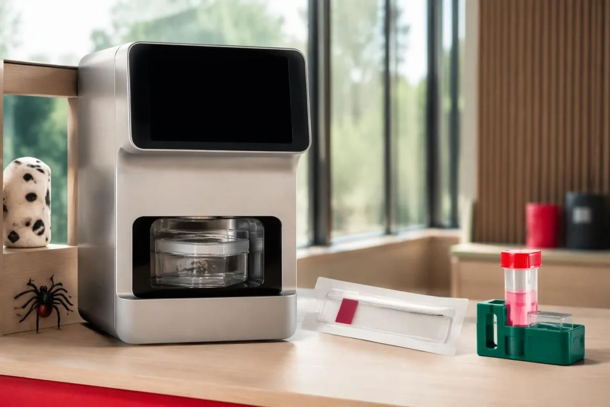
Unlike serology, PCR looks for the organism's genetic material rather than the host's immune response. Its usefulness depends on 3 practical variables: the specimen collected, illness severity and timing, and whether antibiotics have already reduced detectable organisms; there is no single sensitivity percentage applicable to every RMSF PCR order.
A clinician may arrange PCR or immunohistochemical testing of a rash skin biopsy through an appropriate laboratory, because affected tissue can provide evidence unavailable in circulating blood. Even when tissue testing is considered, collecting a specimen before the first doxycycline dose is desirable only if it does not delay urgent treatment.
Ask whether a panel actually includes R. rickettsii and whether it reports species-specific results or a broader rickettsial group. The PCR timing explanation for whooping cough demonstrates why molecular tests have specimen and treatment windows, but its respiratory sampling rules do not apply to RMSF.
Why do RMSF and ehrlichiosis tests behave differently?
RMSF and ehrlichiosis require different diagnostic reasoning even when the same tick-exposure evaluation includes both. Whole-blood PCR is generally more useful early in ehrlichiosis because Ehrlichia organisms infect white cells, whereas R. rickettsii primarily affects vessel-lining cells; 1 negative tick-borne panel is not a universal exclusion test.

Fever, headache, muscle aches, and abnormal liver enzymes can occur in more than 1 tick-borne illness. Exposure location, platelet count, white-cell count, rash, and illness timing guide test selection; clinicians should not assume that an order called a tick panel covers every relevant organism or uses the best specimen for each.
A ehrlichiosis PCR and titer guide explains why acute PCR and later antibodies may disagree in that illness. For a hypothetical day-4 presentation, an Ehrlichia PCR positive and RMSF IgG negative need interpretation as separate findings—not evidence that all other tick-transmitted illnesses were conclusively excluded.
Kantesti egy AI vérvizsgálat-értelmező platform that explains submitted laboratory reports, not a substitute for organism-specific diagnostic assessment. If a report contains 2 negative results from different methods, we emphasize the distinction between the method's target and the clinician's question rather than treating the word negative as a complete answer.
Which routine blood results can support suspicion of RMSF?
Low platelets, mildly elevated AST or ALT, and low sodium can support suspicion of RMSF, but none confirms the diagnosis and early values may be normal. A platelet count of 110 × 10⁹/L and sodium of 130 mmol/L are contextual clues—not organism-specific thresholds or prerequisites for starting treatment.
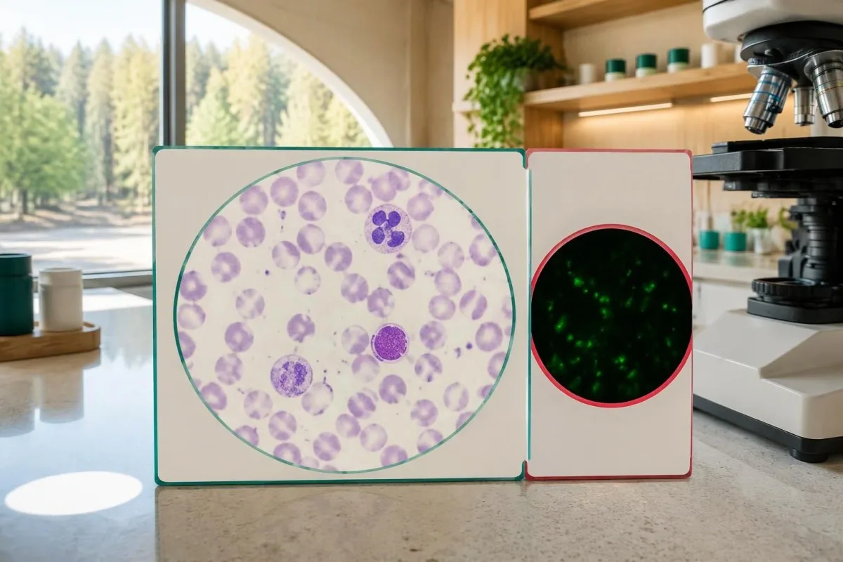
Typical adult platelet reference intervals are approximately 150–450 × 10⁹/L, and serum sodium is commonly 135–145 mmol/L, although local ranges vary. The reason clinicians consider falling platelets together with fever and low sodium is that the combination can suggest systemic illness; it does not distinguish RMSF from several infectious or noninfectious causes.
A hypothetical febrile traveler with platelets 95 × 10⁹/L, AST 82 U/L, and recent outdoor exposure deserves clinical assessment even if white cells are normal. Our Fehérvérsejt és CRP összehasonlításunk explains why normal inflammatory markers cannot independently clear a person with concerning symptoms.
Unexpectedly low platelets may also reflect sample-related clumping, which requires laboratory verification rather than automatic attribution to RMSF; EDTA okozta thrombocyta-aggregáció explains that artifact. If systemic illness is accompanied by abnormal clotting or organ dysfunction, the alvadási vizsgálatok útmutatónk helps explain additional tests, but a repeat specimen must not postpone urgent care.
Why should suspected RMSF treatment start before results?
Suspected RMSF requires prompt empiric doxycycline, because early tests can be negative and delayed treatment increases the risk of severe illness and death. Clinicians should not wait for IgG seroconversion, a PCR report, or a convalescent sample 2–10 weeks later before treating a compatible acute presentation.

Treatment timing is a safety issue, not simply a preference for faster symptom relief. Kirkland et al. reported higher mortality with treatment initiated after the first 5 days of illness in their retrospective study; its observational design limits precise causal estimates, but the finding supports the urgency emphasized in CDC guidance (Kirkland et al., 1995).
CDC recommends doxycycline 100 mg every 12 hours for adults and children weighing at least 45 kg; children under 45 kg receive 2.2 mg/kg every 12 hours, up to 100 mg per dose. These are clinician-prescribed regimens, not instructions for self-treatment; oral versus intravenous therapy depends on severity and the ability to retain medication.
Treatment generally lasts at least 5–7 days and continues until fever has resolved for at least 3 days with clinical improvement; complicated cases may need longer care. Pregnancy or suspected doxycycline allergy requires urgent individualized advice rather than automatic delay, and urgent infection assessment illustrates the same need to coordinate testing and treatment, although Listeria requires different antibiotics.
How should common RMSF test results be interpreted?
RMSF test results must be interpreted using symptoms, illness dates, specimen types, and changes between samples. An early negative result may be uninformative, a single positive may reflect old exposure, and a fourfold paired IgG rise can support recent spotted fever group rickettsial infection without proving the species.

Example 1 is hypothetical: a person develops fever and severe headache after outdoor exposure, tests IgG negative on day 3, and begins clinician-directed doxycycline that day. An acute titer below 1:64 followed by a convalescent titer of 1:256 can provide supportive laboratory evidence, but the laboratory must verify the change against its starting dilution and reporting limits.
Example 2 is also hypothetical: an otherwise well person has IgG 1:256 on 2 specimens obtained several weeks apart, without a compatible intervening illness. Stable positivity does not prove new RMSF, and escalating antibiotics to make the titer disappear is not justified; Kantesti explains this as an interpretation limitation, not a declaration that every stable titer is harmless.
Example 3 involves fever, IgG 1:128, and no properly timed acute comparator, leaving recent versus previous exposure uncertain. The next step depends on clinical assessment and possible additional testing; a Babesia elleni ellenanyagok és a fertőzés demonstrates a related distinction, but babesiosis needs its own organism-detection approach and is not treated with doxycycline alone.
When should symptoms override a negative RMSF result?
A negative RMSF result should not override a concerning acute illness with fever, severe headache, gastrointestinal symptoms, or rash after plausible tick exposure. Symptoms typically begin 3–12 days after an infected tick bite, and neither an unremembered bite nor the absence of an early rash reliably excludes RMSF.

The classic rash often develops 2–4 days after fever begins, so waiting for wrist, ankle, palm, or sole spots can delay care. RMSF can occur without a recognized rash, and its name is geographically misleading: clinicians assess exposure beyond the Rocky Mountain region, including travel and locally circulating ticks.
Confusion, breathing difficulty, fainting, rapidly worsening illness, or a spreading purple or pinpoint rash warrant emergency assessment—not a plan to repeat antibodies in 2 weeks. A fever and severe headache after possible tick exposure merit prompt clinical contact even if an online portal marks the first RMSF test negative.
After a tick bite without symptoms, routine antibody screening and preventive antibiotics are not recommended specifically to prevent RMSF; CDC guidance advises symptom monitoring for approximately 14 days. Tick exposure can also lead to conditions with entirely different testing logic, such as kullancsok után kialakuló alfa-gal-allergia, which involves allergy assessment rather than RMSF IgG interpretation.
Should RMSF antibodies be repeated after treatment?
Repeat RMSF antibodies are useful for diagnostic confirmation, not as a routine test of cure. A convalescent sample collected 2–10 weeks after the acute specimen can document an antibody rise, but persistent IgG after recovery does not by itself mean treatment failed or that more antibiotics are needed.

Fever often improves within 24–48 hours after early appropriate doxycycline treatment, although severe illness may respond more slowly. Failure to improve or clinical deterioration requires reassessment for complications, an alternative diagnosis, inadequate medication absorption, or another illness; it should not be managed by waiting for a titer to fall.
Early antibiotic treatment can sometimes delay or diminish antibody responses in rickettsial illness, so the absence of a fourfold rise is not an absolute retrospective exclusion in every treated case. Do not stop effective treatment to improve test yield; the clinician should interpret the 2 specimens alongside treatment dates, sampling quality, and the original clinical presentation.
Kantesti AI can help organize submitted results for trend review, but a trend graph cannot establish that RMSF is cured or still active. Our vérvizsgálati idővonal-útmutatónk explains why illness and medication dates matter; persistent symptoms after treatment need clinical reassessment, because lasting complications can occur without evidence of chronic ongoing RMSF infection.
What should you check before asking AI to explain an RMSF report?
A useful RMSF report explanation requires the assay method, exact titer, collection date, and symptom-onset date, not just the words positive or negative. Before seeking an AI explanation, identify whether 2 reports represent comparable acute and convalescent specimens; urgent symptoms still require clinical care before any upload or interpretation.

Record at least 5 details: symptom onset, sample collection, IgG versus IgM, IFA titer versus screening index, and the first antibiotic dose. Add recent travel and exposure information for the clinician; a numerical index of 2.1 from one assay cannot be mathematically converted into an IFA titer such as 1:128.
Kantesti egy Mesterséges intelligencia-alapú vérvizsgálat-elemző eszköz that helps explain submitted laboratory information without replacing clinical diagnosis. Our Mesterséges intelligencia technológiai útmutató describes the interpretation approach; for suspected RMSF, 1 negative early result must never become an AI-generated reassurance that postpones medical assessment.
An explanation should separate 3 categories: what the laboratory measured, what that finding can support, and what remains uncertain. Our orvosi jelentés forrásellenőrzéseink describe that safety boundary; useful questions include whether paired IgG is planned, whether PCR covered the suspected species, and whether treatment has already started.
Research publications and the limits of diagnostic evidence
RMSF diagnostic guidance and AI engineering publications answer different questions and should not be treated as interchangeable evidence. The 2016 CDC practical guide supports paired serology and immediate treatment when clinically indicated; 2 Figshare records listed below provide publication context, not proof that an AI system can diagnose or exclude RMSF.
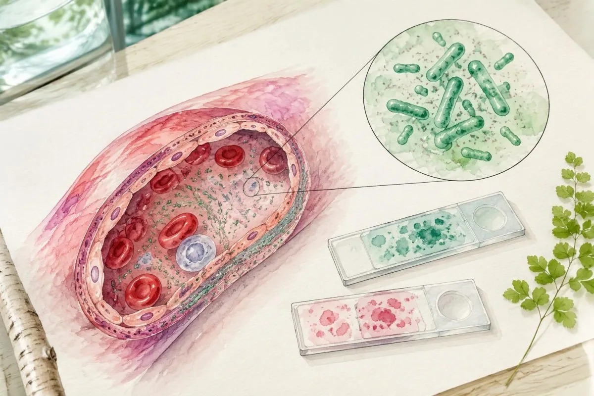
The 1995 therapeutic-delay study and the 2016 CDC recommendations directly address RMSF care, whereas a laboratory's assay validation addresses the particular test it performs. These are 3 different evidence levels—clinical outcomes, diagnostic guidance, and analytical performance—and none makes a first-week negative antibody result a reliable reason to delay treatment.
Kantesti's 2 requested Figshare publication records concern women's health and AI-assisted hantavirus triage, respectively; neither should be cited as an RMSF diagnostic-validation study. A DOI identifies a publication record rather than guaranteeing peer review, clinical accuracy, or applicability to another disease; our orvosi tartalmi szabványok explain the need to distinguish directly relevant sources from contextual material.
For this topic, the practical evidence boundary is straightforward: a 4-fold paired IgG rise supports recent spotted fever group infection, while immediate care depends on clinical suspicion rather than a future titer. Our klinikai validációs keretrendszert provides broader methodology context, but suspected RMSF still requires a clinician who can examine, prescribe, and arrange appropriate confirmatory testing.
Gyakran feltett kérdések
Lehet negatív korai stádiumban a Sziklás-hegységi foltos láz vizsgálata?
Igen, a Sziklás-hegységi foltos láz elleni antitestvizsgálat gyakran negatív a betegség első 7 napján, mert még nem alakultak ki kimutatható antitestek. A korai negatív IgG IFA-eredmény ezért nem zárja ki az RMSF-et. Ha a betegség és az expozíciós előzmények RMSF-re utalnak, a klinikusoknak haladéktalanul el kell kezdeniük a doxiciklin-kezelést, ahelyett hogy megvárnák az ismételt vizsgálat eredményét. Egy későbbi, lábadozás idején vett minta segíthet a diagnózis felállításában.
A pozitív RMSF-IgG aktív fertőzést jelent?
A pozitív RMSF IgG-eredmény önmagában nem bizonyít aktív fertőzést, még 1:256-os titer esetén sem. Az antitestek hónapokig vagy évekig fennmaradhatnak, és más, a foltos láz csoportjába tartozó rickettsiákkal is reakcióba léphetnek. A közelmúltbeli tünetek, az expozíció és az összehasonlítható páros minták közötti négyszeres titeremelkedés valószínűbbé teszi a közelmúltbeli fertőzést. Egy kompatibilis tünetekkel nem rendelkező személynél kapott izolált pozitív eredmény önmagában nem indokolja az antibiotikum-kezelést.
When should the second RMSF antibody sample be collected?
CDC RMSF diagnostic guidance describes acute and convalescent IgG samples collected 2–10 weeks apart. The 2016 CDC practical guide describes a convalescent specimen ideally obtained 2–4 weeks after the acute specimen. The treating clinician and laboratory should select the appropriate interval and use comparable assays. Treatment must not wait for the second sample.
What is a fourfold rise in an RMSF antibody titer?
A fourfold rise means the later IgG titer is four times the earlier titer, such as 1:64 increasing to 1:256. In a common twofold dilution series, this represents two dilution steps. With a compatible illness, that change supports recent spotted fever group rickettsial infection. Routine serology may still be unable to identify Rickettsia rickettsii specifically or establish illness severity.
Does a negative RMSF PCR rule out the disease?
A negative whole-blood RMSF PCR does not rule out the disease because Rickettsia rickettsii primarily affects vessel-lining cells rather than circulating white cells. An illness-day-3 specimen may contain insufficient bacterial DNA for detection, and antibiotics can further reduce test yield. Specialized testing of affected skin tissue may provide additional evidence when appropriate. Clinicians should not delay treatment while obtaining or awaiting PCR results.
Should children receive doxycycline before RMSF results return?
Children of all ages should receive clinician-prescribed doxycycline promptly when RMSF is suspected, without waiting for laboratory confirmation. CDC guidance recommends 2.2 mg/kg every 12 hours for children under 45 kg, with a maximum of 100 mg per dose. Short courses used for rickettsial disease have not been shown to cause the permanent tooth staining associated with older tetracycline concerns. The child's clinician determines route, duration, and whether hospital care is needed.
Why is my RMSF IgG still positive after treatment?
RMSF IgG can remain positive for months and sometimes up to 4 years after illness, so persistent positivity does not by itself indicate treatment failure. Antibody testing is not a routine test of cure. Additional antibiotics should not be prescribed solely to make a titer become negative. Persistent or worsening symptoms need clinical reassessment for complications or another diagnosis rather than interpretation from the antibody number alone.
Érthető magyarázatot kaphat a vérvizsgálat eredményekről
Töltse fel laborleletét, és az Kantesti AI másodpercek alatt közérthetően elmagyarázza az egyes biomarkereket. Világszerte több mint 2 millió ember használja.
Referenciák
- Klein, T., Mitchell, S., & Weber, H. (2026). Női egészségügyi útmutató: ovuláció, menopauza és hormonális tünetek. Kantesti AI orvosi kutatás. DOI
- Klein, T., Mitchell, S., & Weber, H. (2026). Többnyelvű, mesterséges intelligenciával támogatott klinikai döntéstámogatás a hantavírus korai triázsához: tervezés, mérnöki validálás és valós környezetben történő bevezetés 50 000 értelmezett vérvizsgálati lelet alapján. Kantesti AI orvosi kutatás. DOI
- Biggs HM et al. (2016). Diagnosis and Management of Tickborne Rickettsial Diseases: Rocky Mountain Spotted Fever and Other Spotted Fever Group Rickettsioses, Ehrlichioses, and Anaplasmosis — United States. MMWR Ajánlások és jelentések. DOI
- Kirkland KB et al. (1995). Therapeutic delay and mortality in cases of Rocky Mountain spotted fever. Clinical Infectious Diseases.
- Centers for Disease Control and Prevention (n.d.). Clinical and Laboratory Diagnosis for Rocky Mountain Spotted Fever. CDC klinikai útmutató.
- 2 hónapos kortólElemzett vizsgálatok
- 127+Országok
- 100+Nyelvek
Az orvosi csapatról


E-E-A-T bizalmi jelzések
Tapasztalat
Orvosok által vezetett klinikai áttekintés a laboratóriumi értelmezési munkafolyamatokról.
Szakértelem
Laboratóriumi medicina fókusz: hogyan viselkednek a biomarkerek klinikai környezetben.
Tekintélyesség
Orvosi lektor: Dr. Thomas Klein, közreműködött Dr. Sarah Mitchell és Prof. Dr. Hans Weber.
Megbízhatóság
Bizonyítékokon alapuló értelmezés, világos követési útvonalakkal a riadalom csökkentésére.





Orvosi nyilatkozat
A cikk kizárólag oktatási célokat szolgál, és nem minősül orvosi tanácsadásnak. A diagnózisra és a kezelési döntésekre vonatkozóan mindig konzultáljon szakképzett egészségügyi szolgáltatóval.
Közzétéve · Kapcsolat a szerkesztőséggel
Kiadó: Kantesti Ltd, bejegyezve Angliában és Walesben, cégjegyzékszám:. 17090423, London, Egyesült Királyság.
