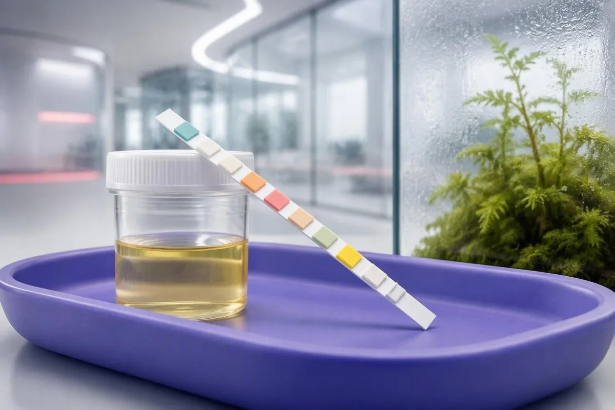
A positive strip is a screening clue, not a diagnosis. Learn what each pad measures, why colors can mislead, and which findings need timely clinical follow-up.


- Read time matters: Most dipstick pads must be compared at a manufacturer-specific time, commonly 30 to 120 seconds; a late reading can create false color changes.
- Nitrite positive: Nitrites strongly support nitrate-reducing bacteria, but a negative nitrite result does not rule out a urinary tract infection.
- Leukocyte esterase: A positive result detects white-cell enzyme activity and can reflect infection, vaginal contamination, stones, or inflammation.
- Blood pad: Trace blood can follow vigorous exercise, menstruation, or a urinary stone; microscopy confirms whether red cells are actually present.
- Protein 1+: A dipstick protein result of 1+ is roughly 30 mg/dL and should usually be repeated with a first-morning urine and urine albumin-creatinine ratio.
- Ketones plus glucose: Glucose with moderate or large ketones needs same-day assessment, especially with vomiting, abdominal pain, rapid breathing, or diabetes.
- Culture threshold: A culture is most useful before antibiotics in pregnancy, recurrent symptoms, fever, flank pain, male patients, and treatment failure.
- Cùram èiginneach: Visible red urine with clots, fever above 38.0°C, one-sided back pain, confusion, or inability to keep fluids down warrants urgent assessment.
What a urine dipstick can and cannot tell you
Urine dipstick results explained simply: each colored pad is a rapid chemical screen for substances such as blood, protein, glucose, ketones, nitrite, and white-cell enzymes; it cannot, by itself, diagnose a UTI, kidney disease, or diabetes. As of September 15, 2026, the safest way to read a positive urine dipstick meaning is to combine the pad pattern with symptoms, collection quality, microscopy, and sometimes culture.
A dipstick is best thought of as a triage tool. It gives a result in roughly 1 to 2 minutes, but it does not identify a bacterial species, quantify albumin accurately, or distinguish menstrual contamination from urinary bleeding. What UA means on reports is also worth checking, because UA means urinalysis here—not uric acid.
In clinic, I have seen a 28-year-old with burning urination, positive nitrite, and 2+ leukocyte esterase whose culture confirmed infection, and a second patient with the identical strip after a poorly collected sample whose culture was negative. Symptoms change the probability: dysuria and frequency make the same flags more meaningful than they are in someone who feels entirely well.
Tha Kantesti na Anailisiche deuchainn fala AI built to interpret laboratory patterns, and it does not treat a urine strip photo as a diagnosis. When urinary findings point to kidney, glucose, liver, or systemic issues, our clinical logic asks for the complementary blood results and the context that a strip cannot supply.
Why the strip is a screen
A negative dipstick also has limits. Early infection, very dilute urine, vitamin C use, and organisms that do not reduce nitrate can all produce reassuring-looking pads despite clinically relevant symptoms.
How to read a urine dipstick color chart safely
A urine dipstick color chart is valid only for the exact strip brand and the exact reading time printed in that strip’s instructions. Colors are semi-quantitative, so lighting, expired strips, and comparing a pad after its time window can turn a small color shift into a misleading flag.
Most pads change color as a chemical reaction occurs, then continue to drift as they dry. Leukocyte esterase is often read at 60 to 120 seconds, while glucose may be read at 30 seconds; those timings vary by manufacturer. Do not compare one manufacturer’s strip to an online chart from another brand.
A result marked trace, 1+, 2+, or 3+ is not a universal concentration. For protein, many strips roughly map trace to 10 to 20 mg/dL, 1+ to 30 mg/dL, 2+ to 100 mg/dL, and 3+ to 300 mg/dL, but urine concentration changes what those categories mean clinically.
The practical habit I recommend is simple: photograph the strip beside its original bottle chart at the correct time, then record symptoms, menstrual status, exercise, and medicines. That context is often more useful than trying to memorise colors from a generic chart; cloudy urine has several non-UTI causes.
Leukocyte esterase and nitrite: the UTI pattern
Positive leukocyte esterase plus positive nitrite is the dipstick pattern most suggestive of bacterial cystitis in a person with burning urination or frequency. Nitrite is fairly specific but not sensitive, while leukocyte esterase detects white-cell activity rather than bacteria themselves.
Nitrite becomes positive when certain bacteria convert dietary nitrate into nitrite while urine remains in the bladder, usually for about 4 hours. Enterobacterales such as Escherichia coli commonly do this; Enterococcus and Staphylococcus saprophyticus often do not. A negative nitrite result therefore cannot exclude a UTI.
Leukocyte esterase may be trace through 3+ and can rise with bladder inflammation, a stone, sexually transmitted urethritis, or vaginal white cells in the sample. A 2004 BMJ systematic review by Devillé et al. found that combining dipstick markers improves diagnostic value, yet no single pad has the accuracy to replace clinical judgment or culture in higher-risk cases.
I am particularly cautious with leukocyte esterase positive, nitrite negative results in people without symptoms. Repeat a properly collected specimen before assuming infection, and review our detailed guide to this mixed strip pattern.
What a positive blood pad actually detects
A positive blood pad detects heme activity, so it can reflect intact red cells, free hemoglobin, or myoglobin—not necessarily visible urinary bleeding. Microscopy is the next test that tells whether red blood cells are present and, if so, how many.
Tha hematuria microscopach air a mhìneachadh gu cumanta mar 3 no barrachd cheallan fola dearga gach raon àrd-chumhachd on a properly collected specimen. Dipstick trace blood after a long run, dehydration, intercourse, or menstruation is common enough that I usually repeat testing after 48 to 72 hours of rest when there are no warning symptoms.
A strip can turn positive after muscle injury because myoglobin reacts on the same pad. Dark brown urine, severe muscle pain, weakness, or a recent crushing workout deserves urgent assessment for creatine kinase, kidney function, and potassium rather than an assumption of UTI; runner’s hematuria is usually transient but still deserves a repeat test.
Blood with protein, red-cell casts, high blood pressure, or swelling has a different meaning from isolated trace blood. That combination can point toward kidney-filter inflammation and needs prompt clinician review; persistent findings are covered in our fuil anns an fhual.
Protein on a dipstick: when a plus sign needs follow-up
A urine protein dipstick primarily detects albumin, and a result of 1+ is often about 30 mg/dL in that single sample. Persistent protein needs confirmation with a urine albumin-creatinine ratio, or ACR, because concentration from dehydration can exaggerate a dipstick result.
The normal urine ACR is below 3 mg/mmol in UK units, equivalent to below 30 mg/g in US units. An ACR of 3 to 30 mg/mmol indicates moderately increased albuminuria, while above 30 mg/mmol is severely increased and generally merits timely assessment with eGFR and blood pressure.
Fever, intense exercise, dehydration, urinary infection, and standing for long periods can cause temporary proteinuria. I often ask patients to provide a clean first-morning sample after avoiding vigorous exercise for 24 hours; that approach avoids turning a marathon weekend into a kidney-disease label.
Protein with diabetes, hypertension, ankle swelling, or reduced eGFR is more concerning than protein alone. Pròtain anns an fhual agus an ACR collection guide explain why a quantitative ratio is the better follow-up test.
Glucose and ketones: when urine results need same-day action
Urine glucose usually appears when blood glucose exceeds the kidney’s reabsorption threshold, often around 180 mg/dL (10 mmol/L), although pregnancy and kidney tubular differences can shift that threshold. Moderate or large ketones with high glucose, illness, or vomiting require same-day clinical advice because diabetic ketoacidosis can develop quickly.
Ketones can be positive after fasting, prolonged vomiting, a low-carbohydrate diet, or poorly controlled diabetes. The strip chiefly detects acetoacetate, not beta-hydroxybutyrate, which is why a blood ketone result can be more informative during suspected ketoacidosis. Large urine ketones are not automatically DKA, but they should never be ignored in someone with diabetes who feels unwell.
Urgent evaluation is appropriate for glucose and ketones with abdominal pain, vomiting, deep or rapid breathing, marked thirst, drowsiness, or confusion. A capillary glucose of 11.1 mmol/L (200 mg/dL) or higher with classic symptoms supports diabetes, but diagnosis still requires formal testing under clinical guidance.
Kantesti AI ’s e àrd-ùrlar mìneachaidh deuchainn fala AI that places urine glucose clues alongside HbA1c, fasting glucose, kidney function, and medications. That matters because an SGLT2 medicine can cause urine glucose even when blood glucose is not dramatically raised; urine ketones and DKA risk needs a separate, symptom-led assessment.
Specific gravity and pH: hydration clues with real limits
Urine specific gravity is commonly 1.005 gu 1.030, while urine pH is usually 4.5 to 8.0. Both values describe the sample more than they diagnose disease: specific gravity estimates concentration, and pH changes with diet, medicines, timing, and some infections.
A specific gravity below 1.005 suggests very dilute urine, often after high fluid intake; above 1.030 commonly reflects dehydration, glucose, or other dissolved substances. Dipstick gravity is only an estimate, and a laboratory refractometer is more reliable when water-balance disorders are being evaluated.
Urine pH below 5.5 can occur after a high-protein diet or overnight fasting, while pH above 7.5 may follow a stale sample, plant-heavy diet, vomiting, or urease-producing organisms. Alkaline urine alone does not diagnose a UTI, and acidic urine does not prove a kidney stone type.
In Thomas Klein’s clinical experience, patients often overinterpret a pH of 5 or 8 when the more useful question is whether it fits symptoms and microscopy. For stone prevention or persistent abnormalities, urine pH interpretation agus specific gravity ranges give more targeted next steps.
Bilirubin and urobilinogen: liver clues hidden on the strip
Urine bilirubin should normally be negative because unconjugated bilirubin is not water-soluble; a confirmed positive result can indicate conjugated bilirubin reaching the urine. Urobilinogen is often reported around 0.2 to 1.0 EU/dL, but strip measurement is unstable and must be read with liver blood tests.
A positive bilirubin pad with dark tea-colored urine, pale stools, yellow eyes, itching, or right-upper abdominal discomfort needs prompt medical assessment. Obstruction of bile flow, hepatitis, and medication-related liver injury are examples, but a strip cannot tell which mechanism is present. A positive urine bilirubin result is a reason for blood bilirubin, ALT, ALP, and GGT testing.
Low or absent urobilinogen can occur when bile does not reach the bowel; elevated values can occur with liver dysfunction or increased red-cell breakdown. The evidence is honestly mixed on how much a lone urobilinogen flag adds in modern outpatient practice because sample light exposure and timing affect it substantially.
Tha Kantesti na Inneal mion-sgrùdaidh deuchainn fala le cumhachd AI that interprets bilirubin alongside liver enzymes and full blood count rather than giving a liver diagnosis from urine chemistry. The pattern of direct bilirubin, ALP, and GGT is usually much more decisive; see our guide to pàtrain bilirubin.
False positives, contamination, and medicines that change pads
False-positive urine dipstick results are common enough that a surprising result should be repeated before it becomes a diagnosis, unless red-flag symptoms are present. Menstrual fluid, vaginal discharge, disinfectant residue, delayed testing, vitamin C, and several medicines can alter pad chemistry.
Vitamin C can cause false-negative blood, glucose, nitrite, and bilirubin results on some strips because ascorbate interferes with oxidation reactions. Tell the clinician if you took high-dose supplements, especially above 500 mg daily, and do not stop prescribed treatment simply to repeat a test.
Phenazopyridine can turn urine orange and interfere with color interpretation; metronidazole may darken urine; levodopa and some antibiotics can affect individual pads. A sample left at room temperature for more than 2 uair can become more alkaline, allow bacterial growth, and give a less trustworthy nitrite or leukocyte result.
For a clean-catch sample, wash hands, separate the labia or retract the foreskin, begin urinating, then collect midstream without touching the container rim. Squamous epithelial cells on microscopy support contamination, a topic explained in our epithelial cells guide.
Why urine microscopy often changes the answer
Urine microscopy can verify red cells, white cells, bacteria, casts, crystals, and epithelial cells that a dipstick cannot identify. A dipstick blood result with zero red cells on microscopy suggests pigment such as hemoglobin or myoglobin, or a technical mismatch, rather than straightforward hematuria.
White blood cells above roughly 5 per high-power field can support urinary inflammation, but are not synonymous with bacterial infection. Bacteria seen with many squamous epithelial cells may represent collection contamination, whereas bacteria plus symptoms and pyuria are more persuasive.
Casts form in kidney tubules, so they add anatomical information. A few hyaline casts may follow dehydration or exercise, while red-cell casts are a nephrology-level warning because they suggest bleeding from the kidney filter; granular casts have a different set of causes.
Crystals are often incidental, especially calcium oxalate crystals in a concentrated specimen, but recurrent crystals with colicky flank pain deserve stone-focused assessment. The same dipstick pH can mean little without seeing the sediment, which is why I do not treat a strip as a complete urinalysis.
When a urine culture is needed rather than another strip
A urine culture is most useful when the bacterial identity and antibiotic susceptibility will change care: pregnancy, fever, flank pain, recurrent UTI, male urinary symptoms, catheter use, immune suppression, or symptoms that persist after treatment. Culture should ideally be collected before the first antibiotic dose when clinically safe.
For uncomplicated cystitis in a non-pregnant adult woman with classic symptoms, some clinicians treat without culture; local guidance differs. NICE’s lower UTI guidance recommends considering culture when symptoms are atypical, recurrent, or not improving, because resistance patterns and diagnostic alternatives matter.
A report of fàs measgaichte often means contamination, particularly if the sample has abundant epithelial cells. It is not automatically proof that several organisms are causing infection; a fresh clean-catch culture is frequently more useful than broader antibiotics. How urine culture counts and mixed growth work can help you prepare questions for the prescriber.
The 2019 IDSA guideline by Nicolle et al. advises against screening or treating asymptomatic bacteriuria in most non-pregnant adults, even when a dipstick or culture is positive. The major exceptions include pregnancy and some urologic procedures, where treatment can prevent meaningful harm.
Which dipstick findings need urgent medical assessment
Urgency depends on symptoms and the pattern, not the darkest pad alone. Fever of 38.0°C no nas àirde, shaking chills, flank pain, vomiting, pregnancy, confusion, visible blood with clots, or reduced urine output need urgent clinical assessment whether the strip is mildly or strongly positive.
Possible kidney infection is more likely when urinary symptoms occur with fever, one-sided back pain, nausea, or systemic illness. A normal-looking nitrite pad does not make this safe to watch at home. Children, older adults, pregnant people, and those with kidney disease may present less typically, which is where a lower threshold for review is sensible.
Glucose with ketones plus rapid breathing, severe thirst, abdominal pain, or drowsiness is an emergency pattern until proven otherwise. Visible red or cola-colored urine after trauma, with clots, or with inability to pass urine also needs prompt evaluation; soidhnichean rabhaidh fual dorcha are broader than dehydration alone.
By contrast, isolated trace protein after strenuous exercise or trace blood during menstruation can often be repeated under better conditions within 1 to 2 weeks. That is not dismissal—it is good diagnostic hygiene, provided there are no red flags or significant kidney-risk factors.
A practical repeat-test plan for uncertain results
Repeat a questionable urine dipstick with a first-morning, midstream clean-catch sample within 1 gu 2 sheachdain if you are well and have no red flags. First-morning urine is usually more concentrated, which improves detection of persistent albumin and reduces random day-to-day dilution noise.
Before repeating, avoid vigorous endurance exercise for 24 to 48 hours, document supplements and medicines, and test the sample promptly. Do not deliberately restrict water: aim for usual hydration, because extreme dilution can hide protein or blood while dehydration can exaggerate them.
Write down five details for the appointment: symptoms, temperature, menstrual timing, recent exercise, and whether antibiotics were started before the sample. This small record frequently explains a discordant strip better than repeating the test several times without context; urinalysis an aghaidh cultar clarifies what each can answer.
Thomas Klein, MD, would also ask whether protein, blood, or glucose has appeared previously. A trend matters: two trace blood results 6 months apart are approached differently from a new 3+ blood result with pain, and our lab trend guidance explains why personal baseline is useful.
Connecting urine flags with blood tests and clinical context
Urine dipstick abnormalities often need blood tests because urine provides a local clue while blood shows kidney filtration, glucose control, liver injury, and systemic illness. Protein or blood in urine commonly prompts creatinine, eGFR, electrolytes, blood pressure measurement, and sometimes a full blood count.
Kantesti AI interprets uploaded blood reports by comparing connected markers rather than treating an out-of-range flag as a diagnosis. For example, urine protein plus declining eGFR and elevated potassium deserves a very different response from transient 1+ protein after a febrile illness; ìrean galar dubhaig leantainneach provide the clinical framework.
Kantesti, na seirbheis eadar-mhìneachaidh deuchainn-lann AI, can help readers organise questions about their blood report, medication list, and repeat-test timing. Our medical review standards and limitations are described in the geàrr-shealladh dearbhaidh clionaigeach, and a clinician remains necessary for diagnosis, examination, prescribing, and urgent care decisions.
If a dipstick suggests bilirubin, glucose, persistent protein, or blood, bring the original report rather than only a typed result. A photo can show the strip brand, timing, and whether the result was trace versus clearly positive—details that change interpretation more often than patients expect.
Research, guideline sources, and questions to bring to your clinician
The strongest evidence on dipstick interpretation supports using strip findings as probability modifiers, then confirming high-stakes or unclear cases with microscopy, culture, quantitative urine testing, and clinical assessment. This is especially true for asymptomatic bacteriuria, where treating a positive result without symptoms can cause more harm than benefit.
Devillé et al. (2004) concluded in BMJ that dipstick testing has useful diagnostic value for UTI but works best in context, not as a stand-alone answer. Nicolle et al. (2019) similarly recommend against treatment of asymptomatic bacteriuria for most non-pregnant adults, a distinction that prevents unnecessary antibiotics and misleading follow-up cultures.
For broader laboratory literacy, Kantesti’s physicians and clinical reviewers publish methods and safety boundaries through the Bòrd Comhairleachaidh Meidigeach. In my experience, the most productive appointment question is not “Is this pad positive?” but “Does this pattern explain my symptoms, and what result would change the plan?”
Kantesti LTD. (2026). Deuchainn Fuil Bhìoras Nipah: Stiùireadh airson Lorg is Breithneachadh Tràth 2026. Zenodo. https://doi.org/10.5281/zenodo.18487418. Clàr ResearchGate agus Clàr Academia.edu. Kantesti LTD. (2026). Stiùireadh Seòrsa Fala B Negative, Deuchainn Fala LDH & Cunntas Reticulocyte. Figshare. https://doi.org/10.6084/m9.figshare.31333819. Clàr ResearchGate agus Clàr Academia.edu.
Ceistean Bitheanta
Dè tha smaoineachadh air dipstick urine deimhinneach a 'ciallachadh?
Tha dipstick urine deimhinneach a’ ciallachadh gun do lorg pada ceimiceach ann an susbaint os cionn ìre na stial; chan eil e a’ stèidheachadh breithneachadh leis fhèin. Mar eisimpleir, tha pròtain 1+ gu tric timcheall air 30 mg/dL, fhad ‘s as urrainn do phada fala freagairt do cheallan dearga, hemoglobin, no myoglobin. Tha comharran, càileachd cruinneachaidh, microscopy, agus uaireannan cultar a’ dearbhadh a bheil an toradh cudromach gu clionaigeach. Tha toraidhean nitrite dearbhach a bharrachd air esterase leukocyte nas coltaiche ri UTI nuair a tha losgadh aig àm urination no tricead an làthair.
An urrainn do dipstick urachdan a UTI a nochdadh ma tha na nitrites àicheil?
Tha, faodaidh galairean anns an t-slighe urinary a bhith an làthair le nitrites àicheil oir chan eil a h-uile bacteria urinary a' tionndadh nitrate gu nitrite agus mar as trice feumaidh fual a bhith anns a' mhalairt airson timcheall air 4 uairean airson nitrite a chruthachadh. Bidh Enterococcus agus Staphylococcus saprophyticus gu cumanta a' toirt a-mach galairean nitrate-àicheil. Faodaidh esterase geal-cheallach adhartach le comharraidhean fhathast a bhith a' fìreanachadh measadh meidigeach agus, ann an suidheachaidhean nas àirde de chunnart, cultar fual. Cha bu chòir fiabhras de 38.0°C no nas àirde, pian air taobh na h-achlais, torrachas, no cur a-mach a bhith air a riaghladh le mìneachadh strìp a-mhàin.
Dè cho fada 's a bu chòir dhomh feitheamh mus leugh mi dipstick urine?
Bu chòir dhut gach pada de stiall deuchainn fual a leughadh aig an dearbh àm a tha air a ràdh leis an neach-dèanamh stiall, mar as trice eadar 30 is 120 diog a rèir a’ phada. Faodaidh leughadh ro thràth a bhith a’ call ath-bhualadh, agus faodaidh leughadh ro fhadalach atharrachaidhean dath co-cheangailte ri tiormachadh agus brataichean meallta a chruthachadh. Is e a’ chairt dath air botal na stiall an aon chairt a tha an dùil airson an toraidh sin. Stòraich stiallan dùinte gu teann agus thoir sùil air a’ cheann-latha crìochnachaidh oir bidh taiseachd a’ milleadh reagents.
An urrainn do dìth uisge pròtain no fuil adhbhrachadh air dipstick urine?
Faodaidh dìth uisge a bhith a' dùnadh na h-ùire agus a' dèanamh air leughaidhean pròtain a bhith a' coimhead nas àirde, gu h-àraid nuair a nì stiall cunntas air lorg no 1+ pròtain. Faodaidh eacarsaich làidir còmhla ri dìth uisge cuideachd adhbhrachadh gum bi brataichean fala no pròtain sealach ann, agus mar sin tha ath-aithris glan-glacadh madainn às dèidh 24 gu 48 uairean às aonais eacarsaich dian gu tric reusanta. Bu chòir pròtain leantainneach a bhith air a sgrùdadh le co-mheas albumin-creatinine san ùir fo 3 mg/mmol a bhith mar an stairsneach àbhaisteach àbhaisteach. Feumaidh fuil le pian, fiabhras, clotan, no lùghdachadh ann an toradh ùire ath-sgrùdadh meidigeach seach ath-dheuchainn dachaigh.
Cuin a tha feum agam air cultar fual às dèidh dipstick dearbhach?
Tha cultar fuail gu sònraichte freagarrach mus cleachd thu antibiotics nuair a tha thu trom, nuair a tha fiabhras ort, pian air do thaobh, comharran a tha a' tilleadh, comharran urinary fireann, catheter, dìonachd lag, no comharran nach leasaich. Tha cultar a' comharrachadh a' meanbh-organism agus a chomas air gabhail ri antibiotics, fhad 's nach urrainn dipstick. Gu tric bidh fàs measgaichte a' nochdadh truailleadh sampall agus dh'fhaodadh gum bi feum air sampall ùr glan seach làimhseachadh nas fharsainge. Anns a' mhòr-chuid de dh'inbhich nach eil trom gun chomharraidhean, chan eil stiall dearbhach no cultar leis fhèin ag iarraidh antibiotics.
Am faod bhiotamain C buaidh a thoirt air toraidhean dipstick urine?
Faodaidh dòsan àrda de bhiotamain C builean dipstick fuaim a dhèanamh ceàrr-àicheil airson fuil, glucose, nitrite, agus bilirubin air cuid de shiostaman strì. Tha an buaidh seo nas dualtaiche le stuthan a tha os cionn mu 500 mg gach latha, ged a tha buaidh an urra ris an neach-dèanamh agus dùmhlachd fual. Innsibh don neach-clionaigeach agus don obair-lann mu stuthan mus dèan iad mìneachadh air toradh iongantach. Na cuir stad air cungaidhean-leigheis òrdaichte a-mhàin gus deuchainn sgrìonaidh atharrachadh gun chomhairle mheidigeach.
Faigh Mion-sgrùdadh Deuchainn Fala le Cumhachd AI an-diugh
Thig còmhla ri còrr is 2 mhillean neach air feadh an t-saoghail a tha a’ earbsa Kantesti airson mion-sgrùdadh sa bhad, ceart air deuchainnean obair-lann. Luchdaich suas na toraidhean deuchainn fala agad agus faigh mìneachadh coileanta air biomarcair 15,000+ ann an diogan.
📚 Foillseachaidhean Rannsachaidh le Iomraidhean
Klein, T., Mitchell, S., & Weber, H. (2026). Deuchainn Fuil Bhìoras Nipah: Stiùireadh airson Lorg is Breithneachadh Tràth 2026. Rannsachadh Leigheis AI Kantesti.
Klein, T., Mitchell, S., & Weber, H. (2026). Stuth fala B àicheil, stiùireadh deuchainn fala LDH & cunntas reticulocyte. Rannsachadh Leigheis AI Kantesti.
📖 Iomraidhean Meidigeach Taobh a-muigh
Institiud Nàiseanta airson Slàinte is Cùram Sàr-mhath (NICE) (2018). Galar slighe urinary (ìosal): òrdachadh antimicrobial. NICE Guideline NG109.
⚕️ Àicheadh Meidigeach
Tha an artaigil seo dìreach airson adhbharan foghlaim agus chan eil e a’ dèanamh comhairle mheidigeach. Cuir fios an-còmhnaidh gu solaraiche cùram slàinte teisteanasach airson co-dhùnaidhean breithneachaidh is leigheis.
Comharran earbsa E-E-A-T
Eòlas
Lèirmheas clionaigeach air a stiùireadh le lighiche air sruthan-obrach mìneachaidh obair-lann.
Eòlas
Fòcas air leigheas obair-lann air mar a bhios bith-chomharraidhean (biomarkers) a’ giùlan ann an co-theacsa clionaigeach.
Ùghdarrasachd
Air a sgrìobhadh le Dr. Thomas Klein le ath-sgrùdadh le Dr. Sarah Mitchell agus Prof. Dr. Hans Weber.
Earbsachd
Mìneachadh stèidhichte air fianais le slighean leanmhainn soilleir gus dragh a lughdachadh.