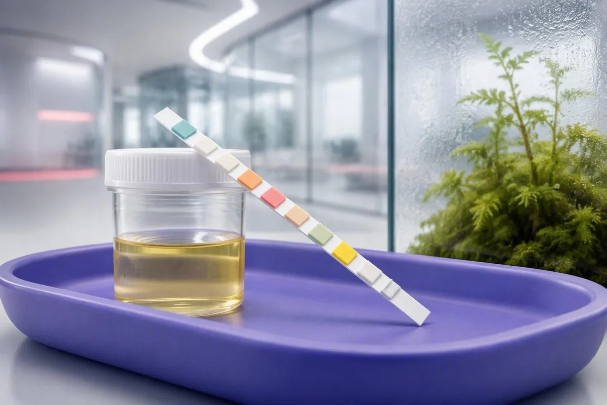
បន្ទះតេស្តដែលបង្ហាញលទ្ធផលវិជ្ជមានគ្រាន់តែជាការសង្ស័យเบื้องต้น មិនមែនជាការวินิจฉัยទេ។ រៀនពីអ្វីដែលបន្ទះនីមួយៗវាស់បាន ហេតុអ្វីពណ៌អាចបោកបញ្ឆោតបាន និងលទ្ធផលណាខ្លះដែលទាមទារការតាមដានពីគ្រូពេទ្យទាន់ពេល។.


- ពេលវេលដែលត្រូវអានមានសារៈសំខាន់: បន្ទះតេស្តភាគច្រើនត្រូវតែត្រូវបានប្រៀបធៀបក្នុងរយៈពេលជាក់លាក់របស់ក្រុមហ៊ុនផលិត ជាទូទៅពី 30 ទៅ 120 វិនាទី; ការអានយឺតអាចបង្កឱ្យមានការផ្លាស់ប្តូរពណ៌មិនពិត។.
- Nitrite វិជ្ជមាន: Nitrites គាំទ្រយ៉ាងខ្លាំងដល់បាក់តេរីដែលកាត់បន្ថយនីត្រាត ប៉ុន្តែលទ្ធផល nitrite អវិជ្ជមានមិនបានបដិសេធការបង្ករោគក្នុងផ្លូវទឹកនោមទេ។.
- Leukocyte esterase: លទ្ធផលវិជ្ជមានរកឃើញសកម្មភាពអង់ស៊ីមកោសិកាឈាមសហើយអាចឆ្ល phản ánh ការបង្ករោគ ការបំពុលទ្វារមាស គ្រួស ឬការរលាក។.
- បន្ទះតេស្តឈាម: ឈាមបន្តិចបន្តួចអាចកើតឡើងបន្ទាប់ពីការហាត់ប្រាណខ្លាំង ការមករដូវ ឬគ្រួសក្នុងផ្លូវទឹកនោម; មីក្រូទស្សន៍នឹងបញ្ជាក់ថាតើកោសិកាឈាមក្រហមមានវត្តមានពិតប្រាកដឬទេ។.
- Protein 1+: លទ្ធផលប្រូតេអ៊ីនលើបន្ទះសាកល្បង១+ គឺប្រហែល ៣០ mg/dL ហើយជាធម្មតាគួរតែធ្វើម្តងទៀតដោយប្រើទឹកនោមពេលព្រឹកដំបូង និងសមាមាត្រអាល់ប៊ុយមីន-ក្រេអាទីនីនក្នុងទឹកនោម។.
- គីតូនរួមជាមួយគ្លុយកូស៖ គ្លុយកូសជាមួយគីតូនកម្រិតមធ្យម ឬច្រើនតម្រូវឲ្យមានការវាយតម្លៃក្នុងថ្ងៃតែមួយ ជាពិសេសក្នុងករណីក្អួត ចុកពោះ ដកដង្ហើមលឿន ឬជំងឺទឹកនោមផ្អែម។.
- កម្រិតវប្បធម៌៖ ការដាំដុះវប្បធម៌មានប្រយោជន៍បំផុតមុនពេលប្រើថ្នាំអង់ទីប៊ីយូទិកក្នុងពេលមានផ្ទៃពោះ រោគសញ្ញាកើតឡើងវិញ គ្រុនក្តៅ ឈឺចង្កេះ អ្នកជំងឺជាបុរស និងការព្យាបាលមិនបានផល។.
- ការថែទាំបន្ទាន់ (Urgent care): ទឹកនោមក្រហមដែលមើលឃើញជាមួយកំណកឈាម គ្រុនក្តៅលើសពី ៣៨.០°C ឈឺចំហៀងមួយចំហៀង ភាន់ច្រឡំ ឬមិនអាចរក្សាជាតិទឹកបានតម្រូវឲ្យមានការវាយតម្លៃបន្ទាន់។.
អ្វីដែលបន្ទះតេស្តទឹកនោមអាចនិងមិនអាចប្រាប់អ្នកបាន
លទ្ធផលបន្ទះសាកល្បងទឹកនោមពន្យល់យ៉ាងសាមញ្ញ៖ បន្ទះពណ៌នីមួយៗគឺជាការធ្វើតេស្តគីមីរហ័សសម្រាប់សារធាតុដូចជាឈាម ប្រូតេអ៊ីន គ្លុយកូស គីតូន នីត្រាត និងអង់ស៊ីមកោសិកាស-ស; វាតែឯងមិនអាចធ្វើរោគវិនិច្ឆ័យការឆ្លងមេរោគផ្លូវនោម ជំងឺតម្រងនោម ឬជំងឺទឹកនោមផ្អែមបានឡើយ។ គិតត្រឹមថ្ងៃទី ១៥ ខែកញ្ញា ឆ្នាំ ២០២៦ វិធីសុវត្ថិភាពបំផុតក្នុងការអានលទ្ធផលបន្ទះសាកល្បងទឹកនោមវិជ្ជមានគឺការបញ្ចូលគ្នានៃលំនាំបន្ទះជាមួយរោគសញ្ញា គុណភាពនៃការប្រមូល មីក្រូទស្សន៍ និងពេលខ្លះវប្បធម៌។.
បន្ទះសាកល្បងត្រូវបានចាត់ទុកជាឧបករណ៍សម្រាប់ការជ្រើសរើសបឋម។ វាផ្តល់លទ្ធផលក្នុងរយៈពេលប្រហែល ១ ទៅ ២ នាទី ប៉ុន្តែវាមិនអាចកំណត់អត្តសញ្ញាណប្រភេទបាក់តេរី បរិមាណអាល់ប៊ុយមីនបានត្រឹមត្រូវ ឬបែងចែកការប្រឡាក់ដោយរដូវពីការហូរឈាមក្នុងទឹកនោមបានឡើយ។. អត្ថន័យ UA ក្នុងរបាយការណ៍ ក៏គួរពិនិត្យផងដែរ ព្រោះ UA មានន័យថា ទឹកនោម-មិនមែនអាស៊ីតអ៊ុយរិកទេ។.
ក្នុងគ្លីនិក ខ្ញុំបានឃើញអ្នកជំងឺអាយុ ២៨ ឆ្នាំម្នាក់មានការរលាកពេលនោម បង្ហាញលទ្ធផលនីត្រាតវិជ្ជមាន និងអង់ស៊ីមកោសិកាស-ស ២+ ដែលវប្បធម៌បញ្ជាក់ការឆ្លងមេរោគ និងអ្នកជំងឺទីពីរដែលមានបន្ទះដូចគ្នាបន្ទាប់ពីសំណាកដែលប្រមូលបានមិនបានល្អ ដែលវប្បធម៌ជាលទ្ធផលអវិជ្ជមាន។. រោគសញ្ញាផ្លាស់ប្តូរភាពទំនង៖ ការឈឺក្នុងពេលនោម និងការនោមញឹកញាប់ធ្វើឲ្យសញ្ញាដូចគ្នាមានសារសំខាន់ជាងវាទៅទៀតក្នុងអ្នកដែលមានអារម្មណ៍សុខស្រួលទាំងស្រុង។.
កាន់តេស្ទី គឺជា ឧបករណ៍វិភាគឈាម AI ត្រូវបានរចនាឡើងដើម្បីបកស្រាយលំនាំមន្ទីរពិសោធន៍ ហើយវាមិនចាត់ទុករូបថតបន្ទះទឹកនោមជារោគវិនិច្ឆ័យទេ។ នៅពេលលទ្ធផលទឹកនោមបង្ហាញពីបញ្ហាតម្រងនោម គ្លុយកូស ថ្លើម ឬប្រព័ន្ធទូទៅ ការគិតតាមគ្លីនិករបស់យើងតម្រូវឲ្យមានលទ្ធផលឈាមបន្ថែម និងបរិបទដែលបន្ទះមិនអាចផ្គត់ផ្គង់បាន។.
ហេតុអ្វីបន្ទះសាកល្បងជាការពិនិត្យបឋម
បន្ទះសាកល្បងអវិជ្ជមានក៏មានដែនកំណត់ដែរ។ ការឆ្លងមេរោគដំណាក់កាលដំបូង ទឹកនោមមានជាតិទឹកខ្លាំងពេក ការប្រើវីតាមីន C និងសារពាង្គកាយដែលមិនកាត់បន្ថយនីត្រាតសុទ្ធតែអាចបង្កើតបន្ទះដែលមើលទៅគួរឲ្យទុកចិត្តបានទោះបីជាមានរោគសញ្ញាទាក់ទងនឹងគ្លីនិកក៏ដោយ។.
របៀបអានតារាងពណ៌បន្ទះតេស្តទឹកនោមដោយសុវត្ថិភាព
A តារាងពណ៌បន្ទះសាកល្បងទឹកនោម មានសុពលភាពតែសម្រាប់ម៉ាកបន្ទះសាកល្បងជាក់លាក់ និងពេលវេលាអានជាក់លាក់ដែលបានបោះពុម្ពក្នុងការណែនាំរបស់បន្ទះនោះប៉ុណ្ណោះ។ ពណ៌គឺជាកម្រិតពាក់កណ្ដាលបរិមាណ ដូច្នេះពន្លឺ បន្ទះដែលផុតកំណត់ និងការប្រៀបធៀបបន្ទះបន្ទាប់ពីរយៈពេលរបស់វាអាចធ្វើឲ្យការផ្លាស់ប្តូរពណ៌តូចទៅជាទង់ដែលធ្វើឲ្យមានការភាន់ច្រឡំ។.
បន្ទះភាគច្រើនផ្លាស់ប្តូរពណ៌នៅពេលមានប្រតិកម្មគីមីកើតឡើង បន្ទាប់មកបន្តរសាត់នៅពេលវាស្ងួត។ អង់ស៊ីមកោសិកាស-ស ត្រូវបានអានជាញឹកញាប់នៅចន្លោះពី ៦០ ទៅ ១២០ វិនាទី ខណៈពេលគ្លុយកូសអាចត្រូវបានអាននៅ៣០វិនាទី។ ពេលវេលាទាំងនោះប្រែប្រួលតាមក្រុមហ៊ុនផលិត។ កុំប្រៀបធៀបបន្ទះរបស់ក្រុមហ៊ុនផលិតមួយជាមួយនឹងតារាងតាមអ៊ីនធឺណិតពីម៉ាកផ្សេង។.
លទ្ធផលដែលសម្គាល់ trace, 1+, 2+, ឬ 3+ មិនមែនជាកំហាប់សកលទេ។ សម្រាប់ប្រូតេអ៊ីន បន្ទះជាច្រើនប្រហាក់ប្រហែលនឹង trace ទៅ១០ ទៅ ២០ mg/dL, 1+ ទៅ ៣០ mg/dL, 2+ ទៅ ១០០ mg/dL, និង 3+ ទៅ ៣០០ mg/dL ប៉ុន្តែកំហាប់ទឹកនោមផ្លាស់ប្តូរនូវអ្វីដែលប្រភេទទាំងនោះមានន័យក្នុងគ្លីនិក។.
The practical habit I recommend is simple: photograph the strip beside its original bottle chart at the correct time, then record symptoms, menstrual status, exercise, and medicines. That context is often more useful than trying to memorise colors from a generic chart; cloudy urine has several non-UTI causes.
Leukocyte esterase និង nitrite: រោគសញ្ញានៃការបង្ករោគក្នុងផ្លូវទឹកនោម
Positive leukocyte esterase plus positive nitrite is the dipstick pattern most suggestive of bacterial cystitis in a person with burning urination or frequency. Nitrite is fairly specific but not sensitive, while leukocyte esterase detects white-cell activity rather than bacteria themselves.
Nitrite becomes positive when certain bacteria convert dietary nitrate into nitrite while urine remains in the bladder, usually for about 4 hours. Enterobacterales such as Escherichia coli commonly do this; Enterococcus and Staphylococcus saprophyticus often do not. A negative nitrite result therefore cannot exclude a UTI.
Leukocyte esterase may be trace through 3+ and can rise with bladder inflammation, a stone, sexually transmitted urethritis, or vaginal white cells in the sample. A 2004 BMJ systematic review by Devillé et al. found that combining dipstick markers improves diagnostic value, yet no single pad has the accuracy to replace clinical judgment or culture in higher-risk cases.
I am particularly cautious with leukocyte esterase positive, nitrite negative results in people without symptoms. Repeat a properly collected specimen before assuming infection, and review our detailed guide to this mixed strip pattern.
អ្វីដែលបន្ទះតេស្តឈាមវិជ្ជមានរកឃើញតាមពិត
A positive blood pad detects heme activity, so it can reflect intact red cells, free hemoglobin, or myoglobin—not necessarily visible urinary bleeding. Microscopy is the next test that tells whether red blood cells are present and, if so, how many.
Microscopic hematuria ជាទូទៅត្រូវបានកំណត់ថាជា កោសិកាឈាមក្រហម ៣ ឬច្រើនជាងនេះក្នុងមួយវាលកម្លាំងខ្ពស់ on a properly collected specimen. Dipstick trace blood after a long run, dehydration, intercourse, or menstruation is common enough that I usually repeat testing after 48 to 72 hours of rest when there are no warning symptoms.
A strip can turn positive after muscle injury because myoglobin reacts on the same pad. Dark brown urine, severe muscle pain, weakness, or a recent crushing workout deserves urgent assessment for creatine kinase, kidney function, and potassium rather than an assumption of UTI; runner’s hematuria is usually transient but still deserves a repeat test.
Blood with protein, red-cell casts, high blood pressure, or swelling has a different meaning from isolated trace blood. That combination can point toward kidney-filter inflammation and needs prompt clinician review; persistent findings are covered in our ឈាមក្នុងទឹកនោម.
ប្រូតេអ៊ីនក្នុងបន្ទះតេស្ត: នៅពេលណាសញ្ញាបូក(+) ទាមទារការតាមដាន
A urine protein dipstick primarily detects អាល់ប៊ុមីន, and a result of 1+ is often about 30 mg/dL in that single sample. Persistent protein needs confirmation with a urine albumin-creatinine ratio, or ACR, because concentration from dehydration can exaggerate a dipstick result.
The normal urine ACR is below 3 mg/mmol in UK units, equivalent to below 30 មីលីក្រាម/ក្រាម in US units. An ACR of 3 to 30 mg/mmol indicates moderately increased albuminuria, while above 30 mg/mmol is severely increased and generally merits timely assessment with eGFR and blood pressure.
Fever, intense exercise, dehydration, urinary infection, and standing for long periods can cause temporary proteinuria. I often ask patients to provide a clean first-morning sample after avoiding vigorous exercise for 24 hours; that approach avoids turning a marathon weekend into a kidney-disease label.
Protein with diabetes, hypertension, ankle swelling, or reduced eGFR is more concerning than protein alone. ប្រូតេអ៊ីនក្នុងទឹកនោម និង ACR collection guide explain why a quantitative ratio is the better follow-up test.
គ្លុយកូស និង ketone: នៅពេលណាលទ្ធផលទឹកនោមទាមទារសកម្មភាពក្នុងថ្ងៃតែមួយ
Urine glucose usually appears when blood glucose exceeds the kidney’s reabsorption threshold, often around 180 mg/dL (10 mmol/L), although pregnancy and kidney tubular differences can shift that threshold. Moderate or large ketones with high glucose, illness, or vomiting require same-day clinical advice because diabetic ketoacidosis can develop quickly.
Ketones can be positive after fasting, prolonged vomiting, a low-carbohydrate diet, or poorly controlled diabetes. The strip chiefly detects acetoacetate, not beta-hydroxybutyrate, which is why a blood ketone result can be more informative during suspected ketoacidosis. Large urine ketones are not automatically DKA, but they should never be ignored in someone with diabetes who feels unwell.
Urgent evaluation is appropriate for glucose and ketones with abdominal pain, vomiting, deep or rapid breathing, marked thirst, drowsiness, or confusion. A capillary glucose of 11.1 mmol/L (200 mg/dL) or higher with classic symptoms supports diabetes, but diagnosis still requires formal testing under clinical guidance.
Kantesti AI គឺជាអ្វីមួយដែល វេទិកាបកស្រាយការធ្វើតេស្តឈាម AI that places urine glucose clues alongside HbA1c, fasting glucose, kidney function, and medications. That matters because an SGLT2 medicine can cause urine glucose even when blood glucose is not dramatically raised; urine ketones and DKA risk needs a separate, symptom-led assessment.
ទំន重ដែលបានបញ្ជាក់និង pH: ការណែនាំអំពីជាតិទឹកក្នុងខ្លួនដែលមានដែនកំណត់ពិតប្រាកដ
Urine specific gravity is commonly 1.005 ទៅ 1.030, while urine pH is usually 4.5 to 8.0. Both values describe the sample more than they diagnose disease: specific gravity estimates concentration, and pH changes with diet, medicines, timing, and some infections.
A specific gravity below 1.005 suggests very dilute urine, often after high fluid intake; above 1.030 commonly reflects dehydration, glucose, or other dissolved substances. Dipstick gravity is only an estimate, and a laboratory refractometer is more reliable when water-balance disorders are being evaluated.
Urine pH below 5.5 can occur after a high-protein diet or overnight fasting, while pH above 7.5 may follow a stale sample, plant-heavy diet, vomiting, or urease-producing organisms. Alkaline urine alone does not diagnose a UTI, and acidic urine does not prove a kidney stone type.
In Thomas Klein’s clinical experience, patients often overinterpret a pH of 5 or 8 when the more useful question is whether it fits symptoms and microscopy. For stone prevention or persistent abnormalities, urine pH interpretation និង specific gravity ranges give more targeted next steps.
Bilirubin និង urobilinogen: ការណែនាំអំពីថ្លើមដែលលាក់ក្នុងបន្ទះតេស្ត
Urine bilirubin should normally be negative because unconjugated bilirubin is not water-soluble; a confirmed positive result can indicate conjugated bilirubin reaching the urine. Urobilinogen is often reported around 0.2 to 1.0 EU/dL, but strip measurement is unstable and must be read with liver blood tests.
A positive bilirubin pad with dark tea-colored urine, pale stools, yellow eyes, itching, or right-upper abdominal discomfort needs prompt medical assessment. Obstruction of bile flow, hepatitis, and medication-related liver injury are examples, but a strip cannot tell which mechanism is present. A positive urine bilirubin result is a reason for blood bilirubin, ALT, ALP, and GGT testing.
Low or absent urobilinogen can occur when bile does not reach the bowel; elevated values can occur with liver dysfunction or increased red-cell breakdown. The evidence is honestly mixed on how much a lone urobilinogen flag adds in modern outpatient practice because sample light exposure and timing affect it substantially.
កាន់តេស្ទី គឺជា ឧបករណ៍វិភាគតេស្តឈាមដោយ AI that interprets bilirubin alongside liver enzymes and full blood count rather than giving a liver diagnosis from urine chemistry. The pattern of direct bilirubin, ALP, and GGT is usually much more decisive; see our guide to លំនាំប៊ីលីរូប៊ីន.
លទ្ធផលវិជ្ជមានក្លែងក្លាយ ការបំពុល និងថ្នាំដែលផ្លាស់ប្តូរបន្ទះ
False-positive urine dipstick results are common enough that a surprising result should be repeated before it becomes a diagnosis, unless red-flag symptoms are present. Menstrual fluid, vaginal discharge, disinfectant residue, delayed testing, vitamin C, and several medicines can alter pad chemistry.
Vitamin C can cause false-negative blood, glucose, nitrite, and bilirubin results on some strips because ascorbate interferes with oxidation reactions. Tell the clinician if you took high-dose supplements, especially above 500 mg daily, and do not stop prescribed treatment simply to repeat a test.
Phenazopyridine can turn urine orange and interfere with color interpretation; metronidazole may darken urine; levodopa and some antibiotics can affect individual pads. A sample left at room temperature for more than 2 ម៉ោង can become more alkaline, allow bacterial growth, and give a less trustworthy nitrite or leukocyte result.
For a clean-catch sample, wash hands, separate the labia or retract the foreskin, begin urinating, then collect midstream without touching the container rim. Squamous epithelial cells on microscopy support contamination, a topic explained in our epithelial cells guide.
ហេតុអ្វីមីក្រូទស្សន៍ទឹកនោមច្រើនតែផ្លាស់ប្តូរចម្លើយ
Urine microscopy can verify red cells, white cells, bacteria, casts, crystals, and epithelial cells that a dipstick cannot identify. A dipstick blood result with zero red cells on microscopy suggests pigment such as hemoglobin or myoglobin, or a technical mismatch, rather than straightforward hematuria.
White blood cells above roughly 5 per high-power field can support urinary inflammation, but are not synonymous with bacterial infection. Bacteria seen with many squamous epithelial cells may represent collection contamination, whereas bacteria plus symptoms and pyuria are more persuasive.
Casts form in kidney tubules, so they add anatomical information. A few hyaline casts may follow dehydration or exercise, while red-cell casts are a nephrology-level warning because they suggest bleeding from the kidney filter; granular casts have a different set of causes.
Crystals are often incidental, especially calcium oxalate crystals in a concentrated specimen, but recurrent crystals with colicky flank pain deserve stone-focused assessment. The same dipstick pH can mean little without seeing the sediment, which is why I do not treat a strip as a complete urinalysis.
នៅពេលណាដែលការដាំទឹកនោមត្រូវបានទាមទារជំនួសឲ្យបន្ទះតេស្តមួយទៀត
A urine culture is most useful when the bacterial identity and antibiotic susceptibility will change care: pregnancy, fever, flank pain, recurrent UTI, male urinary symptoms, catheter use, immune suppression, or symptoms that persist after treatment. Culture should ideally be collected before the first antibiotic dose when clinically safe.
For uncomplicated cystitis in a non-pregnant adult woman with classic symptoms, some clinicians treat without culture; local guidance differs. NICE’s lower UTI guidance recommends considering culture when symptoms are atypical, recurrent, or not improving, because resistance patterns and diagnostic alternatives matter.
A report of ការដុះចម្រុះ often means contamination, particularly if the sample has abundant epithelial cells. It is not automatically proof that several organisms are causing infection; a fresh clean-catch culture is frequently more useful than broader antibiotics. How urine culture counts and mixed growth work can help you prepare questions for the prescriber.
The 2019 IDSA guideline by Nicolle et al. advises against screening or treating asymptomatic bacteriuria in most non-pregnant adults, even when a dipstick or culture is positive. The major exceptions include pregnancy and some urologic procedures, where treatment can prevent meaningful harm.
លទ្ធផលបន្ទះតេស្តណាខ្លះដែលទាមទារការវាយតម្លៃវេជ្ជសាស្រ្តបន្ទាន់
Urgency depends on symptoms and the pattern, not the darkest pad alone. Fever of 38.0°C ឬខ្ពស់ជាងនេះ, shaking chills, flank pain, vomiting, pregnancy, confusion, visible blood with clots, or reduced urine output need urgent clinical assessment whether the strip is mildly or strongly positive.
Possible kidney infection is more likely when urinary symptoms occur with fever, one-sided back pain, nausea, or systemic illness. A normal-looking nitrite pad does not make this safe to watch at home. Children, older adults, pregnant people, and those with kidney disease may present less typically, which is where a lower threshold for review is sensible.
Glucose with ketones plus rapid breathing, severe thirst, abdominal pain, or drowsiness is an emergency pattern until proven otherwise. Visible red or cola-colored urine after trauma, with clots, or with inability to pass urine also needs prompt evaluation; សញ្ញាព្រមានទឹកនោមខ្មៅ are broader than dehydration alone.
By contrast, isolated trace protein after strenuous exercise or trace blood during menstruation can often be repeated under better conditions within 1 to 2 weeks. That is not dismissal—it is good diagnostic hygiene, provided there are no red flags or significant kidney-risk factors.
ផែនការតេស្តឡើងវិញជាក់ស្តែងសម្រាប់លទ្ធផលមិនប្រាកដ
Repeat a questionable urine dipstick with a first-morning, midstream clean-catch sample within 1 ទៅ 2 សប្តាហ៍ if you are well and have no red flags. First-morning urine is usually more concentrated, which improves detection of persistent albumin and reduces random day-to-day dilution noise.
Before repeating, avoid vigorous endurance exercise for 24 to 48 hours, document supplements and medicines, and test the sample promptly. Do not deliberately restrict water: aim for usual hydration, because extreme dilution can hide protein or blood while dehydration can exaggerate them.
Write down five details for the appointment: symptoms, temperature, menstrual timing, recent exercise, and whether antibiotics were started before the sample. This small record frequently explains a discordant strip better than repeating the test several times without context; ការវិភាគទឹកនោម ទល់នឹងការធ្វើវប្បកម្ម clarifies what each can answer.
Thomas Klein, MD, would also ask whether protein, blood, or glucose has appeared previously. A trend matters: two trace blood results 6 months apart are approached differently from a new 3+ blood result with pain, and our lab trend guidance explains why personal baseline is useful.
ការភ្ជាប់សញ្ញាទឹកនោមជាមួយការតេស្តឈាមនិងបរិបទគ្លីនិក
Urine dipstick abnormalities often need blood tests because urine provides a local clue while blood shows kidney filtration, glucose control, liver injury, and systemic illness. Protein or blood in urine commonly prompts creatinine, eGFR, electrolytes, blood pressure measurement, and sometimes a full blood count.
Kantesti AI interprets uploaded blood reports by comparing connected markers rather than treating an out-of-range flag as a diagnosis. For example, urine protein plus declining eGFR and elevated potassium deserves a very different response from transient 1+ protein after a febrile illness; ដំណាក់កាលនៃជំងឺតម្រងនោមរ៉ាំរ៉ៃ provide the clinical framework.
Kantesti ដែលជាអ្នក សេវាកម្មបកស្រាយការធ្វើតេស្តរបស់ AI lab, can help readers organise questions about their blood report, medication list, and repeat-test timing. Our medical review standards and limitations are described in the ទិដ្ឋភាពទូទៅនៃការធ្វើឲ្យមានសុពលភាពផ្នែកគ្លីនិក, and a clinician remains necessary for diagnosis, examination, prescribing, and urgent care decisions.
If a dipstick suggests bilirubin, glucose, persistent protein, or blood, bring the original report rather than only a typed result. A photo can show the strip brand, timing, and whether the result was trace versus clearly positive—details that change interpretation more often than patients expect.
ស្រាវជ្រាវប្រភពការណែនាំនិងសំណួរដែលត្រូវសួរគ្រូពេទ្យរបស់អ្នក
The strongest evidence on dipstick interpretation supports using strip findings as probability modifiers, then confirming high-stakes or unclear cases with microscopy, culture, quantitative urine testing, and clinical assessment. This is especially true for asymptomatic bacteriuria, where treating a positive result without symptoms can cause more harm than benefit.
Devillé et al. (2004) concluded in BMJ that dipstick testing has useful diagnostic value for UTI but works best in context, not as a stand-alone answer. Nicolle et al. (2019) similarly recommend against treatment of asymptomatic bacteriuria for most non-pregnant adults, a distinction that prevents unnecessary antibiotics and misleading follow-up cultures.
For broader laboratory literacy, Kantesti’s physicians and clinical reviewers publish methods and safety boundaries through the ក្រុមប្រឹក្សាប្រឹក្សាវេជ្ជសាស្ត្រ. In my experience, the most productive appointment question is not “Is this pad positive?” but “Does this pattern explain my symptoms, and what result would change the plan?”
Kantesti LTD. (2026)។. ការធ្វើតេស្តឈាមសម្រាប់មេរោគ Nipah៖ ការណែនាំអំពីការរកឃើញ និងធ្វើរោគវិនិច្ឆ័យដំបូង ឆ្នាំ២០២៦. ។ Zenodo។. https://doi.org/10.5281/zenodo.18487418. កំណត់ត្រា ResearchGate និង កំណត់ត្រា Academia.edu. Kantesti LTD. (2026). មគ្គុទេសក៍ប្រភេទឈាម B អវិជ្ជមាន ការពិនិត្យឈាម LDH និងការរាប់កោសិកាឈាមក្រហមវ័យក្មេង (Reticulocyte Count). Figshare។. https://doi.org/10.6084/m9.figshare.31333819. កំណត់ត្រា ResearchGate និង កំណត់ត្រា Academia.edu.
សំណួរដែលសួរញឹកញាប់
លទ្ធផលទឹកនោមវិជ្ជមានលើបន្ទះតេស្តមានន័យដូចម្តេច?
លទ្ធផលវិជ្ជមានលើបន្ទះតេស្តទឹកនោមមានន័យថាបន្ទះគីមីមួយបានរកឃើញសារធាតុលើសពីកម្រិតកំណត់របស់បន្ទះនោះ។ វាមិនបង្កើតការធ្វើរោគវិនិច្ឆ័យដោយខ្លួនឯងទេ។ ឧទាហរណ៍ ប្រូតេអ៊ីន 1+ គឺប្រហែល 30 mg/dL ខណៈពេលដែលបន្ទះឈាមអាចប្រតិកម្មចំពោះកោសិកាឈាមក្រហម អេម៉ូក្លូប៊ីន ឬមីអូក្លូប៊ីន។ រោគសញ្ញា គុALITY នៃការប្រមូល 30 mg/dL និងពេលខ្លះវប្បធម៌កំណត់ថាតើការរកឃើញនោះមានសារសំខាន់ខាងវេជ្ជសាស្ត្រឬទេ។ លទ្ធផលវិជ្ជមានលើនីទ្រីតបូកនឹងលទ្ធផលអេស្ត្រាសកោសិកាសមានភាពប្រហែលនៃ UTI នៅពេលដែលមានការនោមទាស់ ឬការនោមញឹក។.
តើបន្ទះពិនិត្យទឹកនោមអាចបង្ហាញពីការឆ្លងមេរោគក្នុងផ្លូវទឹកនោមបានទេ ប្រសិនបើនីត្រាតអវិជ្ជមាន?
បាទ/ចាស ជំងឺ (UTI) អាចកើតឡើងដោយមានលទ្ធផលតេស្តនីត្រាតអវិជ្ជមាន ដោយសារតែបាក់តេរីក្នុងទឹកនោមមួយចំនួនមិនអាចបំប្លែងនីត្រាតទៅជានីត្រាតបានទេ ហើយជាធម្មតាចាំបាច់ត្រូវទុកទឹកនោមក្នុងប្លោកនោមប្រហែល 4 ម៉ោងដើម្បីបង្កើតនីត្រាត។ Enterococcus និង Staphylococcus saprophyticus ជាធម្មតាបង្កឱ្យមានការឆ្លងមេរោគដោយមានលទ្ធផលនីត្រាតអវិជ្ជមាន។ ការមានលទ្ធផលតេស្ត leukocyte esterase វិជ្ជមានរួមជាមួយនឹងរោគសញ្ញា នៅតែអាចជាការចាំបាច់សម្រាប់ការវាយតម្លៃផ្នែកវេជ្ជសាស្ត្រ ហើយក្នុងស្ថានភាពដែលមានហានិភ័យខ្ពស់ គឺការបញ្ជូនទឹកនោមទៅពិនិត្យ។ ក្តៅខ្លួន 38.0°C ឬខ្ពស់ជាងនេះ ឈឺចំហៀង គភ៌ ឬក្អួត មិនគួរត្រូវបានគ្រប់គ្រងដោយការបកស្រាយលទ្ធផលតេស្តតែមួយមុខនោះទេ។.
ខ្ញុំគួររង់ចាំរយៈពេលប៉ុន្មានទើបអាចអានលទ្ធផលតេស្តទឹកនោមបាន?
អ្នកគួរអានបន្ទះសាកល្បងទឹកនោមរាល់ពេល នៅពេលវេលាពិតប្រាកដដែលអ្នកផលិតបន្ទះបានបញ្ជាក់ ជាទូទៅចន្លោះពី 30 ទៅ 120 វិនាទី អាស្រ័យលើបន្ទះ។ ការអានលឿនពេកអាចខកខានប្រតិកម្ម ខណៈពេលដែលការអានយឺតពេកអាចបង្កើតការផ្លាស់ប្តូរពណ៌ដែលទាក់ទងនឹងការស្ងួត និងទង់ពណ៌ខុស។ តារាងពណ៌នៅលើដបបន្ទះ គឺជាតារាងតែមួយគត់ដែលមានបំណងសម្រាប់ផលិតផលនោះ។ រក្សាទុកបន្ទះឱ្យបិទជិត និងពិនិត្យមើលកាលបរិច្ឆេទផុតកំណត់ ដោយសារសំណើមធ្វើឱ្យសារធាតុគីមីចុះខ្សោយ។.
តើការខ្វះជាតិទឹកអាចបណ្ដាលឲ្យមានប្រូតេអ៊ីន ឬឈាមក្នុងការធ្វើតេស្តទឹកនោមបានទេ?
ការខ្វះជាតិទឹកអាចធ្វើឱ្យទឹកនោមប្រមូលផ្តុំ និងធ្វើឱ្យការអានប្រូតេអ៊ីនមើលទៅខ្ពស់ជាងមុន ជាពិសេសនៅពេលដែលបន្ទះតេស្តបង្ហាញថាមានប្រូតេអ៊ីនក្នុងកម្រិតតិចតួច ឬ 1+។ ការហាត់ប្រាណខ្លាំងក្លារួមជាមួយនឹងការខ្វះជាតិទឹក ក៏អាចបណ្តាលឱ្យមានការបង្ហាញឈាម ឬប្រូតេអ៊ីនជាបណ្តោះអាសន្នផងដែរ ដូច្នេះការយកសំណាកទឹកនោមពីការនោមដំបូងពេលព្រឹកដោយមិនមានការហាត់ប្រាណខ្លាំងក្លាឡើងវិញបន្ទាប់ពី ២៤ ទៅ ៤៨ ម៉ោង ជារឿយៗជាការសមស្រប។ ប្រូតេអ៊ីនដែលនៅតែមានគួរតែត្រូវបានពិនិត្យដោយប្រើសមាមាត្រអាល់ប៊ុយមីន-ក្រេអ៊ Attinine ក្នុងទឹកនោម ដែលមានកម្រិតក្រោម 3 mg/mmol គឺជាដែនកំណត់ធម្មតាជាធម្មតា។ ឈាមដែលមានការឈឺចាប់ ក្តៅខ្លួន កកឈាម ឬការបញ្ចេញទឹកនោមថយចុះ ត្រូវការការពិនិត្យពីគ្រូពេទ្យ ជាជាងការធ្វើតេស្តឡើងវិញនៅផ្ទះ។.
តើខ្ញុំត្រូវការការបណ្ដុះទឹកនោម នៅពេលណា បន្ទាប់ពីមានលទ្ធផលតេស្តវិជ្ជមានលើបន្ទះឆ្នូត?
ការពិនិត្យវប្បធម៌ទឹកនោមគឺសមស្របជាពិសេសមុនពេលប្រើថ្នាំអង់ទីប៊ីយូទិកនៅពេលអ្នកមានផ្ទៃពោះ មានគ្រុនក្តៅ ឈឺចង្កេះ រោគសញ្ញាកើតឡើងវិញ រោគសញ្ញាផ្លូវនោមរបស់បុរស មានបំពង់បង្ហួរនោម ការចុះខ្សោយប្រព័ន្ធភាពស៊ាំ ឬរោគសញ្ញាដែលមិនបានធូរស្រាល។ ការពិនិត្យវប្បធម៌កំណត់អត្តសញ្ញាណមេរោគ និងភាពងាយប្រតិកម្មរបស់វាចំពោះថ្នាំអង់ទីប៊ីយូទិក ខណៈពេលដែលការពិនិត្យដោយបន្ទះតេស្តមិនអាចធ្វើបាន។ ការលូតលាស់ចម្រុះច្រើនតែឆ្លុះបញ្ចាំងពីការចម្លងរោគសំណាក ហើយអាចទាមទារសំណាកស្អាតថ្មីជាជាងការព្យាបាលទូលំទូលាយ។ ក្នុងមនុស្សពេញវ័យភាគច្រើនដែលមិនមានផ្ទៃពោះដោយគ្មានរោគសញ្ញា ការតេស្តបន្ទះវិជ្ជមានតែមួយមុខឬការពិនិត្យវប្បធម៌តែមួយមុខមិនតម្រូវឱ្យប្រើថ្នាំអង់ទីប៊ីយូទិកឡើយ។.
តើវីតាមីន C អាចប៉ះពាល់ដល់លទ្ធផលនៃការធ្វើតេស្តទឹកនោមដោយបន្ទះឈើបានទេ?
វីតាមីន C កម្រិតខ្ពស់អាចបណ្តាលឱ្យលទ្ធផលតេស្តទឹកនោមដោយប្រើក្រដាសត្រាំមានលទ្ធផលអវិជ្ជមានមិនពិត (false-negative) ចំពោះឈាម គ្លុយកូស នីត្រាត និងប៊ីលីរុយប៊ីន នៅលើប្រព័ន្ធតេស្តមួយចំនួន។ បាតុភូតនេះទំនងជានឹងកើតឡើងជាមួយនឹងថ្នាំបំប៉នដែលមានកម្រិតលើសពីប្រហែល 500 mg ក្នុងមួយថ្ងៃ ទោះបីជាការជ្រៀតជ្រែកអាស្រ័យលើអ្នកផលិត និងកំហាប់ទឹកនោម។ សូមប្រាប់គ្រូពេទ្យ និងមន្ទីរពិសោធន៍អំពីថ្នាំបំប៉ន មុនពេលពួកគេបកស្រាយលទ្ធផលដែលធ្វើអោយភ្ញាក់ផ្អើល។ កុំបញ្ឈប់ការប្រើប្រាស់ថ្នាំតាមវេជ្ជបញ្ជា ដោយគ្រាន់តែដើម្បីផ្លាស់ប្តូរលទ្ធផលតេស្តស្គ្រីន ដោយគ្មានដំបូន្មានពីគ្រូពេទ្យ។.
ទទួលការវិភាគឈាមដោយ AI ដែលមានថាមពលថ្ងៃនេះ
ចូលរួមជាមួយអ្នកប្រើប្រាស់ជាង 2 លាននាក់នៅទូទាំងពិភពលោក ដែលទុកចិត្ត Kantesti សម្រាប់ការវិភាគលទ្ធផលតេស្តមន្ទីរពិសោធន៍ភ្លាមៗ និងត្រឹមត្រូវ។ ផ្ទុកឡើងលទ្ធផលពិនិត្យឈាមរបស់អ្នក ហើយទទួលការបកស្រាយយ៉ាងទូលំទូលាយនៃសញ្ញាសម្គាល់ (biomarkers) 15,000+ ក្នុងរយៈពេលប៉ុន្មានវិនាទី។.
📚 ឯកសារស្រាវជ្រាវដែលបានយោង
Klein, T., Mitchell, S., & Weber, H. (2026). ការធ្វើតេស្តឈាមសម្រាប់មេរោគ Nipah៖ ការណែនាំអំពីការរកឃើញ និងធ្វើរោគវិនិច្ឆ័យដំបូង ឆ្នាំ២០២៦.។ ការស្រាវជ្រាវវេជ្ជសាស្ត្រដោយ AI របស់ Kantesti។.
Klein, T., Mitchell, S., & Weber, H. (2026). មគ្គុទេសក៍ប្រភេទឈាម B អវិជ្ជមាន ការពិនិត្យឈាម LDH និងចំនួន Reticulocyte.។ ការស្រាវជ្រាវវេជ្ជសាស្ត្រដោយ AI របស់ Kantesti។.
📖 ឯកសារយោងវេជ្ជសាស្ត្រខាងក្រៅ
Devillé WLJM et al. (2004). ការធ្វើតេស្ត dipstick ទឹកនោម មានប្រយោជន៍ក្នុងការបដិសេធការឆ្លង៖ ការវិភាគមេតា (meta-analysis) នៃភាពត្រឹមត្រូវ.។ BMJ។.
Nicolle LE et al. (2019). គោលការណ៍ណែនាំការអនុវត្តគ្លីនិកសម្រាប់ការគ្រប់គ្រងបាក់តេរី Asymptomatic: ការធ្វើបច្ចុប្បន្នភាពឆ្នាំ 2019 ដោយសមាគមជំងឺឆ្លងអាមេរិក.។ Clinical Infectious Diseases។.
វិទ្យាស្ថានជាតិដើម្បីសុខភាព និងការថែទាំឧត្តមភាព (National Institute for Health and Care Excellence) (2018)។. ការឆ្លងមេរោគផ្លូវទឹកនោម (lower): ការចេញវេជ្ជបញ្ជាថ្នាំប្រឆាំងមេរោគ (antimicrobial prescribing). NICE Guideline NG109.
⚕️ ការបដិសេធផ្នែកវេជ្ជសាស្ត្រ
អត្ថបទនេះមានគោលបំណងសម្រាប់ការអប់រំប៉ុណ្ណោះ ហើយមិនមែនជាការផ្តល់ដំបូន្មានផ្នែកវេជ្ជសាស្ត្រទេ។ សូមពិគ្រោះជាមួយអ្នកផ្តល់សេវាសុខភាពដែលមានសមត្ថភាពជានិច្ច សម្រាប់ការសម្រេចចិត្តអំពីការធ្វើរោគវិនិច្ឆ័យ និងការព្យាបាល។.
សញ្ញាទុកចិត្ត E-E-A-T
បទពិសោធន៍
ការពិនិត្យព្យាបាលដោយវេជ្ជបណ្ឌិតលើដំណើរការការបកស្រាយលទ្ធផលមន្ទីរពិសោធន៍។.
ជំនាញ
ផ្តោតលើវិទ្យាសាស្ត្រមន្ទីរពិសោធន៍ថា សញ្ញាសម្គាល់ (biomarkers) មានឥរិយាបថយ៉ាងដូចម្តេចក្នុងបរិបទព្យាបាល។.
ភាពមានសិទ្ធិអំណាច
សរសេរដោយវេជ្ជបណ្ឌិត Thomas Klein ជាមួយការពិនិត្យឡើងវិញដោយវេជ្ជបណ្ឌិត Sarah Mitchell និងសាស្ត្រាចារ្យវេជ្ជបណ្ឌិត Hans Weber។.
ភាពគួរឱ្យទុកចិត្ត
ការបកស្រាយដោយផ្អែកលើភស្តុតាង ជាមួយផ្លូវបន្តតាមដានច្បាស់លាស់ ដើម្បីកាត់បន្ថយការភ័យខ្លាច។.