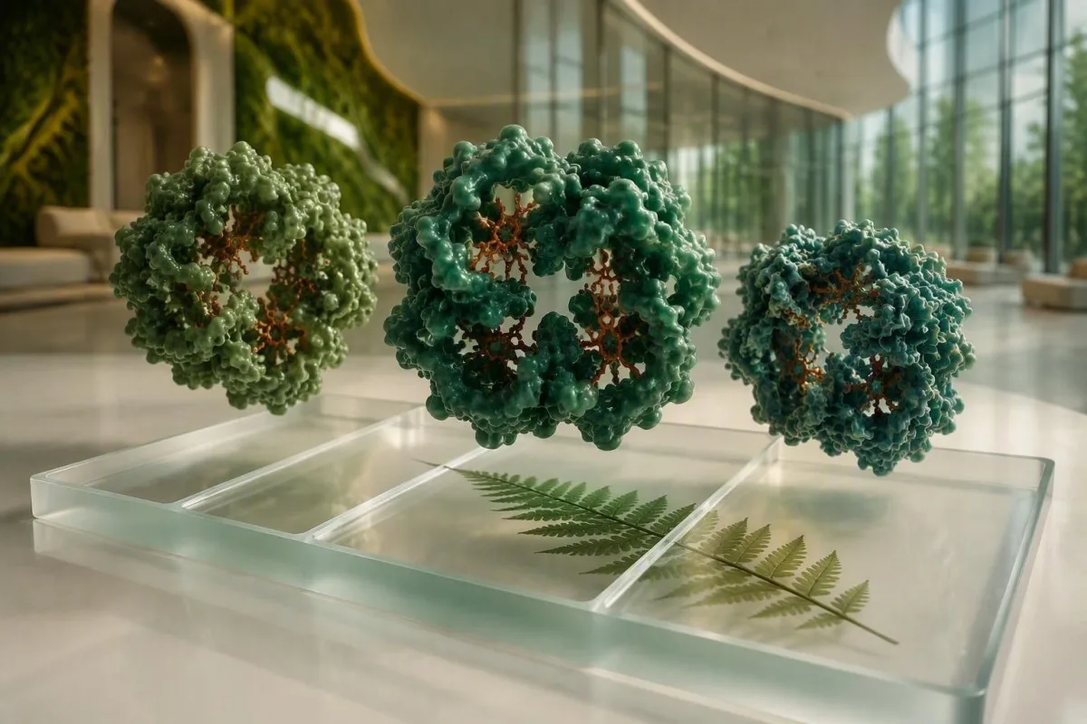
Hemoglobinaren frakzioek garraiatzaile-eredu bat identifikatu dezakete edo hemoglobina-nahaste bat iradoki dezakete, baina portzentajeak bakarrik gutxitan kontatzen du istorio guztia. Adinak, transfusio-historiak, globulu gorrien indizeek eta proba berretsaileek interpretazioa aldatzen dute.


- HbA Helduetan normalean –97 ingurukoa da hemoglobina-aldaera estrukturalik gabeko pertsonengan; nabarmen murriztutako edo falta den HbAk testuingurua behar du.
- HbS trait normalean –65 inguruko HbA eta –43 inguruko HbS erakusten du, bizkarrezurren egoera arruntetan bizitzeko sintomarik gabe garraiatzaile gehienentzat.
- HbC trait normalean –60 inguruko HbA eta –40 inguruko HbC ekoizten du; normalean isildu klinikoa da baina ugalketa-plangintzan garrantzitsua da.
- HbA gabe HbS altuarekin anemia faltazko gaixotasuna edo HbS/beta-zero talasemia iradoki dezake, nahiz eta duela gutxi egindako transfusio batek emailearen HbA gehi dezakeen.
- HbA2 3.5% baino handiagoa beta-thalassemia trait ondoan onartzen du heldu santu batean, baina burdina-gabezia eta metodo analitiko batzuek muga hori lausotu dezakete.
- Jaioberrien emaitzak ez dira helduen ehunekoen arabera interpretatzen, hemoglobina fetala normalean menderatzen baita lehen hilabeteetan.
- Duela gutxiko transfusioa hemoglobina elektroforesi desitxera ditzake 90–120 egun inguru, emaile-zelulen biziraupenaren eta galdera klinikoaren arabera.
- Azterketa genetikoa erabilgarriena da eredua anbiguoa denean, bikote batek haurdunaldia aurreikusten duenean, edo HbS eta beta-thalassemia ezin direnean konfiantzaz bereizi.
Nola Irakurri Hemoglobina Frakzioen Txosten bat Lehenik
Hemoglobina elektroforesiak hemoglobina motak bereizten ditu karga edo migrazioaren arabera, eta gero bakoitza hemoglobina osoaren ehuneko gisa jakinarazten du. Heldu batean, HbA –1 inguru, HbA2 %2,0–3,5 inguru eta HbF %1 inguru azpiko eredua da ohiko erreferentzia, laborategi bakoitzak bere tarteak balidatzen baditu ere.
Lehenengo galdera da ea HbA dagoen, ez ea zatiren bat markatu den. HbA ohiko helduen beta-katea den hemoglobina da; pertsona transfusiorik gabean ez egoteak diferentziala estutzen du, eta 5–30% HbA HbS/beta-plus thalassemiaren aldekoa izan daiteke, HbSS klasikoaren ordez.
Zati baten txostena ez da sintoma-puntuazio bat. HbS eta HbA dituen pertsona batek sarritan izango du anaiaren traza eta guztiz ondo sentituko da, berriz, HbS eta HbA-rik gabeko pertsona batek espezialistaren arreta beharko du, astebete horretan ondo sentitzen bada ere.
Kantesti bat da. AI odol-analisi analizatzailea horrek hemoglobina zatiak CBC, erretikulozito kopurua, adina eta adierazitako transfusio-historiaren ondoan kokatzen ditu, elektroforesiko banda koloreztatu bat diagnostiko gisa hartu beharrean. Ikuspegi parekatu hori bereziki erabilgarria daenean CBCko hemoglobina eta hematokritoa desberdinak dira ustekabeko arrazoi batengatik.
HbA Emaitzaren Esanahia: HbA, HbA2 eta HbF Normala
A normal adult HbA result usually means HbA accounts for roughly 95–98% of total hemoglobin, provided the person has not recently received donor cellular products. HbA2 and HbF are small fractions, but they often carry the diagnostic clue when HbA looks broadly normal.
HbA contains two alpha and two beta globin chains; HbA2 contains alpha and delta chains. HbA2 above 3.5% is a useful clue to beta-thalassemia trait, particularly when MCV is below 80 fL and the red-cell count is relatively preserved, but borderline values from 3.2–3.8% deserve careful review rather than a snap label.
HbF is normally below 1% in many adults, yet 1–5% can occur with hereditary persistence of fetal hemoglobin, pregnancy, marrow recovery, or treatment such as hydroxyurea. In people with sickle cell disease, higher HbF is generally associated with less polymerization of HbS, but it does not erase acute-care planning.
I often see a low MCV attributed to a hemoglobin variant when the actual explanation is iron deficiency. Compare the fraction pattern with low MCV causes and ferritin before assuming thalassemia; iron depletion can lower HbA2 enough to obscure beta-thalassemia trait.
Why an HbA result can be lower than expected
HbA may fall after donor-free marrow disorders, in beta-thalassemia syndromes, and whenever a variant occupies part of the total hemoglobin pool. A result of 88% HbA is not automatically dangerous; it becomes interpretable only alongside the identity and size of the other 12%, the MCV, and the laboratory method.
Zergatik Desadostu Daitezke Hemoglobina Elektroforesiaren Metodoak
Capillary electrophoresis and high-performance liquid chromatography identify many common variants accurately, but neither method alone proves every hemoglobin genotype. Co-elution, migration overlap, altered fractions after treatment, and rare variants can create a plausible but incomplete answer.
HPLC measures retention time, while capillary electrophoresis measures migration in an electrical field. HbS, HbD and HbG can share migration regions in some systems, and HbE may overlap with HbA2 on certain HPLC programs; laboratories resolve these by using a second method, family studies, or molecular analysis.
A surprisingly high HbA2 in a sample containing HbS can be analytical rather than biological because glycated or modified HbS fractions may co-elute near HbA2. That is why HbA2 alone should not diagnose beta-thalassemia when HbS is present.
As of September 15, 2026, Kantesti AI interprets a reported method, fractions and reference interval as separate pieces of evidence, not interchangeable facts. Our baliozkotze klinikoaren ikuspegia is built around flagging method-dependent uncertainty for clinician review.
What the laboratory may add after an unusual band
A laboratory may perform acid electrophoresis, capillary electrophoresis, repeat HPLC under another program, a peripheral cell sample review, or targeted HBB gene testing. These are complementary tests: a second separation clarifies identity, while DNA testing clarifies the inherited sequence change.
HbS Trait Test: HbA gehi HbS Eredua
Sickle cell trait usually produces both HbA and HbS, with HbA around 55–65% and HbS around 35–45% in an untreated adult. This pattern means one usual beta-globin gene and one HbS gene are commonly present; it is not the same as sickle cell disease.
Most people with HbS trait do not have chronic anemia, pain crises, or shortened red-cell survival. Rare complications can arise with severe dehydration, very low oxygen exposure, extreme exertion, or high altitude, and painless visible blood in urine deserves medical assessment rather than being casually blamed on exercise.
An HbS fraction below 35% can occur with co-inherited alpha-thalassemia, while HbS above 45% may occur when HbA is lower for another reason. A 28% HbS result therefore does not rule out trait; the red-cell count, MCV, and whether a transfusion occurred in the prior 3 months matter.
Kantesti AI-k an HbS trait test alongside anemia markers and clinical notes, then explains why carrier status is different from active sickle cell disease. Readers who need symptom and urgent-care context can review our sickle cell overview. DeBaun et al. (2020) describe sickle cell disease as a systemic condition with risks extending beyond episodic pain.
Trait and reproductive risk
When both biological parents carry HbS, each pregnancy has a 25% probability of HbSS, a 50% probability of HbS trait, and a 25% probability of neither inheritance pattern. Those percentages apply independently to each pregnancy, not across four pregnancies as a guaranteed sequence.
HbC Emaitzak: Trait, HbSC Gaixotasuna eta Auzo Arruntenak
HbC trait commonly shows HbA around 50–60% and HbC around 30–40%, whereas HbSC disease usually shows substantial HbS and HbC with no HbA unless transfusion has occurred. The distinction matters because HbSC disease can cause real complications despite sometimes milder anemia than HbSS.
HbC trait is usually asymptomatic and does not ordinarily cause clinically meaningful anemia. In contrast, HbSC disease can lead to vaso-occlusive pain, retinal complications, splenic problems and avascular necrosis; it should not be dismissed because the hemoglobin concentration may sit near 10–12 g/dL in some patients.
With HbSC disease, HbS and HbC are frequently each near 45–50%, but exact percentages vary with HbF, transfusion and assay technique. A small HbA fraction after a recent transfusion reflects donor cells more often than a sudden change in inherited genotype.
A raised reticulocyte count, bilirubin and LDH can support increased red-cell turnover, though none is specific to HbSC. Our guide to reticulocyte changes explains why the absolute count is usually more informative than the percentage when anemia is present.
Why eye symptoms need prompt attention in HbSC
HbSC disease carries a meaningful risk of proliferative retinopathy, sometimes in people whose general symptoms have been modest. New floaters, a curtain-like visual change, or reduced vision warrants same-day eye assessment, not a wait for the next routine hematology appointment.
HbA Ez eta HbS Goi Mailan: Ereduak Berretsi Beharrekoak
Absent HbA with HbS predominance can indicate HbSS disease or HbS/beta-zero thalassemia, but electrophoresis alone may not reliably separate them. HbS often exceeds 80% in HbSS, while HbF and HbA2 vary substantially by age, therapy and co-inherited traits.
HbSS commonly shows no HbA, HbS roughly 80–95%, HbF from under 1% to well above 15%, and HbA2 around 2–4% depending on the method. Hydroxyurea can increase HbF materially, so a 20% HbF value may reflect effective treatment rather than a different inherited diagnosis.
HbS/beta-zero thalassemia also lacks HbA, but persistent microcytosis and HbA2 above 3.5% may support it. Iron deficiency can produce microcytosis too, and HbA2 may be method-sensitive in the presence of HbS—this is exactly where HBB sequencing earns its keep.
A low hemoglobin level around 6–9 g/dL, fever of 38.5°C or higher, chest pain, new breathing difficulty, stroke-like symptoms, or prolonged painful erection needs urgent clinical care in someone known or suspected to have sickle cell disease. Hemolysis-related low haptoglobin can add evidence but never replaces clinical triage.
Why HbF is not a severity score by itself
Higher HbF tends to reduce HbS polymer formation, yet the relationship is not linear from one person to another. Distribution of HbF among red cells, kidney function, infection exposure, access to disease-modifying therapy and prior complications all influence lived risk.
Nola Aldatzen Dituzten Transfusioak eta Tratamenduak Frakzioak
A red-cell transfusion can introduce HbA and dilute HbS or HbC for about 90–120 days, making a post-transfusion electrophoresis unsuitable for definitive carrier diagnosis. The laboratory should know the transfusion date, product type and indication before reporting a pattern as inherited.
A person with HbSS who receives donor red cells may show 20–60% HbA, depending on timing and transfusion intensity. That HbA is not evidence that the person has HbS trait; it is a temporary mixture of donor and recipient cellular populations.
Hydroxyurea raises HbF in many people with sickle cell disease, sometimes from 5% to above 15%, and can increase MCV by 5–15 fL. Missing this medication history can lead to the false impression that the laboratory pattern has changed genetically.
Kantesti bat da. AI laborategiko proba interpretatzeko zerbitzuan that asks about transfusion, hydroxyurea and age before explaining hemoglobin electrophoresis results. The same caution applies to HbA1c transfusioaren ondoren, where donor red cells can make a diabetes marker misleading for weeks.
What to bring to the follow-up visit
Bring the original fraction report, CBC, ferritin or iron studies, newborn screening report if available, medication list, and dates of any transfusions in the prior 4 months. That small timeline often prevents unnecessary repeat testing and gives the hematologist a much cleaner starting point.
HbS edo HbC MCV Baxuarekin: Noiz Sartzen den Talasemia
HbS or HbC plus an MCV below 80 fL should prompt evaluation for iron deficiency and thalassemia, because structural hemoglobin traits alone usually do not cause marked microcytosis. A low MCV can change the expected percentage of HbS or HbC and complicate carrier classification.
Beta-thalassemia trait classically causes MCV around 60–75 fL, MCH below 27 pg, a normal or raised red-cell count, and HbA2 above 3.5% in many untreated adults. Iron deficiency often lowers ferritin below 15–30 ng/mL, but inflammation may keep ferritin deceptively normal or high.
Alpha-thalassemia trait can lower MCV without raising HbA2, and it may reduce the HbS percentage in sickle trait. This is why an HbS result of 30% with MCV 68 fL can represent HbAS plus alpha-thalassemia rather than a laboratory error.
Do not start iron solely because MCV is low. Confirm with ferritin, transferrin saturation and inflammatory context; our burdinaren ikasketen gida explains why transferrin saturation below 20% is supportive but not independently diagnostic of iron deficiency.
A useful CBC pattern
A red-cell count above 5.0 × 10^12/L with hemoglobin near 11–13 g/dL and MCV below 75 fL is more suggestive of thalassemia trait than uncomplicated iron deficiency, though overlap is common. The Mentzer index is a screening clue, not a diagnosis.
Jaioberrien eta Haurren Hemoglobina Emaitzek Adinean Oinarritutako Irakurketa Behar dute
Newborn screening normally shows predominantly HbF, often 60–90%, so an adult-style expectation of high HbA would be incorrect. An “FAS” screen commonly means fetal hemoglobin plus HbA plus HbS, which generally indicates sickle trait, but the newborn program must complete its own confirmatory pathway.
A baby with HbSS may have an “FS” pattern because HbF and HbS are present but HbA is absent. However, FS can also reflect HbS/beta-zero thalassemia or a rare variant, which is why confirmatory testing and early specialist referral are standard rather than optional.
HbF normally falls during the first 6–12 months as adult beta-chain production increases. Testing at 2 months versus 12 months can therefore look radically different even in the same child; the report date is clinically meaningful.
Parents should preserve the original screening letter, even when a child appears well. For wider pediatric result context, see safe blood-test interpretation for children; age-specific reference intervals are not a cosmetic detail.
When family testing helps
Testing biological parents can clarify whether a child’s pattern reflects HbS, HbC, beta-thalassemia or another variant, especially when the initial neonatal fraction is incomplete. It also supports informed discussion before future pregnancies without assigning blame to either parent.
Haurdunaldia, Bikotekideen Testak eta Aholkularitza Genetikoa
A hemoglobin variant result in pregnancy should trigger timely partner testing because inheritance risk depends on the pair of parental variants, not on the pregnant person’s fraction percentage. HbAS plus HbAS gives a 25% chance of HbSS in each pregnancy; HbAS plus HbAC gives a 25% chance of HbSC disease.
The American College of Obstetricians and Gynecologists recommends offering universal hemoglobinopathy testing to people planning pregnancy or at the initial prenatal visit, rather than selecting testing solely by self-identified ancestry (ACOG, 2022). This approach catches mixed or unknown family backgrounds that older ancestry-based algorithms can miss.
If both partners carry clinically relevant variants, genetic counselling can explain diagnostic options, timing and limitations without steering a family toward a particular decision. Cell-free screening is not a substitute for definitive fetal diagnostic testing when a specific hemoglobin genotype is the question.
Pregnancy itself can lower hemoglobin through plasma-volume expansion, but it does not turn HbS trait into sickle cell disease. Review iron stores separately: ferritin ranges in pregnancy change by trimester and should not be judged by a non-pregnant cut-off alone.
A quiet but frequent misconception
Carrier status is inherited, not acquired through pregnancy, diet, travel or a partner. A newly discovered trait feels new because testing was new; the underlying globin gene has been present since conception.
Zergatik eragin diezaioketen Hemoglobina Aldaerek HbA1c Emaitzei
HbS and HbC can affect HbA1c accuracy either by shortening red-cell survival or by interfering with a particular assay method. Even when an assay is analytically unaffected, increased red-cell turnover can make HbA1c read lower than the person’s true average glucose exposure.
The National Glycohemoglobin Standardization Program lists methods that show no clinically significant interference from HbS or HbC trait, but the laboratory must identify its exact assay. In HbSS or HbSC disease, HbA1c is often less reliable because red-cell lifespan is shortened, regardless of the analyzer.
Fructosamine reflects glycation of circulating proteins over roughly 2–3 weeks and can be considered when HbA1c is unreliable, although low albumin, nephrotic protein loss and thyroid disease affect it. A continuous glucose monitor or structured glucose testing may be more actionable for some patients.
An HbA1c of 5.4% cannot be assumed reassuring in a person with hemolysis and hemoglobin disease. Our fructosamine guide details where that alternative helps and where it misleads.
Ask one practical laboratory question
Ask, “Which HbA1c method was used, and is it validated for HbS or HbC trait?” That exact question distinguishes analytical assay interference from a biologically shortened red-cell lifespan, which require different solutions.
Sintomak, Anemiaren Markagailuak eta Noiz Eskatu Zaintza Premiazkoa
Hemoglobin electrophoresis identifies fractions; it does not determine whether current symptoms are an emergency. Chest pain, breathlessness, fever of 38.5°C or higher, focal weakness, confusion, severe headache, or rapidly worsening pain in a person with a sickling disorder warrants urgent assessment.
For HbSS, hemoglobin commonly runs around 6–9 g/dL, while HbSC may be nearer 9–12 g/dL, but an individual’s established baseline is more useful than a single population number. A fall of 2 g/dL from baseline with jaundice, dark urine or fatigue can signal accelerated hemolysis or another complication.
Reticulocytes, bilirubin, LDH and haptoglobin help identify red-cell turnover, yet aplastic crisis may present with a low reticulocyte count rather than a high one. Parvovirus B19 is a classic trigger, and that counterintuitive low-reticulocyte pattern is easy to miss.
Dr. Thomas Klein, our Chief Medical Officer, advises patients not to use a carrier report to explain away symptoms that need urgent evaluation. A low red-cell count has causes beyond hemoglobin variants, including bleeding, kidney disease, B12 deficiency and marrow suppression.
Why oxygen saturation can be deceptively reassuring
A normal finger oxygen reading does not rule out acute chest syndrome, pulmonary embolism or early infection. It measures peripheral saturation, not lung imaging findings, pain severity, evolving anemia or oxygen delivery to tissues.
Noiz Beharrezkoak diren Proba Genetikoak edo Hematologiaren Berrikuspena
Genetic testing is most useful when hemoglobin electrophoresis cannot distinguish HbSS from HbS/beta-zero thalassemia, when a rare variant is suspected, or when reproductive decisions depend on exact genotype. Sequencing or targeted variant analysis answers a different question from fraction separation: it identifies the DNA change.
A hematologist will usually combine the fraction report with CBC indices, ferritin, bilirubin, reticulocytes, a family history and transfusion dates. Traeger-Synodinos et al. (2015) recommend combined hematologic and molecular approaches when carrier identification remains uncertain, especially for prenatal-risk assessment.
Tell the specialist about relatives with anemia, gallstones at a young age, splenectomy, pain episodes, newborn screening results or unexplained infant deaths. Family history is imperfect—many carriers have no known family story—but a single detail can suggest which gene panel is most efficient.
Kantesti works with physician oversight through our Medikuntza Aholku Batzordea when an interpretation needs escalation rather than reassurance. In my experience, the most valuable genetic test is the one ordered after the phenotype and family question have been defined clearly.
What genetic testing can and cannot predict
Genetic testing can identify an HBB variant and many co-inherited globin-gene changes, but it cannot precisely forecast pain frequency, organ complications or treatment response for one individual. Genotype informs risk; ongoing clinical monitoring defines care.
Lan-fluxu Seguruagoa Emaitzak Kargatzeko eta Berraztertzeko
The safest way to review hemoglobin electrophoresis results is to upload the complete report with the CBC and state whether transfusion, pregnancy, hydroxyurea or newborn screening applies. A fraction percentage without these facts can be technically correct and clinically misleading.
Kantesti bat da. AI biomarkatzaileen interpretazio-plataforma that reads HbA, HbS, HbC, HbA2 and HbF together with red-cell indices, reference intervals and longitudinal changes. Our system can organize questions for a clinician, but it does not replace a hematologist’s diagnostic responsibility or urgent-care evaluation.
Check the report for specimen date, methodology, every listed fraction, the laboratory’s reference range and comments such as “variant window” or “recommend confirmation.” Photographing only the bold abnormal line removes information that may explain the entire pattern.
For a transparent explanation of how our model handles laboratory context, visit the AI teknologiaren gida. Dr. Thomas Klein’s practical rule is simple: preserve the original PDF and compare like with like—capillary electrophoresis and HPLC percentages should not be treated as identical assays.
Questions worth asking at follow-up
Ask whether the result is definitive or presumptive, whether recent donor cells could be present, whether iron studies alter interpretation, whether partner testing is indicated, and whether genetic confirmation would change management. Those five questions are usually more productive than asking whether a fraction is merely “high.”
Ikerketa-argitalpenak eta Odol-konteoko Testuinguru Erlazionatua
Red-cell indices such as MCV, MCHC and RDW do not identify HbS or HbC, but they often expose iron deficiency, thalassemia or hemolysis that changes how a fraction pattern should be read. A hemoglobin electrophoresis report is strongest when interpreted as part of a complete hematology picture.
A rising RDW after iron treatment can reflect newly produced, better-hemoglobinized cells mixing with older microcytic cells; it is not automatically treatment failure. Our RDW and red-cell indices review explains why an RDW value above 14.5% is a clue to mixed cell populations, not a diagnosis.
Kantesti supports readers in 127+ countries and more than 75 languages, but local laboratory methods and referral pathways still govern clinical decisions. Learn how our organization approaches privacy and medical information at Guri buruz; personal reports should be shared with a qualified clinician when disease or reproductive risk is possible.
For related publication context, Klein, T. (2026). RDW odol-analisia: RDW-CV, MCV eta MCHCren gida osoa. Zenodo. https://doi.org/10.5281/zenodo.18202598. Klein, T. (2026). BUN/Kreatinina ratioaren azalpena: Giltzurruneko funtzioaren probaren gida. Zenodo. https://doi.org/10.5281/zenodo.18207872.
Bottom line for an unexpected band
A hemoglobin band is a starting point, not a verdict. Confirm the analytical identity, check CBC and iron data, account for age and transfusion, then use genetic testing when the answer will affect treatment, family planning or a child’s follow-up.
Maiz egiten diren galderak
Zer esan nahi du HbA normal batek hemoglobinaren elektroforesian?
Helburu normal batek heldu heldugabe batean normalean esan nahi du HbA hemoglobina osoaren –98% inguru dela, HbA2 %2,0–3,5% eta HbF %1% azpitik. Eredu honek beta-katearen aldaera estruktural handi bat gutxitan gertatzen dela adierazten du, baina ez du alfa-talasemiaren ezaugarria edo oso arraroak diren aldaerak baztertzen. Duela gutxi egindako odol-transfusio batek aldi baterako handitu dezake neurtutako HbA, gutxi gorabehera 90–120 egunez. Laborategiko erreferentzia-tarte eta metodo propioek beti lehentasuna izan beharko lukete gida orokor baten gainetik.
Zein ehuneko HbS esan nahi du anemia faltziformea izatea?
Anemia falciformaren ezaugarriak HbS izan ohi du heldu tratatu gabe batean –40 inguru eta HbA –65 inguru. HbA substantzialaren presentzia da eredu nagusia, normalean beta-globina gene tipiko bat HbA ekoizten ari dela adierazten duelako. HbS baino gutxiagora jaitsi daiteke alfa-talasemia batera gertatzen bada, eta azkenaldiko transfusio batek are gehiago jaitsi dezake. Mediku batek eredua berretsi beharko luke HbA falta bada, MCV txikia bada, edo emaitzak ugalketa-plangintzan lagunduko badu.
Zer esan nahi dute HbA 0% eta HbS 90%?
0%-ko HbA, ia 90%-ko HbS-rekin, anemia faltziformoa dela adierazten du, gehienetan HbSS edo HbS/beta-zero talasemia pertsona batean, duela gutxi emailearen globulu gorririk jaso ez badu. HbF 1% baino gutxiagoko eta 15% baino gehiagoko tartean egon daiteke, bereziki haur txikietan edo hidroxikarbamida hartzen duten pertsonengan. 3,5% baino gehiagoko HbA2 eta mikrozitosi nabarmena HbS/beta-zero talasemiaren aldekoak izan daitezke, baina proba molekularrak beharrezkoak izan daitezke ziurtasunerako. Eredu honek hematologiaren jarraipena behar du, sintomak unean uneko arinak izan arren.
Zer esan nahi du HbC 35%?
HbC – inguruan eta HbA – inguruan izatea HbC ezaugarriaren patroi ohikoa da. HbC ezaugarriak ez du normalean anemia kliniko garrantzitsurik edo falziformetze sintomarik sortzen, nahiz eta garrantzi erreproduktiboa izan dezakeen bikote biologikoak HbS, HbC edo beta-talasemia baldin baditu. HbS eta HbC biak – inguruan baldin badaude eta HbA ez baldin badago, HbSC gaixotasuna litekeena da HbC ezaugarria baino. Transfusio berriek HbA sar dezakete eta bereizketa hori zaildu.
Burdinaren gabeziak hemoglobinaren elektroforesiaren emaitzak alda ditzake?
Burdinaren gabeziak hemoglobinaren elektroforesi-interpretazioa alda dezake HbA2 jaitsiz, batzuetan beta-talasemiaren arrastoa ezkutatuz. 15–30 ng/mL azpiko ferritinak heldu askotan burdin-dendak agortuta daudela adierazten du, baina hanturak ferritinaren maila igotzea eragin dezake burdinak gorputz-zelulen produkzioa mugatu arren. Burdinaren gabeziak ez du HbS edo HbC sortzen, baina MCV 80 fL azpitik egotea eragin dezake eta ehuneko-eredua interpretatzea zaildu. Arduradunek askotan azterketa batzuk errepikatzen dituzte burdina berriz beteta dagoenean, beta-talasemia kezka izaten jarraitzen badu.
Transfusioren ondoren zenbat denbora igaro behar da hemoglobinaren elektroforesi errepikatu aurretik?
Hemoglobinaren proba heredatu iraunkorrak egiteko, mediku askok 90–120 egun inguru itxaron dute globulu gorrien transfusioaren ondoren, dohaintzan hartutako globulu gorriek, HbA izan dezaketenez, tarte horretan neurgarriak izan daitezkeelako. Denbora egokia transfusioaren bolumenaren, etengabeko transfusio-terapiaren eta premiaren araberakoa da, beraz, tratamendua egiten duen hematologoak proba genetikoak lehenago erabil ditzake, erantzuna itxaron ezin bada. Transfusio osteko emaitza batek nahasmendu gaizto bat kudeatzen lagun dezake, baina ez da nahastu behar garbitasun-proba gisa. Bete beti transfusioaren zehaztutako data laborategiko eskaeran.
Lortu gaur AI bidezko odol-analisien analisia
Batu mundu osoko 2 milioi erabiltzaile baino gehiagok Kantesti-n konfiantza dutenak, laborategiko analisiak berehala eta zehaztasunez aztertzeko. Igo zure odol-analisien emaitzak eta jaso 15,000+ biomarkatzaileen interpretazio integrala segundo gutxitan.
📚 Erreferentziatutako ikerketa-argitalpenak
Klein, T., Mitchell, S., & Weber, H. (2026). RDW odol-analisia: RDW-CV, MCV eta MCHC-rako gida osoa. Kantesti AI Medical Research.
Klein, T., Mitchell, S., & Weber, H. (2026). BUN/Kreatinina ratioaren azalpena: Giltzurruneko funtzioaren probaren gida. Kantesti AI Medical Research.
📖 Kanpoko erreferentzia medikoak
American College of Obstetricians and Gynecologists (2022). Hemoglobinopathies in Pregnancy. ACOG Practice Advisory.
⚕️ Ohar medikoa
Artikulu hau hezkuntza-helburuetarako da soilik eta ez du mediku-aholkurik ematen. Diagnostiko- eta tratamendu-erabakietarako, beti kontsultatu osasun-profesional kualifikatu bati.
E-E-A-T Konfiantza-seinaleak
Esperientzia
Medikuek gidatutako berrikuspen klinikoa laborategiko interpretazioaren lan-fluxuei buruz.
Espezializazioa
Laborategiko medikuntzaren ikuspegia biomarkatzaileek testuinguru klinikoan nola jokatzen duten aztertzean.
Autoritatea
Dr. Thomas Klein-ek idatzia, eta Dr. Sarah Mitchell eta Prof. Dr. Hans Weber-ek berrikusia.
Fidagarritasuna
Ebidentzian oinarritutako interpretazioa, alarma murrizteko jarraipen-bide argiekin.