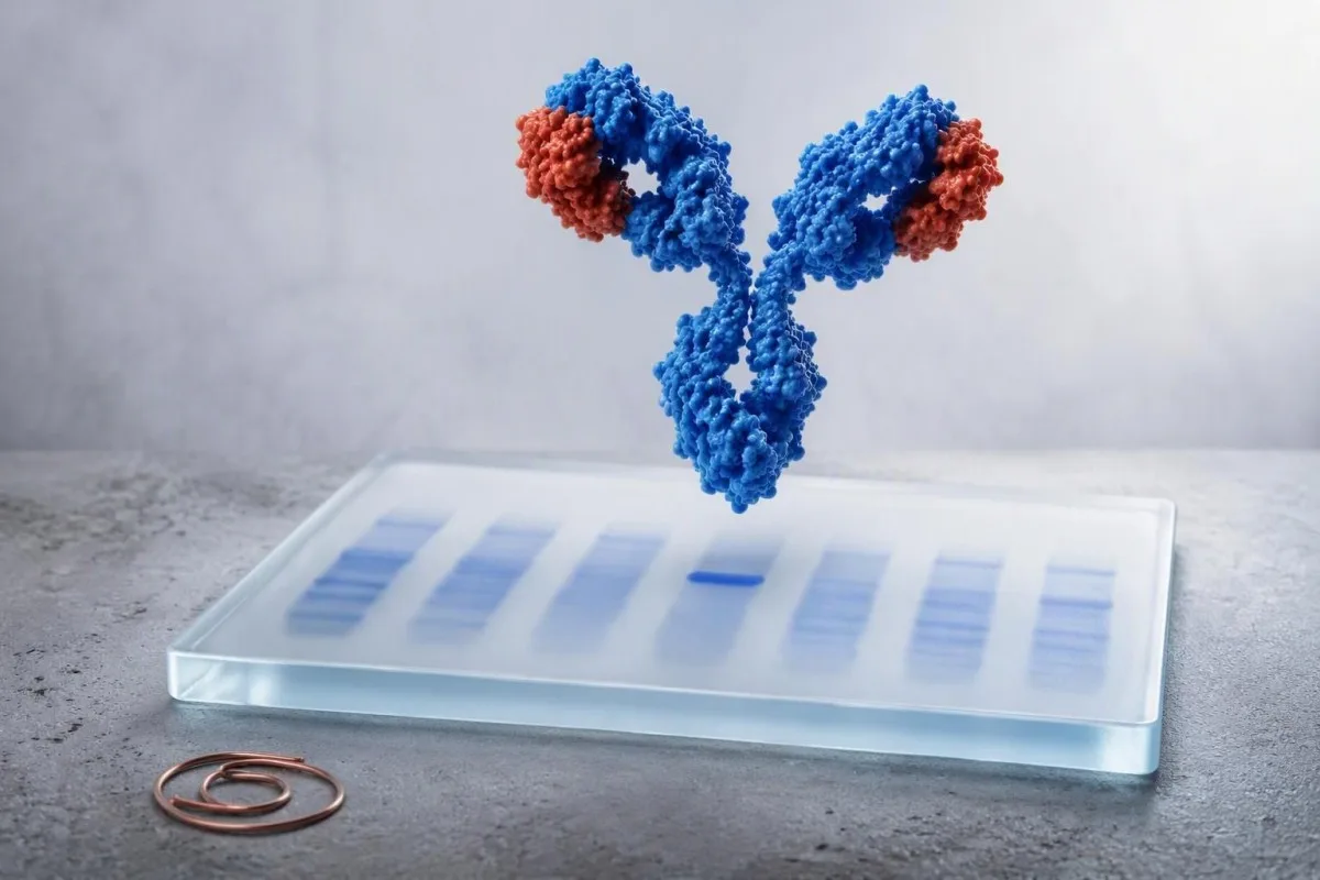
Immunofixation clarifies whether an abnormal SPEP pattern represents one clone of antibodies or a broad immune response. The band type, its persistence, and your kidney, blood-count, calcium, and symptom profile determine what happens next.


- Absent band means no monoclonal protein was detected by the assay; it does not explain every high-protein result.
- Faint band can be transient after immune stimulation, but a repeat serum IFE in 3-6 months is often sensible when clinical concern remains.
- IgG monoclonal bands are the most common MGUS pattern and are usually risk-stratified with M-protein amount and free light-chain ratio.
- IgA monoclonal bands may migrate in the beta region, where an SPEP can underestimate or obscure the measurable protein.
- IgM monoclonal bands call for a different clinical frame because Waldenström macroglobulinemia and some lymphomas enter the differential.
- Free light chains add essential information: a markedly abnormal ratio or involved light chain of 100 mg/L or more needs prompt specialist review.
- Επείπτωτα συμπτώματα include new confusion, severe weakness, reduced urine output, major thirst, bone pain, recurrent infections, or unexplained weight loss.
- One result is not a diagnosis; clinicians interpret IFE beside CBC, creatinine/eGFR, corrected calcium, quantitative immunoglobulins, urine testing, and prior results.
What immunofixation electrophoresis results actually tell you
Immunofixation electrophoresis results identify whether a narrow protein signal comes from one antibody-producing cell clone and, if present, name its heavy chain and light chain. An IFE blood test result such as “IgG kappa monoclonal band” confirms a monoclonal protein; it does not, by itself, diagnose myeloma or lymphoma.
SPEP sorts serum proteins by electrical movement and produces a broad pattern; IFE then applies antisera to IgG, IgA, IgM, kappa, and lambda. A true monoclonal protein appears in the same position in one heavy-chain lane and one light-chain lane. Read the SPEP M-spike guide first if the report began with an abnormal spike.
Το Καντέστι είναι ένα Αναλυτής εξέτασης αίματος AI that reads IFE wording alongside total protein, albumin, globulin gap, kidney markers, calcium, and historical panels rather than treating “positive” as a diagnosis. As of September 28, 2026, that context matters more than an isolated faint band.
In my clinical work, the commonest misunderstanding is assuming that IFE measures the size of a clone. It usually does not. The laboratory may quantify an M-protein on SPEP in g/dL, while IFE mainly establishes identity and can detect proteins too small to quantify.
Why the extra test follows SPEP
IFE is more sensitive and more specific than SPEP for characterizing a small abnormal protein. A normal-looking SPEP can still have a positive IFE when the clone is tiny, migrates poorly, or produces mainly free light chains.
Absent or negative bands after an abnormal SPEP
A negative immunofixation result means the laboratory found no discrete monoclonal immunoglobulin in that serum sample. This commonly reflects polyclonal immune activity, dehydration, liver disease, or a technical SPEP irregularity rather than a plasma-cell disorder.
A broad gamma-region rise is usually polyclonal: many B-cell populations making antibodies at once. Chronic liver disease, autoimmune illness, persistent infection, and recent immune activation can do this, so the Οδηγός για πρωτεΐνες και σφαιρίνες ορού is useful when total protein or globulin is high.
A negative serum IFE does not completely exclude light-chain disease. If there is unexplained anemia, kidney impairment, protein in urine, neuropathy, cardiomyopathy, or suspicious bone symptoms, clinicians generally add serum free light chains and urine electrophoresis with urine IFE.
The practical question is whether there was a clinical reason to look. A gamma gap above 4 g/dL is a clue, not a validated screening diagnosis; I would not pursue broad hematology testing from that number alone in an otherwise well person.
When a negative test is reassuring
A negative IFE with normal hemoglobin, calcium, creatinine, and no concerning symptoms is usually reassuring. Repeating it is generally driven by an evolving laboratory pattern, not by anxiety alone.
Faint or trace restricted bands: repeat, ignore, or refer?
A faint restricted band is an equivocal low-level finding that often merits confirmation rather than immediate treatment or panic. Some disappear on repeat testing, while others represent early MGUS that remains stable for years.
A laboratory may say “faint,” “trace,” “restricted,” “small band,” or “cannot rule out monoclonal protein.” These terms are not interchangeable across laboratories. I usually want the exact report, an SPEP quantity if measurable, serum free light chains, and a repeat specimen from the same laboratory in about 3-6 months.
Transient oligoclonal bands can follow immune stimulation, transplantation, infection, or immune-directed therapy; several narrow bands rather than one matched heavy-light pair often support that explanation. Sample problems matter too—review hemolysis-related result errors before attributing a surprising pattern to disease.
Dr. Thomas Klein would be more concerned about a faint band that persists and coincides with falling hemoglobin, rising creatinine, elevated calcium, or a clearly abnormal free light-chain ratio. The combination changes the pre-test probability far more than the adjective “faint.”
Typed IgG kappa or IgG lambda band results
An IgG kappa or IgG lambda band confirms the most common class of monoclonal protein and is often classified as MGUS when the M-protein is below 3 g/dL and there is no organ injury attributable to the clone. IgG type alone cannot tell whether a person has benign MGUS, smoldering myeloma, or active myeloma.
The International Myeloma Working Group defines MGUS by serum M-protein below 3 g/dL, marrow clonal plasma cells below 10%, and no CRAB organ damage or amyloidosis caused by the clone. Rajkumar et al. (2014) also recognize biomarkers that can define active myeloma before classic organ injury.
For non-IgM MGUS, low-risk features are IgG type, M-protein 1.5 g/dL or lower, and a normal free light-chain ratio. Using those factors, many low-risk patients can avoid immediate marrow sampling, although age, symptoms, and laboratory trajectory still matter.
Kantesti AI interprets typed IgG results by comparing the IFE language with free light-chain ratio findings and prior protein values. A rising quantified M-protein is more informative than a stable typed label.
What an IgA monoclonal band changes in the work-up
An IgA kappa or IgA lambda band is a monoclonal protein that deserves quantification and free light-chain testing because IgA often migrates in the beta region and may be underestimated on SPEP. The next step is usually hematology-guided risk assessment, not an assumption of cancer.
IgA monoclonal proteins may overlap transferrin, complement proteins, or beta-lipoproteins on electrophoresis. That is why a report can say “beta-region restriction” even when no obvious gamma spike appears. Quantitative IgA and serum free light chains help establish the biological burden.
In practice, an IgA band with M-protein 0.4 g/dL, normal CBC, eGFR, calcium, and stable light chains is managed very differently from an IgA band with anemia and a rapidly increasing value. The immunoglobulin results guide explains why uninvolved immunoglobulins may also be measured.
A suppressed uninvolved IgG or IgM—sometimes called immunoparesis—can signal a less diverse antibody response, but it is not diagnostic by itself. Recurrent bacterial infections are a clinical reason to take that finding more seriously.
IgM band results and why they follow a different pathway
An IgM monoclonal band prompts assessment for IgM MGUS, Waldenström macroglobulinemia, and certain B-cell lymphomas rather than the standard non-IgM myeloma pathway. Serum viscosity symptoms and enlarged nodes, spleen, or liver alter the urgency.
IgM is a large pentameric antibody, so high concentrations can increase serum viscosity. Headache, blurred vision, spontaneous nose or gum bleeding, dizziness, confusion, or shortness of breath alongside a substantial IgM result should be assessed urgently; those symptoms are not explained by a faint IgM band alone.
A hematologist may order quantitative IgM, CBC with film, serum viscosity when clinically indicated, imaging, and sometimes marrow testing with molecular studies. A bone-marrow procedure is not automatic for every small, stable IgM protein.
Ο rouleaux formation guide helps explain why high circulating proteins can alter how cellular elements stack on a slide. That observation is supportive, not a substitute for IFE or a diagnosis.
When IFE shows kappa or lambda without a heavy chain
An isolated kappa or lambda restriction can indicate light-chain MGUS, light-chain myeloma, AL amyloidosis, or a small clone below heavy-chain detection. Serum free light-chain values, the kappa/lambda ratio, kidney function, and urine studies determine how concerning it is.
Typical Freelite reference intervals vary by assay, but many laboratories use a kappa/lambda ratio around 0.26-1.65 in people with preserved kidney function. Chronic kidney disease raises both chains and broadens interpretation; the ratio often remains closer to balanced than in a true single-clone process.
An involved/uninvolved free light-chain ratio of 100 or more, with involved chain at least 100 mg/L, is a myeloma-defining biomarker when confirmed in the appropriate setting. That threshold comes from the IMWG criteria summarized by Rajkumar et al. (2014), not from a routine screening cutoff.
Protein in urine or persistent foamy urine makes urine albumin testing and urine protein electrophoresis more relevant. Review επίμονη αφρώδης ούρων rather than assuming the appearance proves a monoclonal disorder.
Results that need prompt hematology or urgent assessment
IFE results need prompt specialist assessment when they accompany unexplained anemia, kidney decline, high calcium, bone pain, fractures, recurrent infections, or rapidly changing protein values. Emergency care is appropriate for confusion, severe dehydration, markedly reduced urine output, or acute neurologic symptoms.
The classic CRAB framework includes calcium elevation, renal impairment, anemia, and bone involvement. A corrected calcium above 11 mg/dL (2.75 mmol/L), hemoglobin more than 2 g/dL below the lower limit of normal or below 10 g/dL, and creatinine above 2 mg/dL are examples that warrant timely clinical evaluation when a clone is present.
A normal CBC does not rule out clinically significant disease, but it lowers the immediate concern substantially. Conversely, anemia from iron deficiency, kidney disease, or bleeding is common and must be evaluated on its own merits; see reticulocyte hemoglobin clues when iron delivery is uncertain.
In our medical review workflow, Kantesti is an υπηρεσία ερμηνείας δοκιμών AI that flags these combinations as follow-up triggers, not diagnoses. Automated interpretation must never delay urgent in-person assessment when symptoms are progressing.
MGUS risk and follow-up intervals after a positive IFE
Most people with MGUS do not develop cancer, but progression risk averages about 1% per year and persists over time. Follow-up frequency is individualized by isotype, M-protein amount, free light-chain ratio, age, symptoms, and whether values are stable.
Kyle et al. (2006) found MGUS in about 3.2% of adults aged 50 years or older in a predominantly White population, with prevalence rising sharply with age. Population estimates vary by ancestry and ascertainment, so an individual’s risk should not be inferred from prevalence alone.
Many clinicians repeat CBC, creatinine, calcium, SPEP, and free light chains at 6 months after a new MGUS diagnosis. If low-risk values remain stable, annual review—or less frequent review in selected patients—may be reasonable; higher-risk patterns are followed more closely.
Το Καντέστι είναι ένα Εργαλείο ανάλυσης αιματολογικών εξετάσεων με AI-powered that can preserve the exact prior M-protein and kidney values for trend review. Comparing like with like matters: a different electrophoresis method or laboratory can create an apparent shift that is analytical, not biological.
When abnormal protein patterns are not monoclonal disease
High total protein or an abnormal electrophoresis contour can occur without a monoclonal protein. Dehydration concentrates albumin and globulins, while liver disease, autoimmune conditions, and chronic immune stimulation commonly create a broad polyclonal pattern.
Albumin and globulin should be read together. A high total protein with normal albumin suggests increased globulins, but dehydration can elevate both; repeat testing after ordinary hydration is often more informative than aggressive fluid loading before the draw.
Polyclonal hypergammaglobulinemia is common in chronic hepatic disease and systemic immune disorders. If liver enzymes, bilirubin, or albumin are abnormal, the MASLD laboratory overview may provide a more plausible explanation than a plasma-cell condition.
Monoclonal and polyclonal patterns can coexist. That nuance is why I do not dismiss a clear small IFE band merely because CRP is high, nor label all high globulins as a clone.
Test limits, medications, and the best repeat-testing plan
Immunofixation is highly specific for identifying an immunoglobulin type, but its detection threshold, reporting language, and interference profile differ by laboratory. Repeating a borderline result at the same laboratory improves comparability.
Therapeutic monoclonal antibodies can appear as bands and may be mistaken for endogenous proteins unless the laboratory knows the treatment. Daratumumab is a familiar example in myeloma care, but other antibody therapies can also complicate interpretation; bring a complete medication list.
Recent intravenous immunoglobulin can produce temporary electrophoretic abnormalities for weeks because infused donor antibodies remain measurable. A lab should know the infusion date, product, and indication before issuing a conclusion about a new band.
Fasting is not usually required for IFE, although a severely lipemic specimen can interfere with some laboratory processes. Our lipemic sample explanation covers when a redraw is more reliable than over-interpreting a compromised sample.
Questions to take to your clinician after an IFE result
The most useful question after a positive IFE is “What is the quantified M-protein, and do my CBC, calcium, kidney function, and free light chains show an effect?” Asking for the actual numbers converts a frightening label into a practical evaluation plan.
Ask whether the result is definite, faint, or equivocal; whether the heavy and light chains match; and whether an M-protein was quantifiable in g/dL. Also ask whether this is new compared with prior SPEP or IFE results. A one-page λίστα ελέγχου επίσκεψης στον γιατρό can keep those details together.
Ask specifically about urine testing if you have swelling, neuropathy, proteinuria, or unexplained kidney changes. In a 68-year-old patient I recently reviewed, a small IgG band mattered less than the newly abnormal urine protein and falling eGFR—which appropriately accelerated referral.
Dr. Thomas Klein’s practical advice is to avoid self-directed supplements marketed for “protein balance.” There is no supplement that removes a monoclonal clone, and unnecessary products can blur kidney or liver assessment.
Using AI to organize IFE blood test results safely
AI can organize IFE blood test results, identify missing companion tests, and show trends, but it cannot determine whether a monoclonal protein is causing symptoms or replace a hematology assessment. Any new typed band should be reviewed by the clinician who ordered the test.
Kantesti AI interprets monoclonal protein immunofixation language by linking it to CBC, calcium, eGFR, total protein, albumin, and prior laboratory reports. It is especially useful when a report is photographed or uploaded from another language, but original laboratory units and reference intervals should always be retained.
Our approach is governed by clinical oversight and source checking; readers can review our technical clinical validation when deciding how to use automated lab interpretation. Privacy-conscious organization is useful, yet an algorithm cannot examine lymph nodes, assess fractures, or judge constitutional symptoms.
If you are gathering reports for a hematology visit, save the full SPEP tracing, IFE interpretation, free light-chain values, quantitative immunoglobulins, and at least 12 months of CBC and renal results. That sequence is often more valuable than repeating a single panel early.
A practical next-step plan for any IFE result
Negative IFE with reassuring companion tests usually means clinical observation; a faint band usually means confirmation; and a definite typed band usually means risk stratification with free light chains, CBC, calcium, creatinine, and often hematology input. The result is a starting point, not a verdict.
For an absent band, clarify whether the original SPEP was broad rather than narrow and address the likely cause of elevated proteins. For a faint band, repeat serum studies in 3-6 months unless symptoms or abnormal companion markers justify earlier referral.
For a typed IgG or IgA band, document M-protein g/dL, free light-chain ratio, immunoglobulins, CBC, corrected calcium, and renal function. For IgM, add a symptom review for hyperviscosity and lymphoproliferative features; for light-chain-only patterns, urine and organ-specific assessment become more central.
Kantesti’s physician-reviewed educational standards are guided by our Ιατρική Συμβουλευτική Επιτροπή. The sensible endpoint is a shared plan with your clinician: what to repeat, where, when, and which symptom should prompt you to call sooner.
Συχνές Ερωτήσεις
Τι σημαίνει ένα θετικό αποτέλεσμα ηλεκτροφόρησης ανοσοσταθεροποίησης;
Ένα θετικό αποτέλεσμα ηλεκτροφόρησης ανοσοσταθεροποίησης σημαίνει ότι το εργαστήριο ανίχνευσε μια μονοκλωνική ανοσοσφαιρίνη και προσδιόρισε τον τύπο της, όπως IgG κάπα, IgA λάμδα ή IgM κάπα. Δεν διαγιγνώσκει από μόνη της το πολλαπλό μυέλωμα, επειδή η MGUS είναι συχνή και συχνά σταθερή. Οι κλινικοί γιατροί ερμηνεύουν το αποτέλεσμα σε συνδυασμό με την ποσότητα της πρωτεΐνης M σε g/dL, τις ελεύθερες αλυσίδες ελαφρών αλυσίδων του ορού, το CBC, την κρεατινίνη, το ασβέστιο, τα συμπτώματα και μερικές φορές εξετάσεις ούρων. Μια πρωτεΐνη M κάτω από 3 g/dL χωρίς βλάβη οργάνου σχετιζόμενη με το κλώνο μπορεί να πληροί τα κριτήρια της MGUS αντί για ενεργή κακοήθεια.
Είναι σοβαρό ένα αχνό διάγραμμο στην ανοσοπροσρόφηση;
Μια αχνή ζώνη στην ανοσοφύξωση δεν είναι αυτόματα σοβαρή, αλλά πρέπει να ερμηνεύεται ως εύρημα χαμηλού επιπέδου ή αμφίβολο παρά να αγνοείται. Πολλοί κλινικοί επαναλαμβάνουν SPEP, IFE και ελεύθερες αλυσίδες ορού σε περίπου 3-6 μήνες για να προσδιορίσουν εάν η ζώνη επιμένει. Πρώιμη ανασκόπηση αιματολογίας είναι κατάλληλη εάν η αιμοσφαιρίνη είναι κάτω από 10 g/dL, το διορθωμένο ασβέστιο υπερβαίνει τα 11 mg/dL, η νεφρική λειτουργία επιδεινώνεται ή υπάρχουν συμπτώματα όπως πόνος στα οστά ή υποτροπιάζουσες λοιμώξεις. Η ακριβής εργαστηριακή διατύπωση και το προηγούμενο μοτίβο εξέτασης έχουν σημασία.
Ποια είναι η διαφορά μεταξύ SPEP και ηλεκτροφόρησης ανοσοσταθεροποίησης;
Η Ηλεκτροφόρηση Σερικού Πρωτεΐνη (SPEP) διαχωρίζει τις πρωτεΐνες του ορού και μπορεί να ποσοτικοποιήσει μια μετρήσιμη αιχμή M σε g/dL, ενώ η ανοσοσταθεροποίηση (immunofixation electrophoresis) αναγνωρίζει την κλάση και την ελαφρά αλυσίδα του αντισώματος που είναι υπεύθυνη για μια στενή ζώνη. Η SPEP είναι χρήσιμη για την παρακολούθηση της ποσότητας μιας πρωτεΐνης με την πάροδο του χρόνου, ενώ η IFE είναι πιο ευαίσθητη για μικρές πρωτεΐνες και πιο ακριβής για τον χαρακτηρισμό τους. Ένα άτομο μπορεί να έχει θετική IFE χωρίς μετρήσιμη αιχμή M SPEP. Και οι δύο εξετάσεις παραγγέλλονται συνήθως μαζί με τη δοκιμασία ελεύθερων ελαφρών αλυσίδων ορού.
Το IgG κάπα σημαίνει πάντα MGUS;
Η IgG κάππα είναι ο πιο συνηθισμένος τύπος μονοκλωνικής πρωτεΐνης και μπορεί να αντιπροσωπεύει MGUS, αθόρυβο μυέλωμα, ενεργό μυέλωμα, αμυλοείδωση AL ή άλλη πάθηση των πλασματοκυττάρων. Η MGUS απαιτεί M-πρωτεΐνη ορού κάτω από 3 g/dL, λιγότερο από 10% πλασματοκύτταρα μυελού των οστών, και καμία σχετιζόμενη με το κλώνο οργανική βλάβη. Η MGUS χαμηλού κινδύνου έχει γενικά τύπο IgG, M-πρωτεΐνη 1,5 g/dL ή χαμηλότερη, και φυσιολογική αναλογία ελεύθερων ελαφρών αλυσίδων. Επομένως, μια ζώνη καθορισμένου τύπου απαιτεί εκτίμηση κινδύνου, όχι συμπέρασμα μόνο από τον τύπο.
Can immunofixation be negative if you have myeloma?
Serum immunofixation can be negative in uncommon nonsecretory or minimally secretory myeloma and may miss cases producing mainly free light chains. Serum free light-chain testing and urine immunofixation improve detection when clinical suspicion remains high. A free light-chain ratio of 100 or more with an involved light chain of at least 100 mg/L is a myeloma-defining biomarker in the proper clinical setting. Normal IFE is reassuring but does not override concerning anemia, kidney decline, high calcium, or imaging findings.
How often should I repeat immunofixation after MGUS?
Many clinicians repeat laboratory testing 6 months after a new MGUS finding, including CBC, creatinine, calcium, SPEP, and serum free light chains. If low-risk values are stable, follow-up may become annual or less frequent depending on age, symptoms, and local hematology practice. Higher-risk findings, including non-IgG type, M-protein above 1.5 g/dL, or an abnormal free light-chain ratio, usually justify closer monitoring. A new symptom or a rising M-protein should prompt review before the scheduled interval.
Λάβετε σήμερα ανάλυση εξετάσεων αίματος με AI
Εγγραφείτε σε πάνω από 2 εκατομμύρια χρήστες παγκοσμίως που εμπιστεύονται το Kantesti για άμεση, ακριβή ανάλυση εργαστηριακών εξετάσεων. Ανεβάστε τα αποτελέσματα εξετάσεων αίματος και λάβετε ολοκληρωμένη ερμηνεία βιοδεικτών του 15,000+ μέσα σε δευτερόλεπτα.
📚 Παραπομπές σε δημοσιεύσεις έρευνας
Klein, T., Mitchell, S., & Weber, H. (2026). Klein, T. (2026). BUN/Creatinine Ratio Explained: Kidney Function Test Guide. Zenodo. https://doi.org/10.5281/zenodo.18207872. ResearchGate: https://www.researchgate.net/ Academia.edu: https://www.academia.edu/. Ιατρική έρευνα του Kantesti με AI.
Klein, T., Mitchell, S., & Weber, H. (2026). Klein, T. (2026). Urobilinogen in Urine Test: Complete Urinalysis Guide 2026. Zenodo. https://doi.org/10.5281/zenodo.18226379. ResearchGate: https://www.researchgate.net/ Academia.edu: https://www.academia.edu/. Ιατρική έρευνα του Kantesti με AI.
📖 Εξωτερικές ιατρικές αναφορές
⚕️ Ιατρική αποποίηση ευθύνης
Αυτό το άρθρο προορίζεται μόνο για εκπαιδευτικούς σκοπούς και δεν αποτελεί ιατρική συμβουλή. Συμβουλεύεστε πάντα έναν κατάλληλο επαγγελματία υγείας για αποφάσεις σχετικά με τη διάγνωση και τη θεραπεία.
Σήματα εμπιστοσύνης E-E-A-T
Εμπειρία
Κλινική ανασκόπηση από ιατρό για τις ροές εργασίας ερμηνείας εργαστηριακών αποτελεσμάτων.
Πραγματογνωμοσύνη
Εστίαση στην εργαστηριακή ιατρική στο πώς συμπεριφέρονται οι βιοδείκτες στο κλινικό πλαίσιο.
Αυθεντικότητα
Γραμμένο από τον Δρ. Thomas Klein με ανασκόπηση από την Δρ. Sarah Mitchell και τον Καθ. Dr. Hans Weber.
Αξιοπιστία
Ερμηνεία βασισμένη σε τεκμηριωμένα δεδομένα με σαφείς οδούς παρακολούθησης για τη μείωση του συναγερμού.