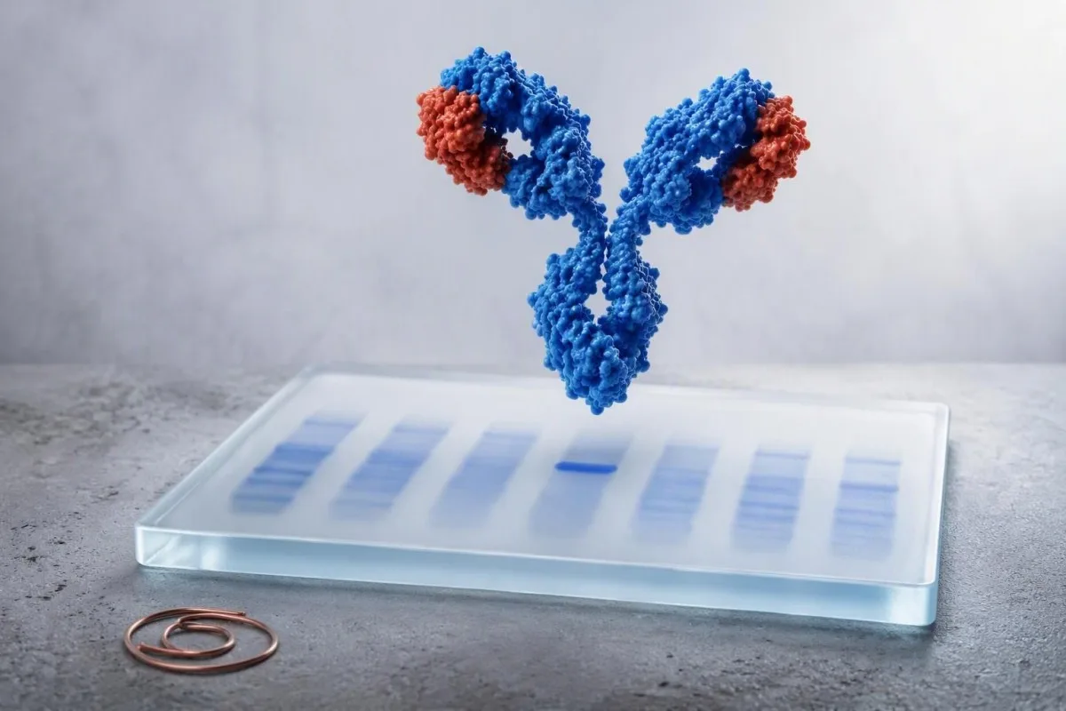
Leasaichidh dearbhadh dùbailteach dè an urrainn do phàtran SPEP neo-àbhaisteach aon chlon antibodies no freagairt dìonach leathann a riochdachadh. Tha an seòrsa còmhlan, a bhuanachd, agus do phroifil dubhaig, cunntas fala, calcium, agus comharraidhean a’ dearbhadh dè a thachras a-nis.


- Còmhlan neo-làthaireach a’ ciallachadh nach deach pròtain monoclonal a lorg leis an assay; chan eil e a’ mìneachadh a h-uile toradh pròtain àrd.
- Còmhlan lag faodaidh e a bhith sealach às dèidh brosnachadh dìonach, ach tha ath-aithris IFE serum ann an 3-6 mìosan gu tric ciallach nuair a tha dragh clionaigeach ann fhathast.
- Còmhlain monoclonal IgG is e am pàtran MGUS as cumanta agus mar as trice bidh iad air an roinn a rèir cunnart le meud pròtain M agus an co-mheas solais saor.
- Còmhlain monoclonal IgA faodaidh iad gluasad anns an raon beta, far am faod SPEP pròtain a ghabhas tomhas a thoirt sìos no fhalach.
- Còmhlain monoclonal IgM gairm air frèam clionaigeach eadar-dhealaichte leis gu bheil mòr-ghalair Waldenström agus cuid de linfomannan a’ dol a-steach don eadar-dhealachadh.
- Slabhraidhean aotrom an-asgaidh cuir fiosrachadh riatanach ris: feumaidh co-mheas a tha gu math neo-àbhaisteach no slabhraidh aotrom air a bheil buaidh de 100 mg/L no barrachd sgrùdadh eòlaiche sgiobalta.
- Comharraidhean èiginneach cuir a-steach troimh-chèile ùr, laigse mhòr, lachdainn fuil na h-ìre nas ìsle, tart mòr, pian cnàimh, galairean ath-chuairteach, no call cuim aig nach eil mìneachadh.
- Chan eil aon toradh na dhearbhadh; bidh luchd-clionaigeach a’ mìneachadh IFE còmhla ri CBC, creatinine/eGFR, calcium ceartach, immunoglobulins tomhasach, deuchainn urine, agus toraidhean roimhe seo.
Na tha toraidhean electrophoresis dearbhaidh dùbailteach ag innse dhut
Tha toraidhean electrophoresis immunofixation a’ comharrachadh a bheil comharra pròtain cumhang a’ tighinn bho aon chlòn cealla a tha a’ dèanamh antibodies agus, ma tha e ann, ag ainmeachadh a shlabhraidh throm agus a shlabhraidh aotrom. Tha toradh deuchainn fala IFE mar “IgG kappa bannan monaclach” a’ dearbhadh pròtain monaclach; chan eil e, leis fhèin, a’ dearbhadh myeloma no lymphoma.
Bidh SPEP a’ sgaradh pròtainean serum a rèir gluas an dealain agus a’ toirt a-mach pàtran farsaing; bidh IFE an uairsin a’ cur antisera ris an IgG, IgA, IgM, kappa, agus lambda. Tha pròtain monaclach fìor a’ nochdadh anns an aon suidheachadh ann an aon shlighe slabhraidh throm agus aon shlighe slabhraidh aotrom. Leugh an treòir M-spike SPEP an toiseach ma thòisich an aithisg le spike neo-àbhaisteach.
Tha Kantesti na Anailisiche deuchainn fala AI a leughas OFE ann an sgriobhadh còmhla ri pròtain iomlan, albumin, beàrn globulin, comharran dubhaig, calcium, agus pannalan eachdraidheil an àite a bhith a“ làimhseachadh ”dearbhach” mar dhearbhadh. Bho 28 Sultain, 2026, tha an co-theacsa sin nas cudromaiche na bannan caolach aonaranach.
Nam obair clionaigeach, is e am mì-thuigse as cumanta a bhith a’ gabhail ris gu bheil IFE a’ tomhas meud cloinne. Mar as trice cha dèan. Faodaidh an obair-lann pròtain M a thomhas air SPEP ann an g/dL, fhad ‘s a tha IFE gu mòr a’ stèidheachadh dearbh-aithne agus faodaidh e pròtainean a lorg a tha ro bheag airson tomhas.
Carson a lean an deuchainn a bharrachd SPEP
Tha IFE nas motha de shoirbheachadh agus nas sònraichte na SPEP airson a bhith a’ toirt cunntas air pròtain beag neo-àbhaisteach. Faodaidh SPEP a tha a’ coimhead àbhaisteach fhathast a bhith le IFE dearbhach nuair a tha an clòchan beag, a’ imrich gu dona, no a’ toirt a-mach prìomh shlabhraidhean aotrom an-asgaidh.
Còmhlain neo-làthaireach no àicheil às deidh SPEP neo-àbhaisteach
Tha toradh immunofixation àicheil a’ ciallachadh nach lorg an obair-lann immunoglobulin monaclach sònraichte sam bith anns an sampall serum sin. Tha seo gu tric a’ nochdadh gnìomhachd dìonachd polyclonach, dìth uisgeachadh, tinneas ae, no mì-rianachd theicnigeach SPEP an àite eas-òrdugh cealla plasma.
Tha àrdachadh farsaing ann an roinn gamma mar as trice polyclonach: mòran bhuidhnean B-cealla a’ dèanamh antibodies aig an aon àm. Faodaidh tinneas ae leantainneach, tinneas fèin-dìon, galar leantainneach, agus brosnachadh dìonachd o chionn ghoirid seo a dhèanamh, mar sin tha an stiùireadh air pròtainean serum agus globulins feumail nuair a tha pròtain iomlan no globulin àrd.
Chan eil IFE serum àicheil a’ dùnadh a-mach gu tur thinneas slabhraidh aotrom. Ma tha anemia aig nach eil mìneachadh, dìth gnìomhachd dubhaig, pròtain ann an urine, neuropathy, cardiomyopathy, no comharran cnàimh a tha amharasach, bidh luchd-clionaigeach mar as trice a’ cur slabhraidhean aotrom serum an-asgaidh agus electrophoresis urine le IFE urine ris.
Is e a’ cheist phractaigeach an robh adhbhar clionaigeach ann airson coimhead. Tha beàrn gamma os cionn 4 g/dL na chomharra, chan e dearbhadh sgàlaidh dearbhte; cha bhithinn a’ leantainn deuchainn hematology farsaing bhon àireamh sin a-mhàin ann an neach fallain eile.
Nuair a tha deuchainn àicheil na fhaochadh
Tha IFE àicheil le hemoglobin, calcium, creatinine àbhaisteach, agus gun chomharran draghail mar as trice na fhaochadh. Mar as trice thèid ath-aithris a dhèanamh air sgàth pàtran obair-lann a tha a’ tighinn am follais, chan ann air sgàth dragh a-mhàin.
Còmhlain chuingealaichte lag no glè bheag: ath-aithris, dearmad, no iomradh?
A faint restricted band is an equivocal low-level finding that often merits confirmation rather than immediate treatment or panic. Some disappear on repeat testing, while others represent early MGUS that remains stable for years.
A laboratory may say “faint,” “trace,” “restricted,” “small band,” or “cannot rule out monoclonal protein.” These terms are not interchangeable across laboratories. I usually want the exact report, an SPEP quantity if measurable, serum free light chains, and a repeat specimen from the same laboratory in about 3-6 months.
Transient oligoclonal bands can follow immune stimulation, transplantation, infection, or immune-directed therapy; several narrow bands rather than one matched heavy-light pair often support that explanation. Sample problems matter too—review hemolysis-related result errors before attributing a surprising pattern to disease.
Dr. Thomas Klein would be more concerned about a faint band that persists and coincides with falling hemoglobin, rising creatinine, elevated calcium, or a clearly abnormal free light-chain ratio. The combination changes the pre-test probability far more than the adjective “faint.”
Toraidhean còmhlan IgG kappa no IgG lambda
An IgG kappa or IgG lambda band confirms the most common class of monoclonal protein and is often classified as MGUS when the M-protein is below 3 g/dL and there is no organ injury attributable to the clone. IgG type alone cannot tell whether a person has benign MGUS, smoldering myeloma, or active myeloma.
The International Myeloma Working Group defines MGUS by serum M-protein below 3 g/dL, marrow clonal plasma cells below 10%, and no CRAB organ damage or amyloidosis caused by the clone. Rajkumar et al. (2014) also recognize biomarkers that can define active myeloma before classic organ injury.
For non-IgM MGUS, low-risk features are IgG type, M-protein 1.5 g/dL or lower, and a normal free light-chain ratio. Using those factors, many low-risk patients can avoid immediate marrow sampling, although age, symptoms, and laboratory trajectory still matter.
Kantesti AI interprets typed IgG results by comparing the IFE language with free light-chain ratio findings and prior protein values. A rising quantified M-protein is more informative than a stable typed label.
Mar a dh’atharraicheas bann monoclonal IgA san obair-ullachaidh
An IgA kappa or IgA lambda band is a monoclonal protein that deserves quantification and free light-chain testing because IgA often migrates in the beta region and may be underestimated on SPEP. The next step is usually hematology-guided risk assessment, not an assumption of cancer.
IgA monoclonal proteins may overlap transferrin, complement proteins, or beta-lipoproteins on electrophoresis. That is why a report can say “beta-region restriction” even when no obvious gamma spike appears. Quantitative IgA and serum free light chains help establish the biological burden.
In practice, an IgA band with M-protein 0.4 g/dL, normal CBC, eGFR, calcium, and stable light chains is managed very differently from an IgA band with anemia and a rapidly increasing value. The immunoglobulin results guide explains why uninvolved immunoglobulins may also be measured.
A suppressed uninvolved IgG or IgM—sometimes called immunoparesis—can signal a less diverse antibody response, but it is not diagnostic by itself. Recurrent bacterial infections are a clinical reason to take that finding more seriously.
Toraidhean còmhlan IgM agus carson a leanas iad slighe eadar-dhealaichte
An IgM monoclonal band prompts assessment for IgM MGUS, Waldenström macroglobulinemia, and certain B-cell lymphomas rather than the standard non-IgM myeloma pathway. Serum viscosity symptoms and enlarged nodes, spleen, or liver alter the urgency.
IgM is a large pentameric antibody, so high concentrations can increase serum viscosity. Headache, blurred vision, spontaneous nose or gum bleeding, dizziness, confusion, or shortness of breath alongside a substantial IgM result should be assessed urgently; those symptoms are not explained by a faint IgM band alone.
A hematologist may order quantitative IgM, CBC with film, serum viscosity when clinically indicated, imaging, and sometimes marrow testing with molecular studies. A bone-marrow procedure is not automatic for every small, stable IgM protein.
An rouleaux formation guide helps explain why high circulating proteins can alter how cellular elements stack on a slide. That observation is supportive, not a substitute for IFE or a diagnosis.
Nuair a nochdas IFE kappa no lambda às aonais slabhraidh trom
An isolated kappa or lambda restriction can indicate light-chain MGUS, light-chain myeloma, AL amyloidosis, or a small clone below heavy-chain detection. Serum free light-chain values, the kappa/lambda ratio, kidney function, and urine studies determine how concerning it is.
Typical Freelite reference intervals vary by assay, but many laboratories use a kappa/lambda ratio around 0.26-1.65 in people with preserved kidney function. Chronic kidney disease raises both chains and broadens interpretation; the ratio often remains closer to balanced than in a true single-clone process.
An involved/uninvolved free light-chain ratio of 100 or more, with involved chain at least 100 mg/L, is a myeloma-defining biomarker when confirmed in the appropriate setting. That threshold comes from the IMWG criteria summarized by Rajkumar et al. (2014), not from a routine screening cutoff.
Protein in urine or persistent foamy urine makes urine albumin testing and urine protein electrophoresis more relevant. Review fual làn-shileach leantach rather than assuming the appearance proves a monoclonal disorder.
Toraidhean a dh’fheumas hematology luath no measadh èiginneach
IFE results need prompt specialist assessment when they accompany unexplained anemia, kidney decline, high calcium, bone pain, fractures, recurrent infections, or rapidly changing protein values. Emergency care is appropriate for confusion, severe dehydration, markedly reduced urine output, or acute neurologic symptoms.
The classic CRAB framework includes calcium elevation, renal impairment, anemia, and bone involvement. A corrected calcium above 11 mg/dL (2.75 mmol/L), hemoglobin more than 2 g/dL below the lower limit of normal or below 10 g/dL, and creatinine above 2 mg/dL are examples that warrant timely clinical evaluation when a clone is present.
A normal CBC does not rule out clinically significant disease, but it lowers the immediate concern substantially. Conversely, anemia from iron deficiency, kidney disease, or bleeding is common and must be evaluated on its own merits; see reticulocyte hemoglobin clues when iron delivery is uncertain.
In our medical review workflow, Kantesti is an seirbheis eadar-mhìneachaidh deuchainn-lann AI that flags these combinations as follow-up triggers, not diagnoses. Automated interpretation must never delay urgent in-person assessment when symptoms are progressing.
Cunnart MGUS agus amannan leantainn às deidh IFE dearbhach
Most people with MGUS do not develop cancer, but progression risk averages about 1% per year and persists over time. Follow-up frequency is individualized by isotype, M-protein amount, free light-chain ratio, age, symptoms, and whether values are stable.
Kyle et al. (2006) found MGUS in about 3.2% of adults aged 50 years or older in a predominantly White population, with prevalence rising sharply with age. Population estimates vary by ancestry and ascertainment, so an individual’s risk should not be inferred from prevalence alone.
Many clinicians repeat CBC, creatinine, calcium, SPEP, and free light chains at 6 months after a new MGUS diagnosis. If low-risk values remain stable, annual review—or less frequent review in selected patients—may be reasonable; higher-risk patterns are followed more closely.
Tha Kantesti na Inneal mion-sgrùdaidh deuchainn fala le cumhachd AI that can preserve the exact prior M-protein and kidney values for trend review. Comparing like with like matters: a different electrophoresis method or laboratory can create an apparent shift that is analytical, not biological.
Nuair nach eil pàtranan pròtain neo-àbhaisteach nan tinneas monoclonal
High total protein or an abnormal electrophoresis contour can occur without a monoclonal protein. Dehydration concentrates albumin and globulins, while liver disease, autoimmune conditions, and chronic immune stimulation commonly create a broad polyclonal pattern.
Albumin and globulin should be read together. A high total protein with normal albumin suggests increased globulins, but dehydration can elevate both; repeat testing after ordinary hydration is often more informative than aggressive fluid loading before the draw.
Polyclonal hypergammaglobulinemia is common in chronic hepatic disease and systemic immune disorders. If liver enzymes, bilirubin, or albumin are abnormal, the MASLD laboratory overview may provide a more plausible explanation than a plasma-cell condition.
Monoclonal and polyclonal patterns can coexist. That nuance is why I do not dismiss a clear small IFE band merely because CRP is high, nor label all high globulins as a clone.
Crìochan deuchainn, cungaidhean-leigheis, agus an plana ath-dheuchainn as fheàrr
Immunofixation is highly specific for identifying an immunoglobulin type, but its detection threshold, reporting language, and interference profile differ by laboratory. Repeating a borderline result at the same laboratory improves comparability.
Therapeutic monoclonal antibodies can appear as bands and may be mistaken for endogenous proteins unless the laboratory knows the treatment. Daratumumab is a familiar example in myeloma care, but other antibody therapies can also complicate interpretation; bring a complete medication list.
Recent intravenous immunoglobulin can produce temporary electrophoretic abnormalities for weeks because infused donor antibodies remain measurable. A lab should know the infusion date, product, and indication before issuing a conclusion about a new band.
Fasting is not usually required for IFE, although a severely lipemic specimen can interfere with some laboratory processes. Our lipemic sample explanation covers when a redraw is more reliable than over-interpreting a compromised sample.
Ceistean ri thoirt don dotair agad às deidh toradh IFE
The most useful question after a positive IFE is “What is the quantified M-protein, and do my CBC, calcium, kidney function, and free light chains show an effect?” Asking for the actual numbers converts a frightening label into a practical evaluation plan.
Ask whether the result is definite, faint, or equivocal; whether the heavy and light chains match; and whether an M-protein was quantifiable in g/dL. Also ask whether this is new compared with prior SPEP or IFE results. A one-page liosta-sgrùdaidh tadhal air an dotair can keep those details together.
Ask specifically about urine testing if you have swelling, neuropathy, proteinuria, or unexplained kidney changes. In a 68-year-old patient I recently reviewed, a small IgG band mattered less than the newly abnormal urine protein and falling eGFR—which appropriately accelerated referral.
Dr. Thomas Klein’s practical advice is to avoid self-directed supplements marketed for “protein balance.” There is no supplement that removes a monoclonal clone, and unnecessary products can blur kidney or liver assessment.
A’ cleachdadh AI gus toraidhean deuchainn fala IFE a chuir air dòigh gu sàbhailte
AI can organize IFE blood test results, identify missing companion tests, and show trends, but it cannot determine whether a monoclonal protein is causing symptoms or replace a hematology assessment. Any new typed band should be reviewed by the clinician who ordered the test.
Kantesti AI interprets monoclonal protein immunofixation language by linking it to CBC, calcium, eGFR, total protein, albumin, and prior laboratory reports. It is especially useful when a report is photographed or uploaded from another language, but original laboratory units and reference intervals should always be retained.
Our approach is governed by clinical oversight and source checking; readers can review our technical clinical validation when deciding how to use automated lab interpretation. Privacy-conscious organization is useful, yet an algorithm cannot examine lymph nodes, assess fractures, or judge constitutional symptoms.
If you are gathering reports for a hematology visit, save the full SPEP tracing, IFE interpretation, free light-chain values, quantitative immunoglobulins, and at least 12 months of CBC and renal results. That sequence is often more valuable than repeating a single panel early.
Plana gnìomh practaigeach airson toradh IFE sam bith
Negative IFE with reassuring companion tests usually means clinical observation; a faint band usually means confirmation; and a definite typed band usually means risk stratification with free light chains, CBC, calcium, creatinine, and often hematology input. The result is a starting point, not a verdict.
For an absent band, clarify whether the original SPEP was broad rather than narrow and address the likely cause of elevated proteins. For a faint band, repeat serum studies in 3-6 months unless symptoms or abnormal companion markers justify earlier referral.
For a typed IgG or IgA band, document M-protein g/dL, free light-chain ratio, immunoglobulins, CBC, corrected calcium, and renal function. For IgM, add a symptom review for hyperviscosity and lymphoproliferative features; for light-chain-only patterns, urine and organ-specific assessment become more central.
Kantesti’s physician-reviewed educational standards are guided by our Bòrd Comhairleachaidh Meidigeach. The sensible endpoint is a shared plan with your clinician: what to repeat, where, when, and which symptom should prompt you to call sooner.
Ceistean Bitheanta
Dè tha toradh dearbhach de electrophoresis immunofixation a' ciallachadh?
Tha toradh dearbhach air electrofòrais immunofixation a’ ciallachadh gun do lorg an obair-lann immunoglobulin monoclònal agus gun do dh’aithnich e a sheòrsa, leithid IgG kappa, IgA lambda, no IgM kappa. Chan eil e a’ dearbhadh myeloma iomadach leis fhèin, leis gu bheil MGUS cumanta agus gu tric seasmhach. Bidh luchd-clionaigeach a’ mìneachadh an toraidh le meud an pròtain M ann an g/dL, sreathan aotrom an-asgaidh serum, CBC, creatinine, calcium, comharran, agus uaireannan deuchainn urine. Is dòcha gun coinnich pròtain M fo 3 g/dL às aonais milleadh organ co-cheangailte ris a’ chròn na slatan-tomhais MGUS an àite droch-dhroch ghalair.
A bheil còmhlan caol air an sgrìobhadh-dhearbhaidh dona?
Chan fuaimneach air immunofixation chan eil e gu fèin-obrachail dona, ach bu chòir a mhìneachadh mar lorg ìosal-ìre no co-ionann seach a bhith air a mhì-fhortanachadh. Mòran de luchd-clionaigeach ath-aithris SPEP, IFE, agus slabhraidhean solais an-asgaidh serum ann an timcheall air 3-6 mìosan gus dearbhadh am bi an raon a’ leantainn. Tha ath-sgrùdadh hematology nas tràithe freagarrach ma tha an haemoglobin nas ìsle na 10 g/dL, tha an calcium ceartachaidh nas àirde na 11 mg/dL, tha obair nan dubhagan a’ fàs nas miosa, no gu bheil comharran mar pian cnàimh no galairean ath-chuairteach ann. Tha cànan cruinn an obair-lann agus am pàtran deuchainn roimhe cudromach.
Dè an diofarachd eadar SPEP agus immunofixation electrophoresis?
SPEP a' sgaradh pròtainean searum agus faodaidh e tomhas M-spike tomhaiste a dhèanamh ann an g/dL, fhad 's a tha electrophoresis ceartachaidh ag aithneachadh clas an aghaidh agus an t-slabhraidh aotrom a tha an urra ris an loidhne cumhang. Tha SPEP feumail airson a bhith a' lorg meud pròtain thar ùine, fhad 's a tha IFE nas motha na sin airson pròtainean beaga agus nas miosa airson gan seòrsa. Faodaidh neach IFE dearbhach fhaighinn gun M-spike SPEP tomhaiste. Tha an dà dheuchainn air an òrdachadh gu cumanta le deuchainn slabhraidh aotrom an-asgaidh ann an searum.
A bheil IgG kappa a' ciallachadh MGUS an-còmhnaidh?
Is e IgG kappa an seòrsa pròtain monoclònach as cumanta agus dh’fhaodadh e a bhith a’ riochdachadh MGUS, myeloma a’ smocadh, myeloma gnìomhach, AL amyloidosis, no suidheachadh eile de cheallan plasma. Tha feum air MGUS air pròtain M serum fo 3 g/dL, nas lugha na 10% ceallan plasma clònach sa smior, agus gun mhilleadh organ co-cheangailte ris a’ chlòn. Mar as trice tha IgG mar sheòrsa air MGUS cunnart ìseal, pròtain M 1.5 g/dL no nas ìsle, agus co-mheas solais an-asgaidh àbhaisteach. Mar sin, tha feum air measadh cunnairt airson còmhlan clò-bhuailte, chan e co-dhùnadh a-mhàin bhon t-seòrsa.
An urrainn do immunofixation a bhith àicheil ma tha myeloma agad?
Faodaidh suathadh dìonachaidh serum a bhith àicheil ann an myeloma neo-dhìomhaireachd no dìomhaireachd ainneamh agus faodaidh e a bhith ag ionndrainn chùisean a tha a’ dèanamh prìomh slabhraidhean aotrom an-asgaidh. Leasaich deuchainn airson slabhraidhean aotrom an-asgaidh serum agus suathadh dìonachaidh fual an lorg nuair a tha amharas clionaigeach fhathast àrd. Tha co-mheas slabhraidh aotrom an-asgaidh de 100 no barrachd leis an t-slabhraidh aotrom a tha an sàs aig co-dhiù 100 mg/L na chomharra a tha a’ mìneachadh myeloma san t-suidheachadh clionaigeach cheart. Tha IFE àbhaisteach a’ toirt misneachd ach cha bhith e a’ cur às do anemia draghail, crìonadh dubhaig, calcium àrd, no toraidhean ìomhaigh.
Cia tric bu chòir dhomh ath-aithris immunofixation às dèidh MGUS?
Bidh mòran lighichean-cuimse a' dèanamh dheuchainnean obair-lann a-rithist 6 mìosan an dèidh toraidhean ùra MGUS a lorg, a' gabhail a-steach CBC, creatinine, calcium, SPEP, agus sreathan aotrom an-asgaidh ann an serum. Ma tha luachan cunnairt ìosal seasmhach, faodaidh an sgrùdadh a bhith bliadhnail no nas trice a rèir aois, comharraidhean, agus cleachdadh hematology ionadail. Mar as trice bidh toraidhean nas cunnartach, a' gabhail a-steach seòrsa neo-IgG, pròtain M os cionn 1.5 g/dL, no neo-riaghailteachd ann an co-mheas sreathan aotrom an-asgaidh, a' toirt adhbhar airson sgrùdadh nas dlùithe. Bu chòir do chomharra ùr no pròtain M a tha a' sìor fhàs adhbhar a thoirt airson ath-sgrùdadh ro àm a' chlàir.
Faigh Mion-sgrùdadh Deuchainn Fala le Cumhachd AI an-diugh
Thig còmhla ri còrr is 2 mhillean neach air feadh an t-saoghail a tha a’ earbsa Kantesti airson mion-sgrùdadh sa bhad, ceart air deuchainnean obair-lann. Luchdaich suas na toraidhean deuchainn fala agad agus faigh mìneachadh coileanta air biomarcair 15,000+ ann an diogan.
📚 Foillseachaidhean Rannsachaidh le Iomraidhean
Klein, T., Mitchell, S., & Weber, H. (2026). Klein, T. (2026). BUN/Creatinine Ratio Explained: Kidney Function Test Guide. Zenodo. https://doi.org/10.5281/zenodo.18207872. ResearchGate: https://www.researchgate.net/ Academia.edu: https://www.academia.edu/. Rannsachadh Leigheis AI Kantesti.
Klein, T., Mitchell, S., & Weber, H. (2026). Klein, T. (2026). Urobilinogen in Urine Test: Complete Urinalysis Guide 2026. Zenodo. https://doi.org/10.5281/zenodo.18226379. ResearchGate: https://www.researchgate.net/ Academia.edu: https://www.academia.edu/. Rannsachadh Leigheis AI Kantesti.
📖 Iomraidhean Meidigeach Taobh a-muigh
⚕️ Àicheadh Meidigeach
Tha an artaigil seo dìreach airson adhbharan foghlaim agus chan eil e a’ dèanamh comhairle mheidigeach. Cuir fios an-còmhnaidh gu solaraiche cùram slàinte teisteanasach airson co-dhùnaidhean breithneachaidh is leigheis.
Comharran earbsa E-E-A-T
Eòlas
Lèirmheas clionaigeach air a stiùireadh le lighiche air sruthan-obrach mìneachaidh obair-lann.
Eòlas
Fòcas air leigheas obair-lann air mar a bhios bith-chomharraidhean (biomarkers) a’ giùlan ann an co-theacsa clionaigeach.
Ùghdarrasachd
Air a sgrìobhadh le Dr. Thomas Klein le ath-sgrùdadh le Dr. Sarah Mitchell agus Prof. Dr. Hans Weber.
Earbsachd
Mìneachadh stèidhichte air fianais le slighean leanmhainn soilleir gus dragh a lughdachadh.