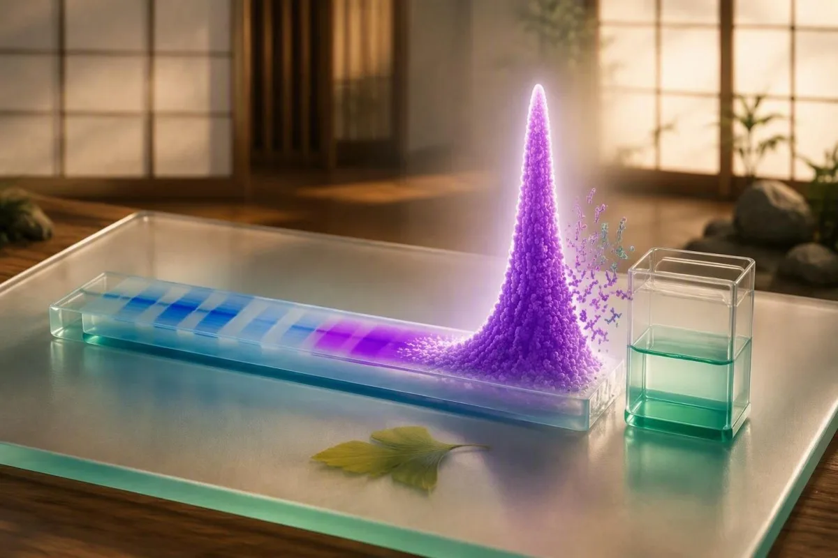
Tha M-spike a' comharrachadh pàtran pròtain antibody dlùth, chan e breithneachadh leis fhèin. Tha meud, seòrsa, gluasad, comharran, gnìomh dubhaig agus cunntasan fala a' dearbhadh dè thachras a-rèir.


- M-spike: Tha mullach cumhang SPEP a' comharrachadh immunoglobulin monoclonal a dh'fhaodadh a bhith ann, ach chan urrainn do SPEP leis fhèin a sheòrsa antibody a chomharrachadh no neo-òrdugh cheallan plasma a dhearbhadh.
- Tha aonadan cudromach: Mar as trice bidh obair-lann na SA a' clàradh an M-protein ann an g/dL; bidh mòran obair-lann san Rìoghachd Aonaichte agus san Roinn Eòrpa a' cleachdadh g/L, far a bheil 10 g/L co-ionann ri 1.0 g/dL.
- Ìre MGUS: Serum M-protein fo 3 g/dL, ceallan plasma smior fo 10%, agus gun mhilleadh organach a' freagairt air slatan-tomhais MGUS a-mhàin an dèidh measadh clionaigeach iomchaidh.
- Dearbhadh: Tha immunofixation serum agus deuchainnean slabhraidh aotrom an-asgaidh serum nan deuchainnean a tha cumanta an dèidh M-spike ùr.
- Chan eil neart beag gun chunnart: Faodaidh M-spike de 0.2 g/dL fhathast dearbhadh a chosnadh, ach chan eil toradh nas motha a’ ciallachadh aillse gu fèin-ghluasadach.
- Pàtran èiginneach: Feumaidh troimh-chèile ùr, laigse mhòr, lùghdachadh luath air toradh fual, calcium os cionn 14 mg/dL, no dùmhlachd mhòr anail a bhith air a mheasadh gu clionaigeach sa bhad.
- Bidh gluasad an treand nas cudromaiche na aon thoradh: Tha àrdachadh ath-riochdachail de 0.5 g/dL no barrachd nas ciallaiche san fharsaingeachd na atharrachadh anailis bheag eadar obairlann.
- Chan eil a h-uile raon gamma neo-àbhaisteach monoclònach: Faodaidh leudachadh polyclònach bho thinneas grùthan, gnìomhachd fèin-ghluasadach, no brosnachadh dìonachd leantainneach a bhith a’ coimhead neo-àbhaisteach gun a bhith a’ cruthachadh fìor M-spike.
Dè tha M-spike a' ciallachadh air aithisg SPEP
Toraidhean SPEP M-spike de phròtain serum a’ sealltainn dùmhlachd cumhang de aon immunoglobulin no fragment antibody; chan eil iad, leotha fhèin, a’ dearbhadh myeloma no aillse sam bith. Is e a’ chiad obair a bhith a’ dearbhadh a bheil an cruinneachadh dha-rìribh monoclònach agus a’ cur a thomhas tomhaiste ann an co-theacsa clionaigeach.
Bidh SPEP a’ sgaradh phròtainean serum a rèir cosgais dealain agus gluasad tro shiostam gel no capillary. Mar as trice bidh albumin a’ cruthachadh a’ chruinneachaidh as motha; tha bloighean alpha-1, alpha-2, beta agus gamma a’ leantainn, agus faodaidh cruinneachadh monoclònach suidhe anns an raon gamma no, gu sònraichte le IgA, anns a’ beta.
An M-spike a“ ciallachadh gu bheil pròtain co-ionann air cruinneachadh gu leòr gus cruinneachadh geur a chruthachadh. Nam chleachdadh clionaigeach, tha am facal ”spike” a’ cur eagal air daoine nas motha na bu chòir dha: is e pàtran obairlann a th’ ann, chan e breithneachadh. A iùl pròtainean serum a’ cuideachadh le bhith a’ sgaradh pròtain iomlan, bloighean globulin agus an A/G ratio.
Tha Kantesti na Anailisiche deuchainn fala AI a leughas M-protein a chaidh aithris còmhla ri pròtain iomlan, albumin, creatinine, calcium agus an cunntas fala iomlan seach a bhith a’ làimhseachadh a’ chruinneachaidh grafaigeach mar diagnios neo-eisimeileach. Bho 12 Sultain, 2026, tha an co-theacsa sin fhathast nas fheumail gu clionaigeach na àireamh SPEP singilte sam bith.
Carson a dh’ fhaodadh an cruinneachadh a bhith a dhìth bhon raon gamma
Bidh pròtainean monoclònach IgA gu tric a“ gluasad anns an raon beta, far a bheil transferrin agus pròtainean co-phàirteach mar-thà a” cruthachadh cùl-raon farsaing. Mar sin faodaidh obairlann aithris a dhèanamh air “còmhlan cuibhrichte” no “com-pàirt monoclònach a dh’ fhaodadh a bhith ann” eadhon nuair nach nochd cruinneachadh gamma follaiseach.
Mar a leughas tu bannan albumin, alpha, beta agus gamma
Bidh bannan SPEP a’ nochdadh bhuidhnean phròtain, chan e galairean fa leth. Mar as trice tha àrdachadh gamma farsaing a’ moladh mòran loidhnichean cealla a’ dèanamh antibody a’ freagairt aig aon àm, fhad ‘s a tha cruinneachadh cumhang, sònraichte a’ togail a’ chomas air aon clone dominant.
Albumin gu tric a’ dèanamh suas mu 55% gu 65% de phròtain serum agus a’ tuiteam le diligeadh, synthesis grùthan lùghdaichte, call pròtain, agus tinneas siostamach. Faodaidh albumin ìosal a dhèanamh gum bi an cuibhreann globulin a’ nochdadh gu neo-chothromach eadhon nuair a tha an dùmhlachd immunoglobulin iomlan gun atharrachadh.
Tha na bloighean alpha a’ toirt a-steach pròtainean pròiseas geur, fhad ‘s a tha am beta mar as trice a’ toirt a-steach transferrin, co-phàirt agus cuid de IgA. Faodaidh àrdachadh gamma farsaing a dhol còmhla ri cirrhosis, brosnachadh dìonachd cronach no galar fèin-ghluasadach; faodaidh luchd-leughaidh le neo-riaghailteachdan grùthan a lorg ar mìneachadh deuchainn grùthan a lorg feumail.
Tha bannan cumhang airidh air dearbhadh, ach tha leud a’ cunntadh. Chunnaic mi 67-bliadhna le cruinneachadh beta-region a bha a’ coimhead àrd a dh’ fhàs gu bhith na IgA aig 0.6 g/dL; rinn a shuidheachadh gum biodh e a’ coimhead dràma lèirsinneach, ach bha an obair-obrach a leanas aig cunnart ìosal.
Dè as urrainn agus nach urrainn SPEP innse dhut
SPEP can estimate the quantity and location of an abnormal protein peak, but it cannot reliably identify the antibody type, determine bone-marrow percentage, or establish whether organs are affected. Those limits are why an M-spike should not be interpreted as a cancer diagnosis from a portal result.
SPEP may miss low-concentration proteins, free light-chain-only disease, and proteins hidden within beta fractions. Serum immunofixation is more sensitive for identifying IgG, IgA, IgM, kappa or lambda components, while free-light-chain testing measures light chains circulating outside intact antibodies.
An M-spike does not tell us whether anemia is from marrow involvement, iron deficiency, kidney disease, medication, or another cause. That distinction matters because iron deficiency can be subtle before hemoglobin falls; see our guide to early low ferritin symptoms.
Tha Kantesti na àrd-ùrlar mìneachaidh deuchainn fala AI that can organize this uncertainty into questions for a clinician, including whether immunofixation was performed and whether creatinine, calcium, hemoglobin and urine protein have changed together. It does not replace hematology review or diagnostic testing.
The false reassurance problem
A normal SPEP does not exclude all clinically relevant monoclonal proteins. When symptoms or laboratory findings raise concern, clinicians often add serum free light chains, immunofixation and sometimes urine testing rather than relying on electrophoresis alone.
An ann a' meud M-spike a' ro-innse mar a tha e dona?
A larger M-spike generally increases the likelihood of a clinically significant plasma-cell disorder, but size alone cannot grade danger. A 0.3 g/dL peak needs confirmation, and a 2.5 g/dL peak may still be stable MGUS if organ assessment is reassuring.
For non-IgM MGUS, a serum monoclonal protein below 3 g/dL, nas lugha na 10% clonal marrow plasma cells, and no attributable organ injury are conventional diagnostic criteria. The 3 g/dL threshold is a classification boundary, not a biological cliff edge (Rajkumar et al., 2014).
Risk is lower when the protein is IgG, the M-protein is under 1.5 g/dL, and the free-light-chain ratio is normal; it rises when these features accumulate. In the Mayo cohort, MGUS progressed at roughly 1% gach bliadhna overall, but individual risk varied substantially over time (Kyle et al., 2006).
A result reported as 15 g/L equals 1.5 g/dL. Before comparing reports, check units and whether the same laboratory measured both samples; our article on fìor atharrachaidhean eadar deuchainnean fala explains why small numerical shifts can be analytical noise.
Nuair nach e aillse a th' ann an pàtran pròtain neo-àbhaisteach
Not all abnormal SPEP patterns are monoclonal or malignant. Liver disease, autoimmune conditions, chronic infections, dehydration and recent immune stimulation can alter protein fractions, usually producing a broad-based rather than sharply restricted gamma pattern.
Polyclonal hypergammaglobulinemia means many B-cell populations are making antibodies, so the gamma region looks wide and diffuse. Chronic liver disease can produce beta-gamma bridging, while autoimmune disorders can raise several immunoglobulin classes at once rather than one discrete component.
Dehydration can increase total protein and albumin concentration without creating a true monoclonal band. Conversely, intravenous immunoglobulin given within days to weeks of testing can transiently create a band-like appearance; laboratories need that medication history to interpret a surprising result accurately.
A high total protein result is not interchangeable with an M-spike. Review our practical discussion of high total protein causes before assuming that one finding explains the other.
Why infections complicate the picture
Recent immune activation can broaden the gamma fraction and occasionally obscure a small restricted component. If the clinical question is not urgent, repeating the panel after recovery may be reasonable, but that timing should come from the ordering clinician rather than a fixed internet schedule.
An t-slighe dearbhaidh a tha cumanta an dèidh M-spike ùr
A newly reported M-spike is usually confirmed with serum immunofixation, serum free-light-chain assay, quantitative IgG/IgA/IgM, CBC, calcium and kidney testing. The order is designed to identify the protein, assess its burden and check for complications without presuming cancer.
Immunofixation electrophoresis (IFE) identifies the heavy-chain and light-chain type, such as IgG-kappa. Serum free light chains provide a kappa value, lambda value and ratio; renal impairment can raise both values, so the ratio often carries more diagnostic weight than either concentration alone.
Clinicians commonly add a full blood count, creatinine or eGFR, corrected calcium, albumin and quantitative immunoglobulins. A urine protein assessment is especially useful when there is foamy urine, edema, a falling eGFR or an abnormal light-chain result; persistent bubbles merit the focused approach in our foamy urine guide.
Kantesti AI interprets monoclonal protein test results by checking whether the relevant companion data are present, then flags missing confirmations for discussion rather than inventing a diagnosis. Our stiùireadh teicneòlais AI explains the principles behind this contextual workflow.
Why urine testing is selective
Urine electrophoresis and immunofixation can detect light chains excreted by the kidneys, but collection quality affects a 24-hour sample. Some clinicians use a spot urine protein-to-creatinine ratio for renal assessment alongside serum free-light-chain testing, depending on the presentation.
Dè na co-fhreagairtean a nì M-spike nas èiginniche
An M-spike warrants faster assessment when it occurs with unexplained anemia, kidney impairment, high calcium, persistent focal bone pain, recurrent severe infections, or rapidly rising protein concentration. These findings matter as a pattern because they may represent organ effects, although each also has common non-myeloma explanations.
The familiar CRAB features are calcium elevation, renal impairment, anemia and bone disease. IMWG criteria consider calcium above 11 mg/dL, creatinine os cionn 2 mg/dL or creatinine clearance below 40 mL/min, and hemoglobin more than 2 g/dL below the lower limit of normal among relevant thresholds when attributable to the plasma-cell disorder (Rajkumar et al., 2014).
Numbers must be interpreted carefully. A calcium of 10.8 mg/dL with albumin 5.1 g/dL may correct downward, while calcium of 11.2 mg/dL with albumin 2.5 g/dL may correct upward; clinicians may repeat ionized calcium if the answer changes management. Our explanation of slightly high calcium covers this trap.
Dr. Thomas Klein’s practical rule is that new anemia plus falling kidney function deserves attention sooner than either result alone. A creatinine rise after dehydration or heavy exercise can be temporary, so compare prior values and consider the nuances in creatinine crìoch-chuingealaichte.
Tha MGUS, myeloma smoldering agus myeloma gnìomhach eadar-dhealaichte
MGUS is a precursor condition with a low average annual progression risk, smoldering myeloma has a higher burden without organ injury, and active myeloma requires treatment-directed specialist care. An M-spike is only one part of separating these categories.
MGUS usually has an M-protein below 3 g/dL and no myeloma-defining event. Smoldering myeloma includes an M-protein of 3 g/dL or more and/or 10% to 60% clonal marrow plasma cells, provided there is no attributable organ damage or defining biomarker.
Active myeloma can be diagnosed before classic CRAB injury if marrow clonal plasma cells reach 60%, involved-to-uninvolved free-light-chain ratio reaches 100 or more with involved light chain at least 100 mg/L, or MRI shows more than one focal marrow lesion at least 5 mm. These specific biomarkers were incorporated to prevent avoidable end-organ injury (Rajkumar et al., 2014).
Terms can sound like a conveyor belt, but they are not. Many people with MGUS never progress, and surveillance intensity is individualized; for a broader map of blood cancer test pathways, focus on how clinicians sequence evidence rather than labels alone.
Carson a tha gluasad M-spike tric a' buntainn barrachd na aon luach
A confirmed M-spike trend is more informative than a single measurement because monoclonal proteins can remain stable for years or rise gradually. The cleanest comparison uses the same assay, laboratory and unit system whenever possible.
A change of 0.1 g/dL may be meaningful for a tiny baseline protein but can also reflect integration differences when the peak overlaps other fractions. In many follow-up protocols, a rise of 0.5 g/dL or more triggers closer review, especially when accompanied by a changing light-chain ratio or new anemia.
Illness, hydration and assay technique can shift total proteins without changing the clone. When I review longitudinal reports, I look first for the exact phrase “M-protein,” then the isotype, then whether albumin and total protein moved in the same direction—a small habit that prevents many misleading comparisons.
Kantesti’s àrd-ùrlar mìneachaidh biomarcadairean AI can place sequential reports side by side and preserve units, dates and related kidney markers. A useful companion is our guide to mion-sgrùdadh obair-lann fad-ùine, particularly when reports come from more than one country.
Mar a dh'atharraicheas slabhraidhean aotrom an-asgaidh agus gnìomh dubhaig mìneachadh
Kidney impairment raises circulating kappa and lambda free light chains because renal clearance falls, so an abnormal absolute value does not automatically show a clone. A disproportionately abnormal ratio, an identified monoclonal band, and kidney findings together are more concerning than either result alone.
The usual standard serum kappa/lambda ratio reference interval is roughly 0.26 to 1.65, although laboratories may apply a wider renal interval in chronic kidney disease. Ask which assay and interval your laboratory used; free-light-chain assays are not perfectly interchangeable.
Protein in urine, declining eGFR and an abnormal light-chain ratio may lead to hematology and nephrology input because monoclonal proteins can affect kidneys even when the M-spike is modest. A normal creatinine does not always exclude early protein loss, which is why urine albumin or protein testing can add useful information.
The practical question is not “Is my light chain high?” but “Are both chains high in proportion to reduced clearance, or is one clearly dominant?” For preparation and interpretation details, review our iùl ìrean tinneas nan dubhagan.
Nuair a dh'fhaodar deuchainnean ìomhaigh no smior cnàimh a mheas
Imaging or bone-marrow testing is considered when the M-protein type, concentration, free-light-chain ratio, symptoms, or companion tests suggest higher risk. Most people with a small, low-risk confirmed MGUS do not need immediate invasive testing.
A hematologist may request low-dose whole-body CT, MRI or PET-CT for unexplained focal bone pain, fractures without sufficient trauma, substantial protein burden or other concerning findings. Conventional skeletal surveys are less sensitive for early lesions than modern cross-sectional imaging, although access differs across health systems.
Bone-marrow sampling measures the proportion and genetic features of plasma cells. It answers a different question from SPEP: a 1.0 g/dL M-spike cannot reliably predict marrow percentage because secretion rates vary greatly among clones.
I tell patients that a referral is a sorting step, not a prediction. Our bòrd comhairleachaidh meidigeach supports clinical oversight standards at Kantesti, while the treating hematology team decides whether imaging or marrow assessment is justified for an individual.
Symptoms that change the threshold
Persistent localized bone pain, unexplained weight loss, repeated infections, new neuropathy, or an abrupt reduction in exercise tolerance should be reported rather than waiting for a routine recheck. These symptoms are nonspecific, but their presence changes how quickly clinicians usually investigate.
Carson a dh'fheumas M-proteins IgM agus IgA dòigh-obrach sònraichte
IgM and IgA M-proteins follow different clinical pathways from typical IgG MGUS. IgM may be associated with lymphoplasmacytic disorders and hyperviscosity symptoms, while IgA may hide in the beta fraction and be underestimated visually on SPEP.
An antibodies IgM M-protein can be associated with neuropathy, cold-related circulation symptoms, enlarged lymph nodes or hyperviscosity when concentrations are high. Blurred vision, headache, mucosal bleeding or marked breathlessness require prompt medical review, not a wait-and-see response to an online result.
IgA iomlan may migrate in the beta region and overlap other proteins, making densitometric quantification less precise. Quantitative immunoglobulin measurements and immunofixation help reconcile an SPEP graph that does not seem to match the reported immunoglobulin concentration.
Immune testing is broader than SPEP. If a report also shows high or low immunoglobulin classes, our article on reading IgG, IgA and IgM explains what those values add—and what they cannot establish.
Mar a nì thu ullachadh airson SPEP ath-aithris agus deuchainnean co-cheangailte
Most people do not need to fast for SPEP, but repeat testing is most useful when performed under comparable conditions and with complete medication information. Do not stop prescribed medicines, immune treatments or supplements solely to improve a laboratory result.
Bring or upload prior SPEP, immunofixation and free-light-chain reports, including units and collection dates. Tell the clinician about intravenous immunoglobulin, monoclonal-antibody treatments, recent infection, major fluid loss, and any imaging contrast exposure; each can alter the interpretation or timing of companion tests.
Hydration should be ordinary rather than forced. Drinking several litres immediately before testing can dilute albumin and total protein, while arriving dehydrated can concentrate them; a normal morning routine gives the fairest longitudinal comparison.
Kantesti can extract values from a PDF or photo and organize dated results, but source verification still matters when formatting is unclear. Use our liosta-sgrùdaidh cruinneas airson luchdachadh suas PDF before relying on a transcribed M-protein value.
What to record after each test
Record illness in the prior 2 weeks, new medicines, hydration problems, laboratory name and whether immunofixation was done. Those details can explain why two reports with nearly identical totals carry very different clinical messages.
Ceistean a dh'fhaighneachd an dèidh toraidhean electrophorasis pròtain serum
The most useful questions after an M-spike are what protein was identified, how much is present, whether the free-light-chain ratio is abnormal, and whether kidney function, calcium or hemoglobin suggest organ involvement. Asking these four questions converts a frightening label into a practical next-step conversation.
Ask: “Was the result confirmed by immunofixation, and what is the isotype?” Then ask whether the laboratory quantified the M-protein or only described a restricted band. A stated “faint band” may be below reliable quantification yet still deserve follow-up, depending on the isotype and symptoms.
Ask for a written monitoring interval and the specific change that should prompt earlier contact. For low-risk MGUS, some specialists repeat testing at 6 mìosan and then extend to every 1 to 3 years if stable; higher-risk patterns are monitored more closely, and local practice varies.
Dr. Thomas Klein recommends taking a one-page summary to appointments: current M-protein, previous value and date, isotype, free-light-chain ratio, hemoglobin, calcium, creatinine/eGFR, symptoms and medicines. Our liosta-gnàthachaidh deuchainn-fala makes that discussion more efficient.
Nuair a dh'fheumas toradh M-spike cùram èiginneach seach cùram àbhaisteach
An M-spike alone rarely requires emergency care, but urgent assessment is appropriate for confusion, severe dehydration, markedly reduced urine output, acute shortness of breath, new severe weakness, uncontrolled vomiting, or severe focal bone pain with neurological symptoms. The urgency comes from possible organ dysfunction, not the printed peak alone.
Seek same-day medical advice for new confusion, fainting, severe drowsiness, rapidly worsening breathlessness, or urine output that has fallen sharply. Calcium above 14 mg/dL (3.5 mmol/L), potassium abnormalities, and abrupt kidney injury need prompt clinician-led assessment regardless of the M-spike amount.
For a stable small spike with no red-flag symptoms, arrange the confirmation pathway rather than self-treating with supplements or restrictive diets. There is no diet or over-the-counter product proven to remove a monoclonal clone, and indiscriminate supplements can complicate kidney and calcium results.
Kantesti is an AI lab test interpretation service built to make results understandable while directing potentially urgent patterns toward clinical care. Readers assessing reliability and physician oversight can review our dòigh-obrach dearbhaidh meidigeach againn before using any AI-generated laboratory explanation.
Ceistean Bitheanta
A bheil M-spike a' ciallachadh aillse an-còmhnaidh?
Chan eil. Tha M-spike a' ciallachadh gu bheil aon phròtain immunoglobulin no antibody an làthair ann an pàtran dlùth, ach cha bhith e a' dearbhadh aillse leis fhèin. Tha mòran de phròtainean monoclònach dearbhte nan MGUS, aig a bheil ìre deasachaidh cuibheasach timcheall air 1% gach bliadhna san fharsaingeachd seach deasachadh do-sheachanta. Tha immunofixation, free light chains, CBC, calcium agus gnìomh dubhaig a' cuideachadh le bhith a' dearbhadh an roinn cheart agus an leantainn.
Dè an ìre M-spike a thathas a’ meas àrd?
Spìc M de 3.0 g/dL, co-ionann ri 30 g/L, is e aon stairsneach a thathas a' cleachdadh nuair a thathar a' measadh air myeloma smoldering, ach chan eil e a' cumail a-mach leis fhèin. Faodaidh luach 1.5 g/dL cunnart nas àirde a thoirt ma tha e neo-IgG no air a chàradh le co-mheas neo-àbhaisteach de shlabhraidhean aotrom an-asgaidh, fhad 's a dh'fhaodadh luach os cionn 3.0 g/dL fhathast feumach air grunn eileamaidean a bharrachd mus tèid beachdachadh air làimhseachadh. Tha an seòrsa pròtain, an gluasad agus dearbhadh buaidhean organ nas cudromaiche na aon ghearradh.
Dè na deuchainnean a dhearbhas pròtain monaclònal às dèidh SPEP?
Dearbhaichidh serum immunofixation electrophoresis pròtain monoclònach agus roinnidh e mar IgG, IgA, IgM, kappa no lambda i. Tomhaisidh deuchainn serum free-light-chain kappa, lambda agus an co-mheas aca; mar as trice bidh co-mheas àbhaisteach mu 0.26 gu 1.65, ged a dh’ fhaodadh obair-lann raointean atharraichte le dubhan a chleachdadh. Bidh luchd-clionaigeach gu tric a’ cur immunoglobulins tomhasach, CBC, creatinine/eGFR, calcium agus deuchainn pròtain nan urine ris nuair a thèid an comharrachadh.
An urrainn dìth uisgeachadh adhbhrachadh M-spike?
Faodaidh dìth uisgeachadh pròtain iomlan agus albumin àrdachadh le bhith a’ dùmhlachadh an t-seileach, ach mar as trice cha bhith e a’ cruthachadh fìor M-spike monoclònach air immunofixation. Faodaidh e gnàthachd a th’ ann mar-thà nochdadh nas motha gu àireamhach no toirt air earrannan pròtain farsaing a bhith a’ coimhead nas fhollaisiche. Faodaidh sampall ath-aithris às deidh uisgeachadh àbhaisteach soilleireachadh a dhèanamh air buaidhean dùmhlachd, ach feumaidh còmhlan cuibhrichte a chaidh aithris fhathast na deuchainnean dearbhaidh a chaidh a mholadh leis an neach-clionaigeach.
An urrainn do M-spike a dhol à bith?
Faodaidh còmhlan lag a dhol à bith air deuchainn ath-aithris nuair a bha e sealach, fo na crìochan tomhais, co-cheangailte ri làimhseachadh dìonach o chionn ghoirid, no gun a bhith riochdaireil monoclonaidh. Faodaidh pròtain monoclonaidh dearbhte cuideachd atharrachadh beagan, gu sònraichte aig dùmhlachdan fo 0.5 g/dL, leis gu bheil atharrachadh tomhais agus aontachaidh nas motha a rèir sin. Tha eadar-mhìneachadh ciallach a' feumachdainn an aon dòigh obair-lann far a bheil sin comasach agus coimeas le toraidhean immunofixation agus free-light-chain.
Dè cho tric a bu chòir MGUS a bhith air a chumail sùil?
Thathas a' sgrùdadh mòran de chùisean MGUS a tha ann an cunnart ìosal mu 6 mìosan agus, ma tha iad seasmhach, gach 1 gu 3 bliadhna às a sin, ach tha clàran ag atharrachadh a rèir isotype, dùmhlachd M-protein, co-mheas slabhraidh aotrom, aois agus comharraidhean. Gu tric feumaidh MGUS nas àirde na cunnart leanmhainn nas dlùithe oir chan eil an cunnart rèidh am measg euslaintich. Bu chòir anemia ùr, lughdachadh eGFR, àrdachadh calcium, pian dian bhochd no àrdachadh M-protein a bhrosnachadh fios clionaigeach nas tràithe an àite feitheamh ris an ùine a chaidh a phlanadh.
Faigh Mion-sgrùdadh Deuchainn Fala le Cumhachd AI an-diugh
Thig còmhla ri còrr is 2 mhillean neach air feadh an t-saoghail a tha a’ earbsa Kantesti airson mion-sgrùdadh sa bhad, ceart air deuchainnean obair-lann. Luchdaich suas na toraidhean deuchainn fala agad agus faigh mìneachadh coileanta air biomarcair 15,000+ ann an diogan.
📚 Foillseachaidhean Rannsachaidh le Iomraidhean
Klein, T., Mitchell, S., & Weber, H. (2026). RDW Blood Test: Complete Guide to RDW-CV, MCV & MCHC. Kantesti LTD. Zenodo. https://doi.org/10.5281/zenodo.18202598. ResearchGate and Academia.edu record links available through the DOI landing page.. Rannsachadh Leigheis AI Kantesti.
Klein, T., Mitchell, S., & Weber, H. (2026). BUN/Creatinine Ratio Explained: Kidney Function Test Guide. Kantesti LTD. Zenodo. https://doi.org/10.5281/zenodo.18207872. ResearchGate and Academia.edu record links available through the DOI landing page.. Rannsachadh Leigheis AI Kantesti.
📖 Iomraidhean Meidigeach Taobh a-muigh
⚕️ Àicheadh Meidigeach
Tha an artaigil seo dìreach airson adhbharan foghlaim agus chan eil e a’ dèanamh comhairle mheidigeach. Cuir fios an-còmhnaidh gu solaraiche cùram slàinte teisteanasach airson co-dhùnaidhean breithneachaidh is leigheis.
Comharran earbsa E-E-A-T
Eòlas
Lèirmheas clionaigeach air a stiùireadh le lighiche air sruthan-obrach mìneachaidh obair-lann.
Eòlas
Fòcas air leigheas obair-lann air mar a bhios bith-chomharraidhean (biomarkers) a’ giùlan ann an co-theacsa clionaigeach.
Ùghdarrasachd
Air a sgrìobhadh le Dr. Thomas Klein le ath-sgrùdadh le Dr. Sarah Mitchell agus Prof. Dr. Hans Weber.
Earbsachd
Mìneachadh stèidhichte air fianais le slighean leanmhainn soilleir gus dragh a lughdachadh.