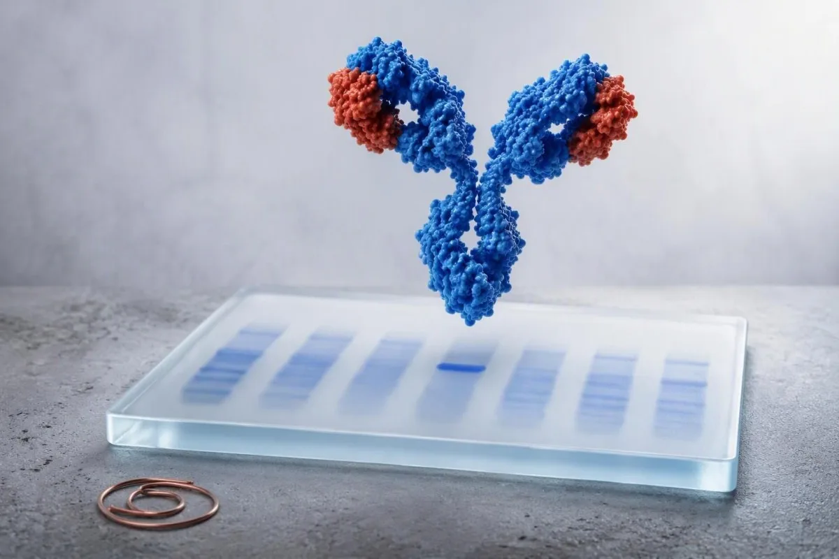
فیکسیی ئەمنیی دەربارەی ئەوە ڕووندەکاتەوە کە ئایا شێوازێکی نائاسایی SPEP نوێنەرایەتی یەک کلۆنی ئەنتیبۆدی دەکات یان وەڵامێکی بەرگری فراوان. جۆری باندەکە، بەردەوامییەکەی، و ڕەوشی گورچیلە، خوێن، کالسیۆم، و نیشانەکانت دیاری دەکات کە چی دواتر ڕوودەدات.


- باند دەرنەکەوتوو واتە هیچ پرۆتینی مۆنۆکلۆناڵ لەلایەن تاقیکردنەوەکە otkراون نەكراوە؛ ئەمەش هەموو ئەنجامە بەرزەکانی پرۆتین ڕووناک ناکاتەوە.
- باندێکی کاڵ دەتوانێت کاتی بێت دوای هاندانی بەرگری، بەڵام دووبارەکردنەوەی IFE سیرم لە ماوەی 3-6 مانگدا زۆرجار دروستە کاتێک نیگەرانی کلینیکی دەمێنێتەوە.
- باندە مۆنۆکلۆناڵەکانی IgG باوترین شێوازی MGUSن و زۆرجار مەترسییان لەگەڵ بڕی پرۆتینی M و ڕێژەی خانەسووری ئازاددا ڕیزبەندی دەکرێت.
- باندە مۆنۆکلۆناڵەکانی IgA دەکرێت لە ناوچەی بێتا کۆچ بکەن، کە SPEP دەکرێت کەمبværى بکات یان پرۆتینی پێوانەکراو بشارێتەوە.
- باندە مۆنۆکلۆناڵەکانی IgM داوای چوارچێوەیەکی کلینیکی جیاواز دەکات چونکونکە Waldenström macroglobulinemia و هەندێک لیمفۆما دەگرێتە خۆی لە نێوان دیاریکراوەکاندا.
- زنجیرە سووکەکانی ئازاد زانیاری گرنگ زیاد دەکات: ڕێژەیەکی ناجێگیری بەرچاو یان زنجیرە سووکی تێدا بەشداربوو بە بڕی ١٠٠ ملگم/لتر یان زیاتر پێویستی بە پێداچوونەوەی خێرای پسپۆڕانە هەیە.
- نەخۆشی/ئەلامەتە فوریتەکان بگرێتە خۆی تێکچوونی نوێ، لاوازییەکی سەخت، کەمبوونەوەی میز، تینووێتییەکی زۆر، ئازاری ئێسک، هەوکردنی دووبارە، یان دابەزینی کێشی بێ هۆکار.
- یەک ئەنجام دەستنیشانکردن نییە; ؛ پزیشکان IFE لەگەڵ CBC، کرێاتینین/eGFR، کالسیۆمی چاککراوە، ئیمونوگڵوبولینی ڕێژەیی، پشکنینی میز، و ئەنجامە پێشووەکان لێکدەدەنەوە.
ئەنجامەکانی ئەلیکترۆفۆڕێسی فیکسیی ئەمنی لە ڕاستیدا چی پێ دەڵێن
ئەنجامەکانی Electrophoresis-ی ئیمونۆفیکساسیۆن دیاری دەکەن کە ئایا هێڵێکی پڕۆتینی تەسک لە یەک کلۆنی خانەی بەرگرییەوە هاتووە و، ئەگەر بوونی هەبێت، ناوی زنجیرە قورس و زنجیرە سووکی دیاری دەکات. ئەنجامێکی پشکنینی خوێنی IFE وەک “IgG kappa monoclonal band” پڕۆتینی مۆنۆکلۆناڵ پشتڕاست دەکاتەوە؛ بە تەنیا، مایەلۆما یان لیمفۆما دیاری ناکات.
SPEP پڕۆتینەکانی سیرم بەپێی جوڵەی کارەبایی ڕیزبەندی دەکات و شێوازێکی فراوان دروست دەکات؛ پاشان IFE دژەبەرەکان بۆ IgG، IgA، IgM، کاپا، و لامبدا بەکاردەهێنێت. پڕۆتینێکی ڕاستەقینەی مۆنۆکلۆناڵ لە یەک شوێندا لە ڕێڵی زنجیرە قورسێک و ڕێڵی زنجیرە سووکێکدا دەردەکەوێت. بخوێنەرەوە ڕێبەری SPEP M-spike یەکەمجار ئەگەر ڕاپۆرتەکە بە سەرکەوتووبوونێکی ناجێگیر دەستی پێکردبێت.
Kantestî yek e Analyzerê testa xwînê ya AI کە IFE ووشەی ئەنجامەکە لەگەڵ پڕۆتینی گشتی، ئەلبومین، جیاوازی گڵوبولین، ئاماژەکانی گورچیلە، کالسیۆم، و پەنێڵە مێژووییەکاندا دەخوێنێتەوە لەبری مامەڵەکردن لەگەڵ “ئەرێنی” وەک دەستنیشانکردن. لە ٢٨ی ئەیلولی ٢٠٢٦ەوە، ئەو ڕاستییە زیاتر لە هێڵێکی تەنک و بچووک گرنگە.
لە کارە کلینیکییەکەمدا، زۆرترین هەڵەی تێگەیشتن بریتییە لە دانانی ئەوەی کە IFE قەبارەی کلۆنێک دەپێوێت. بەزۆری ئەمە ناکات. تاقیگە ڕەنگە M-protein لە SPEP بە g/dL دیاری بکات، لە کاتێکدا IFE زیاتر ناسنامەی دادەمەزرێنێت و دەتوانێت پڕۆتینەکان زۆر بچووک دیاری بکات کە نەتوانرێت ڕێژەیان دیاری بکرێت.
بۆچی پشکنینی زیادە دوای SPEP دێت
IFE هەستیارتر و تایبەتمەندترە لە SPEP بۆ دیاریکردنی پڕۆتینێکی بچووک و ناجێگیر. SPEPـێکی ئاسایی ڕەنگە هێشتا IFEـی ئەرێنی هەبێت کاتێک کلۆنەکە بچووکە، خراپ دەجوڵێت، یان زیاتر زنجیرەی سووکی ئازاد بەرهەم دەهێنێت.
باندە دەرنەکەوتووەکان یان نەرێنییەکان دوای SPEP نائاسایی
ئەنجامێکی نەرێنی ئیمونۆفیکساسیۆن واتە تاقیگە هیچ ئیمونوگڵوبولینی مۆنۆکلۆناڵی دیاریکراوی لەو نموونەی سیرمەدا نەدۆزیوەتەوە. ئەمە بە شێوەیەکی گشتی ڕەنگدانەوەی چالاکی بەرگری پۆلیکلۆناڵ، وشکبوونەوە، نەخۆشی جگەر، یان ناتەواویەکی تەکنیکی SPEP ە نەک کێشەیەکی خانەی پلاسما.
بەرزبوونەوەیەکی فراوانی گاما بە گشتی پۆلیکلۆناڵە: زۆر کۆمەڵەی خانەی B لە یەک کاتدا ئەنتی بادی دروست دەکەن. نەخۆشی درێژخایەنی جگەر، نەخۆشی ئۆتۆ ئیون، هەوکردنی بەردەوام، و چالاکبوونی نوێی بەرگری دەتوانێت ئەمە بکات، بۆیە rêbernameya proteînên serum û globulinan کاتێک پڕۆتینی گشتی یان گڵوبولین بەرزە بەکارهێنراوە.
IFEـی نەرێنی سیرم ناتوانێت نەخۆشی زنجیرەی سووک بە تەواوی لابەرێت. ئەگەر کەمخوێنی بێ هۆکار، کێشەی گورچیلە، پڕۆتین لە میزدا، نوورۆپاتی، کاردیۆمایۆپاتی، یان نیشانەکانی گوماناوی ئێسک هەبوو، پزیشکان بە گشتی زنجیرە سووکەکانی ئازادی سیرم و ئەلیکترۆفۆڕێزی میز لەگەڵ IFEـی میز زیاد دەکەن.
پرسیاری پراکتیکی ئەوەیە کە ئایا هۆکارێکی کلینیکی هەبووە بۆ لێگەڕین. جیاوازی گاما لە سەرووی ٤ گ/دل کلۆمە، نەک دەستنیشانکردنی پشتڕاستکراوەی پشکنین؛ من پشکنینی فراوانی خوێن لەو ژمارەیەی تەنیا لە کەسێکی تەواو تەندروستدا بەدوایدا ناگەڕێم.
کاتێک پشکنینێکی نەرێنی دڵنیایی دەبەخشێت
IFEـی نەرێنی لەگەڵ هیمۆگڵوبینی ئاسایی، کالسیۆم، کرێاتینین، و بێ نیشانەی دڵتەزێن بە گشتی دڵنیایی دەبەخشێت. دووبارەکردنەوەی بە گشتی بەهۆی شێوازێکی گەشەسەندوی تاقیگەوەیە، نەک تەنها بەهۆی ترسی بێجگە.
باندە سنووردارەکانی کاڵ یان ترایس: دووبارە بکەرەوە، پشتگوێ بخە، یان ئاراستە بکە؟
A faint restricted band is an equivocal low-level finding that often merits confirmation rather than immediate treatment or panic. Some disappear on repeat testing, while others represent early MGUS that remains stable for years.
A laboratory may say “faint,” “trace,” “restricted,” “small band,” or “cannot rule out monoclonal protein.” These terms are not interchangeable across laboratories. I usually want the exact report, an SPEP quantity if measurable, serum free light chains, and a repeat specimen from the same laboratory in about 3-6 months.
Transient oligoclonal bands can follow immune stimulation, transplantation, infection, or immune-directed therapy; several narrow bands rather than one matched heavy-light pair often support that explanation. Sample problems matter too—review hemolysis-related result errors before attributing a surprising pattern to disease.
Dr. Thomas Klein would be more concerned about a faint band that persists and coincides with falling hemoglobin, rising creatinine, elevated calcium, or a clearly abnormal free light-chain ratio. The combination changes the pre-test probability far more than the adjective “faint.”
ئەنجامەکانی باند IgG کاپا یان IgG لامبدا
An IgG kappa or IgG lambda band confirms the most common class of monoclonal protein and is often classified as MGUS when the M-protein is below 3 g/dL and there is no organ injury attributable to the clone. IgG type alone cannot tell whether a person has benign MGUS, smoldering myeloma, or active myeloma.
The International Myeloma Working Group defines MGUS by serum M-protein below 3 g/dL, marrow clonal plasma cells below 10%, and no CRAB organ damage or amyloidosis caused by the clone. Rajkumar et al. (2014) also recognize biomarkers that can define active myeloma before classic organ injury.
For non-IgM MGUS, low-risk features are IgG type, M-protein 1.5 g/dL or lower, and a normal free light-chain ratio. Using those factors, many low-risk patients can avoid immediate marrow sampling, although age, symptoms, and laboratory trajectory still matter.
Kantesti AI interprets typed IgG results by comparing the IFE language with free light-chain ratio findings and prior protein values. A rising quantified M-protein is more informative than a stable typed label.
گۆڕانکارییەکانی باند مۆنۆکلۆناڵی IgA لە پشکنیندا
An IgA kappa or IgA lambda band is a monoclonal protein that deserves quantification and free light-chain testing because IgA often migrates in the beta region and may be underestimated on SPEP. The next step is usually hematology-guided risk assessment, not an assumption of cancer.
IgA monoclonal proteins may overlap transferrin, complement proteins, or beta-lipoproteins on electrophoresis. That is why a report can say “beta-region restriction” even when no obvious gamma spike appears. Quantitative IgA and serum free light chains help establish the biological burden.
In practice, an IgA band with M-protein 0.4 g/dL, normal CBC, eGFR, calcium, and stable light chains is managed very differently from an IgA band with anemia and a rapidly increasing value. The immunoglobulin results guide explains why uninvolved immunoglobulins may also be measured.
A suppressed uninvolved IgG or IgM—sometimes called immunoparesis—can signal a less diverse antibody response, but it is not diagnostic by itself. Recurrent bacterial infections are a clinical reason to take that finding more seriously.
ئەنجامەکانی باند IgM و هۆکارێک کە بۆچی ڕێگەیەکی جیاوازیان هەیە
An IgM monoclonal band prompts assessment for IgM MGUS, Waldenström macroglobulinemia, and certain B-cell lymphomas rather than the standard non-IgM myeloma pathway. Serum viscosity symptoms and enlarged nodes, spleen, or liver alter the urgency.
IgM is a large pentameric antibody, so high concentrations can increase serum viscosity. Headache, blurred vision, spontaneous nose or gum bleeding, dizziness, confusion, or shortness of breath alongside a substantial IgM result should be assessed urgently; those symptoms are not explained by a faint IgM band alone.
A hematologist may order quantitative IgM, CBC with film, serum viscosity when clinically indicated, imaging, and sometimes marrow testing with molecular studies. A bone-marrow procedure is not automatic for every small, stable IgM protein.
Ew rouleaux formation guide helps explain why high circulating proteins can alter how cellular elements stack on a slide. That observation is supportive, not a substitute for IFE or a diagnosis.
کاتێک IFE کاپا یان لامبدا پیشان دەدات بە بێ زنجیرەی قورس
An isolated kappa or lambda restriction can indicate light-chain MGUS, light-chain myeloma, AL amyloidosis, or a small clone below heavy-chain detection. Serum free light-chain values, the kappa/lambda ratio, kidney function, and urine studies determine how concerning it is.
Typical Freelite reference intervals vary by assay, but many laboratories use a kappa/lambda ratio around 0.26-1.65 in people with preserved kidney function. Chronic kidney disease raises both chains and broadens interpretation; the ratio often remains closer to balanced than in a true single-clone process.
An involved/uninvolved free light-chain ratio of 100 or more, with involved chain at least 100 mg/L, is a myeloma-defining biomarker when confirmed in the appropriate setting. That threshold comes from the IMWG criteria summarized by Rajkumar et al. (2014), not from a routine screening cutoff.
Protein in urine or persistent foamy urine makes urine albumin testing and urine protein electrophoresis more relevant. Review میزە لووسی بەردەوام rather than assuming the appearance proves a monoclonal disorder.
ئەنجامەکان کە پێویستیان بە هەماناتۆلۆجی خێرا یان هەڵسەنگاندنی بەپەلە هەیە
IFE results need prompt specialist assessment when they accompany unexplained anemia, kidney decline, high calcium, bone pain, fractures, recurrent infections, or rapidly changing protein values. Emergency care is appropriate for confusion, severe dehydration, markedly reduced urine output, or acute neurologic symptoms.
The classic CRAB framework includes calcium elevation, renal impairment, anemia, and bone involvement. A corrected calcium above 11 mg/dL (2.75 mmol/L), hemoglobin more than 2 g/dL below the lower limit of normal or below 10 g/dL, and creatinine above 2 mg/dL are examples that warrant timely clinical evaluation when a clone is present.
A normal CBC does not rule out clinically significant disease, but it lowers the immediate concern substantially. Conversely, anemia from iron deficiency, kidney disease, or bleeding is common and must be evaluated on its own merits; see reticulocyte hemoglobin clues when iron delivery is uncertain.
In our medical review workflow, Kantesti is an خزمەتگوزاری تێکست/تێگەیشتنی تاقیکردنی لابراتۆریی AI that flags these combinations as follow-up triggers, not diagnoses. Automated interpretation must never delay urgent in-person assessment when symptoms are progressing.
مەترسی MGUS و ماوەی چاودێریکردن دوای IFE ئەرێنی
Most people with MGUS do not develop cancer, but progression risk averages about 1% per year and persists over time. Follow-up frequency is individualized by isotype, M-protein amount, free light-chain ratio, age, symptoms, and whether values are stable.
Kyle et al. (2006) found MGUS in about 3.2% of adults aged 50 years or older in a predominantly White population, with prevalence rising sharply with age. Population estimates vary by ancestry and ascertainment, so an individual’s risk should not be inferred from prevalence alone.
Many clinicians repeat CBC, creatinine, calcium, SPEP, and free light chains at 6 months after a new MGUS diagnosis. If low-risk values remain stable, annual review—or less frequent review in selected patients—may be reasonable; higher-risk patterns are followed more closely.
Kantestî yek e ئامێری توێژینەوەی تاقیکردنەوەی خوێن بە پشتبەستن بە AI that can preserve the exact prior M-protein and kidney values for trend review. Comparing like with like matters: a different electrophoresis method or laboratory can create an apparent shift that is analytical, not biological.
کاتێک شێوازە نائاساییەکانی پرۆتین نەخۆشی مۆنۆکلۆناڵ نین
High total protein or an abnormal electrophoresis contour can occur without a monoclonal protein. Dehydration concentrates albumin and globulins, while liver disease, autoimmune conditions, and chronic immune stimulation commonly create a broad polyclonal pattern.
Albumin and globulin should be read together. A high total protein with normal albumin suggests increased globulins, but dehydration can elevate both; repeat testing after ordinary hydration is often more informative than aggressive fluid loading before the draw.
Polyclonal hypergammaglobulinemia is common in chronic hepatic disease and systemic immune disorders. If liver enzymes, bilirubin, or albumin are abnormal, the MASLD laboratory overview may provide a more plausible explanation than a plasma-cell condition.
Monoclonal and polyclonal patterns can coexist. That nuance is why I do not dismiss a clear small IFE band merely because CRP is high, nor label all high globulins as a clone.
سنوورەکانی تاقیکردنەوە، دەرمانەکان، و باشترین پلانی دووبارە تاقیکردنەوە
Immunofixation is highly specific for identifying an immunoglobulin type, but its detection threshold, reporting language, and interference profile differ by laboratory. Repeating a borderline result at the same laboratory improves comparability.
Therapeutic monoclonal antibodies can appear as bands and may be mistaken for endogenous proteins unless the laboratory knows the treatment. Daratumumab is a familiar example in myeloma care, but other antibody therapies can also complicate interpretation; bring a complete medication list.
Recent intravenous immunoglobulin can produce temporary electrophoretic abnormalities for weeks because infused donor antibodies remain measurable. A lab should know the infusion date, product, and indication before issuing a conclusion about a new band.
Fasting is not usually required for IFE, although a severely lipemic specimen can interfere with some laboratory processes. Our lipemic sample explanation covers when a redraw is more reliable than over-interpreting a compromised sample.
پرسیارەکان بۆ بردن بۆ لای پزیشکەکەت دوای ئەنجامی IFE
The most useful question after a positive IFE is “What is the quantified M-protein, and do my CBC, calcium, kidney function, and free light chains show an effect?” Asking for the actual numbers converts a frightening label into a practical evaluation plan.
Ask whether the result is definite, faint, or equivocal; whether the heavy and light chains match; and whether an M-protein was quantifiable in g/dL. Also ask whether this is new compared with prior SPEP or IFE results. A one-page لیستی ڕێنمایی ویزیت لە دکتۆر can keep those details together.
Ask specifically about urine testing if you have swelling, neuropathy, proteinuria, or unexplained kidney changes. In a 68-year-old patient I recently reviewed, a small IgG band mattered less than the newly abnormal urine protein and falling eGFR—which appropriately accelerated referral.
Dr. Thomas Klein’s practical advice is to avoid self-directed supplements marketed for “protein balance.” There is no supplement that removes a monoclonal clone, and unnecessary products can blur kidney or liver assessment.
بەکارهێنانی AI بۆ ڕێکخستنی ئەنجامەکانی تاقیکردنەوەی خوێن IFE بە سەلامەتی
AI can organize IFE blood test results, identify missing companion tests, and show trends, but it cannot determine whether a monoclonal protein is causing symptoms or replace a hematology assessment. Any new typed band should be reviewed by the clinician who ordered the test.
Kantesti AI interprets monoclonal protein immunofixation language by linking it to CBC, calcium, eGFR, total protein, albumin, and prior laboratory reports. It is especially useful when a report is photographed or uploaded from another language, but original laboratory units and reference intervals should always be retained.
Our approach is governed by clinical oversight and source checking; readers can review our technical clinical validation when deciding how to use automated lab interpretation. Privacy-conscious organization is useful, yet an algorithm cannot examine lymph nodes, assess fractures, or judge constitutional symptoms.
If you are gathering reports for a hematology visit, save the full SPEP tracing, IFE interpretation, free light-chain values, quantitative immunoglobulins, and at least 12 months of CBC and renal results. That sequence is often more valuable than repeating a single panel early.
پلانی کرداریی قۆناغی داهاتوو بۆ هەر ئەنجامێکی IFE
Negative IFE with reassuring companion tests usually means clinical observation; a faint band usually means confirmation; and a definite typed band usually means risk stratification with free light chains, CBC, calcium, creatinine, and often hematology input. The result is a starting point, not a verdict.
For an absent band, clarify whether the original SPEP was broad rather than narrow and address the likely cause of elevated proteins. For a faint band, repeat serum studies in 3-6 months unless symptoms or abnormal companion markers justify earlier referral.
For a typed IgG or IgA band, document M-protein g/dL, free light-chain ratio, immunoglobulins, CBC, corrected calcium, and renal function. For IgM, add a symptom review for hyperviscosity and lymphoproliferative features; for light-chain-only patterns, urine and organ-specific assessment become more central.
Kantesti’s physician-reviewed educational standards are guided by our Lijneya Şêwirmendiya Bijîşkî. The sensible endpoint is a shared plan with your clinician: what to repeat, where, when, and which symptom should prompt you to call sooner.
Pirsên Pir tên Pirsîn
مانای ئەنجامی ئییەکستراشنی ئیمونۆفیکساسیۆن ئەرێنی چییە؟
مانای ئەنجامی پۆزەتیفی ئەلیکترۆفۆڕێزی ئییۆنۆفۆڕێزی مانای ئەوەیە کە تاقیگە ئییۆنۆگڵۆبوڵینێکی مۆنۆکلۆنالی دەستنیشان کردووە و جۆری دیاریکردووە، وەک IgG کاپا، IgA لامبدا، یان IgM کاپا. بە تەنها مایەلۆمای فرەقوڵپین دیاری ناکات، چونکە MGUS بڵاوە و زۆرجار جێگیرە. پزیشکان ئەنجامەکە لێکدەدەنەوە لەگەڵ بڕی M-protein بە g/dL، زنجیرە کاڵی ئازادی سیرۆم، CBC، کریاتینین، کالسیۆم، نیشانەکان، و هەندێکجار پشکنینی میز. M-protein کەمتر لە 3 g/dL بێ زیانی ئەندام پەیوەست بە کلۆن لەوانەیە لەبری شێرپەنجەی چالاک، ببێتە هۆی دەرکەوتنی مەرجەکانی MGUS.
ئایا ناودارێکی لاواز لەسەر ئیمونۆفیکسیشن مەترسیدارە؟
بانەیەکی لاواز لەسەر ئیمونۆفیکسایشن بە شێوەیەکی خۆکار مەترسیدار نییە، بەڵام دەبێت وەک نیشانەیەکی کەم یان گومانلێکراو لێکبدرێتەوە نەک پشتگوێ بخرێت. زۆرێک لە پزیشکان SPEP، IFE، و زنجیرە سووکەکانی سیرۆم بە ئازادی دووبارە دەکەنەوە لە ماوەی نزیکەی 3-6 مانگدا بۆ دیاریکردنی ئەوەی ئایا بانەکە بەردەوامە. پێداچوونەوەی زوو بە هیماتۆلۆجی گونجاوە ئەگەر هیمۆگڵۆبین کەمتر بێت لە 10 گ/دڵ، کالسیۆمی چاککراوە زیاتر بێت لە 11 ملگم/دڵ، چالاکی گورچیلە خراپتر بێت، یان نیشانەکانی وەک ئازاری ئێسک یان هەوکردنی دووبارەبووەوە بوونی هەبێت. وشەسازی وردی تاقیگە و شێوازی پێشووی تاقیکردنەوەکان گرنگن.
جیاوازی نێوان SPEP و electrophoresis ی ئیمونۆفیکسیشن چییە؟
SPEP پ tientekانی سیرم جیا دەکاتەوە و دەتوانێت M-spike ی پێوانەکراو بە g/dL بکاتە بڕێک، لەکاتێکدا ئەلیکترۆفۆڕێزیی یەکگرتنی وەرەمناسین پۆل و زنجیرە سووکییەکە کە بەرپرسیارن بۆ پشتێنەیەکی تەسک دیاری دەکات. SPEP بۆ چاودێریکردنی بڕی پ tientێک لەگەڵ تێپەڕبوونی کاتدا بەسوودە، لەکاتێکدا IFE هەستیارترە بۆ پ tientەکانی بچووک و وردترە بۆ جۆرەکەیان. کەسێک دەتوانێت IFE ی ئەرێنی هەبێت بەبێ M-spike ی قیاسکراوی SPEP. هەردوو پشکنینەکە زۆربەی جار لەگەڵ پشکنینی زنجیرە سووکییەکانی سیرمدا داوا دەکرێن.
ئایا IgG کاپا هەمیشە مانای MGUS دەگەیەنێت؟
IgG کاپا باهاترین جۆری پرۆتینی مۆنۆکلۆنالە و لەوانەیە نوێنەرایەتی MGUS، مایلۆمای سمنین، مایلۆمای چالاک، ئەمیلۆیدۆسی AL، یان بارێکی تری خانەی پلاسما بکات. MGUS پێویستی بە M-پروتینی سیرۆم کەمتر لە 3 گ/دڵ، کەمتر لە 10% خانەی پلاسما لە مۆخی ئێسکدا، و هیچ زیانێکی ئەندام کە پەیوەندی بە کلۆنەوە هەبێت، هەیە. MGUS ی مەترسی کەم بە گشتی جۆری IgG، M-پروتینی 1.5 گ/دڵ یان کەمتر، و ڕێژەیەکی ئاسایی لامبدای ئازادی هەیە. بۆیە بەندی دیاریکراو پێویستی بە هەڵسەنگاندنی مەترسی هەیە، نەک دەرئەنجامێک تەنها لە جۆرەکەوە.
ئایا ئیمونۆفیکسیشن دەکرێت نەرێنی بێت ئەگەر مایلۆمات هەبێت؟
لە کاتی میەلۆمای نا-ئەسرەوتوو یان بە کەمترین ئەسرەوتوو، و هەروەها میەلۆمایەک کە تەنها زنجیرە سووکی ئازاد بەرھەم دەھێنێت، تاقیکردنەوەی وەرچەرخانی ناوپۆشەری ئامێزی (serum immunofixation) لەوانەیە نەرێنی بێت و حاڵەتەکان لەدەست بچن. تاقیکردنەوەی زنجیرە سووکی ئازادی ناوپۆشەر (serum free light-chain) و وەرچەرخانی ناوپۆشەری میز (urine immunofixation) دەستنیشانکردن باشتر دەکەن کاتێک گومانی کلینیکی بەرز دەمێنێتەوە. ڕێژەی زنجیرە سووکی ئازادی ١٠٠ یان زیاتر، لەگەڵ زنجیرەیەکی سووکی تێوەگلانی لانیکەم ١٠٠ ملگم/ل، نیشانەیەکی دیاریکەری میەلۆمەیە لە چوارچێوە کلینیکی گونجاودا. IFE ی ئاسایی دڵنیایی دەبەخشێت، بەڵام جێگرەوەی ئەنیمیا، دابەزینی گورچیلە، کالسیۆمی بەرز، یان دەرئەنجامەکانی وێنەگرتن ناگرێتەوە.
چەند جارێک پێویستە پشکنینی ئێمونۆفێکسیشن دوای MGUS دووبارە بکەمەوە؟
زۆرێک لە کلینیککاران تاقیکردنەوەی تاقیگەیی دووپات دەکەنەوە 6 مانگ دوای دۆزینەوەیەکی نوێی MGUS، لەوانە CBC، کراتینین، کالسیۆم، SPEP، و زنجیرە سووکەکانی ئازادی سیرم. ئەگەر بەها نزمەکان جێگیر بن، چاودێریکردن دەکرێت ساڵانە یان کەمتر بێت بەپێی تەمەن، نیشانەکان، و پراکتیزەی هیماتۆلۆجی ناوچەکە. دۆزینەوە بەرزەکان، لەوانە جۆرەکانی نا-IgG، پرۆتینی M زیاتر لە 1.5 گ/دڵ، یان ڕێژەیەکی نائاسایی زنجیرە سووکەکانی ئازادی، بەزۆری چاودێری وردتر جێگایە. نیشانەیەکی نوێ یان زیادبوونی پرۆتینی M دەبێت هان بدرێت بۆ پێداچوونەوە پێش ماوەی دیاریکراو.
ئەمڕۆ AI-پاوەرد لەسەر تاقیکردنەوەی خوێن بەدەست بهێنە
بە یارمەتی زیاتر لە 2 ملیۆن بەکارهێنەر لە هەموو جیهاندا کە Kantesti دەستپێدەکەن بۆ تاقیکردنەوەی لابراتۆری ڕاست و بەهێز لە کاتێکی کەم. ڕەخنەی تاقیکردنەوەی خوێنت بنێرە و تفسیرێکی تەواو لە 15,000+ نیشانەی زیستی (biomarkers) لە ماوەی چرکەکاندا وەرگرە.
📚 توێژینەوە سەرچاوە پەیوەندیدارەکان
کلاین، ت.، میچێڵ، س.، & وێبەر، ه. (2026). Klein, T. (2026). BUN/Creatinine Ratio Explained: Kidney Function Test Guide. Zenodo. https://doi.org/10.5281/zenodo.18207872. ResearchGate: https://www.researchgate.net/ Academia.edu: https://www.academia.edu/. Kantesti توێژینەوەی پزیشکی AI.
کلاین، ت.، میچێڵ، س.، & وێبەر، ه. (2026). Klein, T. (2026). Urobilinogen in Urine Test: Complete Urinalysis Guide 2026. Zenodo. https://doi.org/10.5281/zenodo.18226379. ResearchGate: https://www.researchgate.net/ Academia.edu: https://www.academia.edu/. Kantesti توێژینەوەی پزیشکی AI.
📖 سەرچاوەی پزیشکی دەرەکی
⚕️ Daxuyaniya Bijîşkî
ئەم مادەیە تەنها بۆ. I think I must continue but user expects all items.
سەنگەری باوەڕپێکردن E-E-A-T
Tecribe
ڕەوی پشکنینی کلینیکی لەلایەن پزیشکەوە بۆ ڕێکخستنی ڕووداوەکانی لێکدانەوەی ئازمایش.
Pisporî
گرێدانی لابراتۆرییەی پزیشکی بەوەی چۆن بایۆمارکەرەکان لە کۆنتێکستی کلینیکی دەگۆڕن.
Desthilatdarî
نووسراوە لەلایەن د. توماس کلاین بە پشکنینی لەلایەن د. سارا میچێڵ و پڕۆف. د. هانس وێبەر.
Bawerî
لێکدانەوە بە بنەمای دڵنیابوون (Evidence-based) بە ڕێڕەوی دوایینەوەی ڕوون بۆ کەمکردنەوەی هەست بە ترس/هەڵوەشاندن.