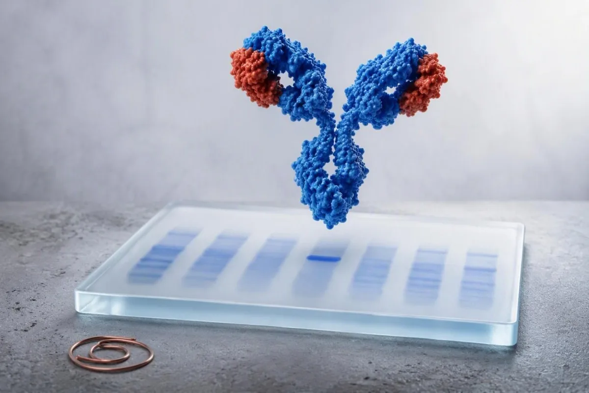
Իմունոֆիքսացիան պարզաբանում է, արդյոք SPEP-ի անսովոր նախշը ներկայացնում է հակամարմինների մեկ կլոն, թե հակված իմունային պատասխան: Խմբի տեսակը, դրա կայունությունը, ինչպես նաև երիկամների, արյան պատկերի, կալցիումի և ախտանիշների ձեր պրոֆիլը որոշում են հետագա քայլերը:.


- Բացակա խումբ նշանակում է, որ անալիզի միջոցով մոնոկլոնալ սպիտակուց չի հայտնաբերվել; այն չի բացատրում բոլոր բարձր սպիտակուցային արդյունքները:.
- Աննշան խումբ կարող է լինել անցողիկ՝ իմունային խթանումից հետո, սակայն 3-6 ամիս հետո շիճուկային IFE-ի կրկնությունը հաճախ իմաստալից է, երբ կլինիկական մտահոգությունը մնում է:.
- IgG մոնոկլոնալ խմբեր MGUS-ի ամենատարածված նախշն են և սովորաբար ռիսկային խմբավորվում են M-սպիտակուցի քանակի և ազատ թեթև շղթայի հարաբերակցության միջոցով:.
- IgA մոնոկլոնալ խմբեր կարող են տեղափոխվել բետա շրջանում, որտեղ SPEP-ը կարող է թերագնահատել կամ խաթարել չափելի սպիտակուցը:.
- IgM մոնոկլոնալ խմբեր տարբեր կլինիկական շրջանակ են պահանջում, քանի որ Վալդենստրյոմի մակրոգլոբուլինեմիան և որոշ լիմֆոմաներ մտնում են տարբերակիչ ախտորոշման մեջ:.
- Ազատ ծանր շղթաներ ավելացրեք էական տեղեկատվություն. 100 մգ/լ կամ ավելի արտահայտված ախտաբանական հարաբերակցությունը կամ ներգրավված ծանր շղթան պահանջում է անհապաղ մասնագիտական վերանայում:.
- Արտակարգ ախտանիշներ ներառեք նոր շփոթություն, ուժեղ թուլություն, միզարձակման նվազում, ուժեղ ծարավ, ոսկրային ցավ, կրկնվող վարակներ կամ անբացատրելի քաշի կորուստ:.
- Մեկ արդյունքը ախտորոշում չէ; բժիշկները մեկնաբանում են IFE-ն CBC-ի, կրեատինինի/eGFR-ի, շտկված կալցիումի, քանակական իմունոգլոբուլինների, մեզի թեստավորման և նախորդ արդյունքների կողքին:.
Իմունոֆիքսացիոն էլեկտրաֆորեզի արդյունքները, ինչ են իրականում ասում
Իմունաֆիքսացիոն էլեկտրաֆորեզի արդյունքները որոշում են, արդյոք նեղ սպիտակուցային ազդանշանը գալիս է մեկ հակամարմին արտադրող բջիջային կլոնից, և եթե այն առկա է, նշում են դրա ծանր և թեթև շղթան:. IFE արյան թեստի արդյունքը, ինչպիսին է “IgG կապպա մոնոկլոնալ գիծը”, հաստատում է մոնոկլոնալ սպիտակուցը; այն ինքնին չի ախտորոշում միելոմա կամ լիմֆոմա:.
SPEP-ը շիճուկային սպիտակուցները բաժանում է ըստ էլեկտրական շարժունակության և արտադրում է լայն պատկեր; IFE-ն այնուհետև կիրառում է հակաճիճուներ IgG-ի, IgA-ի, IgM-ի, կապպայի և լյամբդայի վրա: Իրական մոնոկլոնալ սպիտակուցը հայտնվում է նույն դիրքում մեկ ծանր շղթայի և մեկ թեթև շղթայի շերտում: Կարդացեք SPEP M-սպիկ ուղեցույց նախ, եթե հաշվետվությունն սկսվել է ախտաբանական սպիկով:.
Կանտեստին մի է AI արյան անալիզատոր որը կարդում է IFE բառապաշարը ընդհանուր սպիտակուցի, ալբումինի, գլոբուլինային բացի, երիկամային մարկերների, կալցիումի և պատմական պանելների կողքին, այլ ոչ թե “դրական”-ը համարելով ախտորոշում: 2026 թվականի սեպտեմբերի 28-ի դրությամբ, այդ կոնտեքստը ավելի կարևոր է, քան մեկուսացված մթագնած գիծը:.
Իմ կլինիկական աշխատանքում ամենատարածված թյուրիմացությունն այն է, որ ենթադրվում է, թե IFE-ն չափում է կլոնի չափը: Այն սովորաբար չի անում: Լաբորատորիան կարող է քանակականորեն որոշել M-սպիտակուցը SPEP-ում g/dL-ով, մինչդեռ IFE-ն հիմնականում հաստատում է նույնականությունը և կարող է հայտնաբերել չափից ավելի փոքր սպիտակուցներ, որոնք չեն կարող քանակականորեն որոշվել:.
Ինչու է լրացուցիչ թեստը հաջորդում SPEP-ին
IFE-ն ավելի զգայուն է և ավելի յուրահատուկ, քան SPEP-ը փոքր ախտաբանական սպիտակուցը բնութագրելու համար: Նորմալ թվացող SPEP-ը կարող է դեռ ունենալ դրական IFE, երբ կլոնը փոքր է, վատ է միգրացվում, կամ հիմնականում արտադրում է ազատ թեթև շղթաներ:.
Աննորմալ SPEP-ից հետո բացակայող կամ բացասական խմբեր
Բացասական իմունաֆիքսացիոն արդյունքը նշանակում է, որ լաբորատորիան չի հայտնաբերել առանձին մոնոկլոնալ իմունոգլոբուլին այդ շիճուկային նմուշում:. Սա սովորաբար արտացոլում է պոլիկլոնալ իմունային ակտիվություն, դեհիդրատացիա, լյարդի հիվանդություն կամ տեխնիկական SPEP անկանոնություն, այլ ոչ թե պլազմային բջիջների խանգարում:.
Լայն գամմա-շրջանի բարձրացումը սովորաբար պոլիկլոնալ է. բազմաթիվ B-բջիջների պոպուլյացիաներ միաժամանակ արտադրում են հակամարմիններ: Քրոնիկ լյարդի հիվանդություն, աուտոիմուն հիվանդություն, մշտական վարակ և վերջին իմունային ակտիվացումը կարող են սա պատճառել, ուստի Արյան շիճուկի սպիտակուցների և գլոբուլինների ուղեցույց օգտակար է, երբ ընդհանուր սպիտակուցը կամ գլոբուլինը բարձր է:.
Բացասական շիճուկային IFE-ն լիովին չի բացառում թեթև շղթայի հիվանդությունը: Եթե կա անբացատրելի անեմիա, երիկամային անբավարարություն, սպիտակուց մեզի մեջ, նեյրոպաթիա, սրտամկանային հիվանդություն կամ կասկածելի ոսկրային ախտանշաններ, բժիշկները սովորաբար ավելացնում են շիճուկային ազատ թեթև շղթաներ և մեզի էլեկտրաֆորեզ՝ մեզի IFE-ով:.
Գործնական հարցն այն է, արդյոք եղել է կլինիկական պատճառ նայելու: 4 գ/դլ-ից բարձր գամմա բացը հուշում է, ոչ թե հաստատված սկրինինգային ախտորոշում; Ես չէի ձեռնարկի լայնարար արյունաբանական թեստավորում այդ թվից միայն մեկուսացված, առողջ մարդու մոտ:.
Երբ բացասական թեստը հանգստացնող է
Բացասական IFE՝ նորմալ հեմոգլոբինով, կալցիումով, կրեատինինով և առանց անհանգստացնող ախտանշանների սովորաբար հանգստացնող է: Կրկնելը սովորաբար պայմանավորված է լաբորատոր պատկերի էվոլյուցիայով, այլ ոչ թե միայն անհանգստությամբ:.
Աննշան կամ հետքի չափով սահմանափակ խմբեր. կրկնե՞լ, անտեսե՞լ, թե՞ ուղղորդե՞լ:
Մթագնած սահմանափակ գիծը երկիմաստ ցածր մակարդակի գտածո է, որը հաճախ արժանի է հաստատման, այլ ոչ թե անմիջական բուժման կամ խուճապի:. Some disappear on repeat testing, while others represent early MGUS that remains stable for years.
A laboratory may say “faint,” “trace,” “restricted,” “small band,” or “cannot rule out monoclonal protein.” These terms are not interchangeable across laboratories. I usually want the exact report, an SPEP quantity if measurable, serum free light chains, and a repeat specimen from the same laboratory in about 3-6 months.
Transient oligoclonal bands can follow immune stimulation, transplantation, infection, or immune-directed therapy; several narrow bands rather than one matched heavy-light pair often support that explanation. Sample problems matter too—review hemolysis-related result errors before attributing a surprising pattern to disease.
Dr. Thomas Klein would be more concerned about a faint band that persists and coincides with falling hemoglobin, rising creatinine, elevated calcium, or a clearly abnormal free light-chain ratio. The combination changes the pre-test probability far more than the adjective “faint.”
Տիպավորված IgG կապպա կամ IgG լյամբդա խմբի արդյունքներ
An IgG kappa or IgG lambda band confirms the most common class of monoclonal protein and is often classified as MGUS when the M-protein is below 3 g/dL and there is no organ injury attributable to the clone. IgG type alone cannot tell whether a person has benign MGUS, smoldering myeloma, or active myeloma.
The International Myeloma Working Group defines MGUS by serum M-protein below 3 g/dL, marrow clonal plasma cells below 10%, and no CRAB organ damage or amyloidosis caused by the clone. Rajkumar et al. (2014) also recognize biomarkers that can define active myeloma before classic organ injury.
For non-IgM MGUS, low-risk features are IgG type, M-protein 1.5 g/dL or lower, and a normal free light-chain ratio. Using those factors, many low-risk patients can avoid immediate marrow sampling, although age, symptoms, and laboratory trajectory still matter.
Kantesti AI interprets typed IgG results by comparing the IFE language with free light-chain ratio findings and prior protein values. A rising quantified M-protein is more informative than a stable typed label.
Ինչպես է IgA մոնոկլոնալ խումբը փոխում հետազոտությունը
An IgA kappa or IgA lambda band is a monoclonal protein that deserves quantification and free light-chain testing because IgA often migrates in the beta region and may be underestimated on SPEP. The next step is usually hematology-guided risk assessment, not an assumption of cancer.
IgA monoclonal proteins may overlap transferrin, complement proteins, or beta-lipoproteins on electrophoresis. That is why a report can say “beta-region restriction” even when no obvious gamma spike appears. Quantitative IgA and serum free light chains help establish the biological burden.
In practice, an IgA band with M-protein 0.4 g/dL, normal CBC, eGFR, calcium, and stable light chains is managed very differently from an IgA band with anemia and a rapidly increasing value. The immunoglobulin results guide explains why uninvolved immunoglobulins may also be measured.
A suppressed uninvolved IgG or IgM—sometimes called immunoparesis—can signal a less diverse antibody response, but it is not diagnostic by itself. Recurrent bacterial infections are a clinical reason to take that finding more seriously.
IgM խմբի արդյունքներ և ինչու են դրանք տարբեր ուղի ընտրում
An IgM monoclonal band prompts assessment for IgM MGUS, Waldenström macroglobulinemia, and certain B-cell lymphomas rather than the standard non-IgM myeloma pathway. Serum viscosity symptoms and enlarged nodes, spleen, or liver alter the urgency.
IgM is a large pentameric antibody, so high concentrations can increase serum viscosity. Headache, blurred vision, spontaneous nose or gum bleeding, dizziness, confusion, or shortness of breath alongside a substantial IgM result should be assessed urgently; those symptoms are not explained by a faint IgM band alone.
A hematologist may order quantitative IgM, CBC with film, serum viscosity when clinically indicated, imaging, and sometimes marrow testing with molecular studies. A bone-marrow procedure is not automatic for every small, stable IgM protein.
Այն rouleaux formation guide helps explain why high circulating proteins can alter how cellular elements stack on a slide. That observation is supportive, not a substitute for IFE or a diagnosis.
Երբ IFE-ն ցույց է տալիս կապպա կամ լյամբդա առանց ծանր շղթայի
An isolated kappa or lambda restriction can indicate light-chain MGUS, light-chain myeloma, AL amyloidosis, or a small clone below heavy-chain detection. Serum free light-chain values, the kappa/lambda ratio, kidney function, and urine studies determine how concerning it is.
Typical Freelite reference intervals vary by assay, but many laboratories use a kappa/lambda ratio around 0.26-1.65 in people with preserved kidney function. Chronic kidney disease raises both chains and broadens interpretation; the ratio often remains closer to balanced than in a true single-clone process.
An involved/uninvolved free light-chain ratio of 100 or more, with involved chain at least 100 mg/L, is a myeloma-defining biomarker when confirmed in the appropriate setting. That threshold comes from the IMWG criteria summarized by Rajkumar et al. (2014), not from a routine screening cutoff.
Protein in urine or persistent foamy urine makes urine albumin testing and urine protein electrophoresis more relevant. Review մշտական փրփրուն մեզի մասին rather than assuming the appearance proves a monoclonal disorder.
Արդյունքներ, որոնք պահանջում են հեմատոլոգիայի հրատապ կամ շտապ գնահատում
IFE results need prompt specialist assessment when they accompany unexplained anemia, kidney decline, high calcium, bone pain, fractures, recurrent infections, or rapidly changing protein values. Emergency care is appropriate for confusion, severe dehydration, markedly reduced urine output, or acute neurologic symptoms.
The classic CRAB framework includes calcium elevation, renal impairment, anemia, and bone involvement. A corrected calcium above 11 mg/dL (2.75 mmol/L), hemoglobin more than 2 g/dL below the lower limit of normal or below 10 g/dL, and creatinine above 2 mg/dL are examples that warrant timely clinical evaluation when a clone is present.
A normal CBC does not rule out clinically significant disease, but it lowers the immediate concern substantially. Conversely, anemia from iron deficiency, kidney disease, or bleeding is common and must be evaluated on its own merits; see reticulocyte hemoglobin clues when iron delivery is uncertain.
In our medical review workflow, Kantesti is an ԱԻ լաբորատոր թեստի մեկնաբանման ծառայությունում that flags these combinations as follow-up triggers, not diagnoses. Automated interpretation must never delay urgent in-person assessment when symptoms are progressing.
MGUS ռիսկ և հետագա դիտարկման ժամկետներ դրական IFE-ից հետո
Most people with MGUS do not develop cancer, but progression risk averages about 1% per year and persists over time. Follow-up frequency is individualized by isotype, M-protein amount, free light-chain ratio, age, symptoms, and whether values are stable.
Kyle et al. (2006) found MGUS in about 3.2% of adults aged 50 years or older in a predominantly White population, with prevalence rising sharply with age. Population estimates vary by ancestry and ascertainment, so an individual’s risk should not be inferred from prevalence alone.
Many clinicians repeat CBC, creatinine, calcium, SPEP, and free light chains at 6 months after a new MGUS diagnosis. If low-risk values remain stable, annual review—or less frequent review in selected patients—may be reasonable; higher-risk patterns are followed more closely.
Կանտեստին մի է AI-ով աշխատող արյան թեստի վերլուծության գործիք that can preserve the exact prior M-protein and kidney values for trend review. Comparing like with like matters: a different electrophoresis method or laboratory can create an apparent shift that is analytical, not biological.
Երբ աննորմալ սպիտակուցային նախշերը մոնոկլոնալ հիվանդություն չեն
High total protein or an abnormal electrophoresis contour can occur without a monoclonal protein. Dehydration concentrates albumin and globulins, while liver disease, autoimmune conditions, and chronic immune stimulation commonly create a broad polyclonal pattern.
Albumin and globulin should be read together. A high total protein with normal albumin suggests increased globulins, but dehydration can elevate both; repeat testing after ordinary hydration is often more informative than aggressive fluid loading before the draw.
Polyclonal hypergammaglobulinemia is common in chronic hepatic disease and systemic immune disorders. If liver enzymes, bilirubin, or albumin are abnormal, the MASLD laboratory overview may provide a more plausible explanation than a plasma-cell condition.
Monoclonal and polyclonal patterns can coexist. That nuance is why I do not dismiss a clear small IFE band merely because CRP is high, nor label all high globulins as a clone.
Փորձարկման սահմանափակումներ, դեղորայք և կրկնակի փորձարկման լավագույն պլան
Immunofixation is highly specific for identifying an immunoglobulin type, but its detection threshold, reporting language, and interference profile differ by laboratory. Repeating a borderline result at the same laboratory improves comparability.
Therapeutic monoclonal antibodies can appear as bands and may be mistaken for endogenous proteins unless the laboratory knows the treatment. Daratumumab is a familiar example in myeloma care, but other antibody therapies can also complicate interpretation; bring a complete medication list.
Recent intravenous immunoglobulin can produce temporary electrophoretic abnormalities for weeks because infused donor antibodies remain measurable. A lab should know the infusion date, product, and indication before issuing a conclusion about a new band.
Fasting is not usually required for IFE, although a severely lipemic specimen can interfere with some laboratory processes. Our lipemic sample explanation covers when a redraw is more reliable than over-interpreting a compromised sample.
IFE-ի արդյունքից հետո բժշկին տալիք հարցեր
The most useful question after a positive IFE is “What is the quantified M-protein, and do my CBC, calcium, kidney function, and free light chains show an effect?” Asking for the actual numbers converts a frightening label into a practical evaluation plan.
Ask whether the result is definite, faint, or equivocal; whether the heavy and light chains match; and whether an M-protein was quantifiable in g/dL. Also ask whether this is new compared with prior SPEP or IFE results. A one-page բժշկի այցի ստուգաթերթիկը can keep those details together.
Ask specifically about urine testing if you have swelling, neuropathy, proteinuria, or unexplained kidney changes. In a 68-year-old patient I recently reviewed, a small IgG band mattered less than the newly abnormal urine protein and falling eGFR—which appropriately accelerated referral.
Dr. Thomas Klein’s practical advice is to avoid self-directed supplements marketed for “protein balance.” There is no supplement that removes a monoclonal clone, and unnecessary products can blur kidney or liver assessment.
AI-ի օգտագործումը IFE արյան անալիզի արդյունքները անվտանգ կերպով կազմակերպելու համար
AI can organize IFE blood test results, identify missing companion tests, and show trends, but it cannot determine whether a monoclonal protein is causing symptoms or replace a hematology assessment. Any new typed band should be reviewed by the clinician who ordered the test.
Kantesti AI interprets monoclonal protein immunofixation language by linking it to CBC, calcium, eGFR, total protein, albumin, and prior laboratory reports. It is especially useful when a report is photographed or uploaded from another language, but original laboratory units and reference intervals should always be retained.
Our approach is governed by clinical oversight and source checking; readers can review our technical clinical validation when deciding how to use automated lab interpretation. Privacy-conscious organization is useful, yet an algorithm cannot examine lymph nodes, assess fractures, or judge constitutional symptoms.
If you are gathering reports for a hematology visit, save the full SPEP tracing, IFE interpretation, free light-chain values, quantitative immunoglobulins, and at least 12 months of CBC and renal results. That sequence is often more valuable than repeating a single panel early.
Ցանկացած IFE արդյունքի համար գործնական հաջորդ քայլերի պլան
Negative IFE with reassuring companion tests usually means clinical observation; a faint band usually means confirmation; and a definite typed band usually means risk stratification with free light chains, CBC, calcium, creatinine, and often hematology input. The result is a starting point, not a verdict.
For an absent band, clarify whether the original SPEP was broad rather than narrow and address the likely cause of elevated proteins. For a faint band, repeat serum studies in 3-6 months unless symptoms or abnormal companion markers justify earlier referral.
For a typed IgG or IgA band, document M-protein g/dL, free light-chain ratio, immunoglobulins, CBC, corrected calcium, and renal function. For IgM, add a symptom review for hyperviscosity and lymphoproliferative features; for light-chain-only patterns, urine and organ-specific assessment become more central.
Kantesti’s physician-reviewed educational standards are guided by our Բժշկական խորհրդատվական խորհուրդ. The sensible endpoint is a shared plan with your clinician: what to repeat, where, when, and which symptom should prompt you to call sooner.
Հաճախակի տրվող հարցեր
Ի՞նչ է նշանակում իմունաֆիքսացիոն էլեկտրաֆորեզի դրական արդյունքը։
Իմունաֆիքսացիոն էլեկտրոֆորեզի դրական արդյունքը նշանակում է, որ լաբորատորիան հայտնաբերել է մոնոկլոնալ իմունոգլոբուլին և բացահայտել է դրա տեսակը, ինչպիսին է IgG կապպա, IgA լյամբդա կամ IgM կապպա: Այն ինքնին չի ախտորոշում մուլտիպլ միելոմա, քանի որ MGUS-ը տարածված է և հաճախ կայուն: Կլինիցիստները մեկնաբանում են արդյունքը M-պրոտեինի քանակի (գ/դլ), շիճուկի ազատ թեթև շղթաների, CBC, կրեատինինի, կալցիումի, ախտանշանների և երբեմն միզի թեստավորման հետ մեկտեղ: M-պրոտեինը, որը 3 գ/դլ-ից ցածր է, առանց կլոնի հետ կապված օրգանների վնասի, կարող է համապատասխանել MGUS չափանիշներին, այլ ոչ թե ակտիվ չարորակ ուռուցքին:.
Իմունոֆիքսացիայի վրա բարակ գոտին լուրջ է։
Իմունոֆիքսացիայի վրա թույլ զոլիկն ինքնաբերաբար լուրջ չէ, սակայն այն պետք է մեկնաբանել որպես ցածր մակարդակի կամ երկդիմի արդյունք, այլ ոչ թե անտեսել: Շատ կլինիցիստներ կրկնում են SPEP, IFE և շիճուկային ազատ թեթև շղթաները մոտ 3-6 ամիս հետո՝ որոշելու համար, արդյոք զոլիկը պահպանվում է: Ավելի վաղ հեմատոլոգիական վերանայումը հարմար է, եթե հեմոգլոբինը 10 գ/դլ-ից ցածր է, շտկված կալցիումը գերազանցում է 11 մգ/դլ-ը, երիկամների ֆունկցիան վատթարանում է, կամ առկա են ախտանիշներ, ինչպիսիք են ոսկրային ցավը կամ կրկնվող վարակները: Լաբորատոր ճշգրիտ ձևակերպումը և նախորդ թեստի նախշը կարևոր են:.
Ի՞նչ տարբերություն կա SPEP-ի և իմունաֆիքսացիոն էլեկտրաֆորեզի միջև։
SPEP-ն շիճուկային սպիտակուցները բաժանում է և կարող է քանակականորեն որոշել չափելի M-սպիկը գ/դլ-ով, մինչդեռ իմունաֆիքսացիոն էլեկտրաֆորեզը հայտնաբերում է նեղաշերտի համար պատասխանատու հակամարմնի դասը և թեթև շղթան։ SPEP-ն օգտակար է ժամանակի ընթացքում սպիտակուցի քանակը հետևելու համար, մինչդեռ IFE-ն ավելի զգայուն է փոքր սպիտակուցների համար և ավելի ճշգրիտ է դրանց տեսակավորման համար։ Մարդը կարող է ունենալ դրական IFE առանց քանակական SPEP M-սպիկի։ Երկու թեստերն էլ սովորաբար պատվիրվում են շիճուկային ազատ թեթև շղթաների հետազոտության հետ միասին։.
IgG կապան միշտ նշանակո՞ւմ է MGUS։
IgG կապան ամենատարածված մոնոկլոնալ սպիտակուցի տեսակն է և կարող է ներկայացնել MGUS, smoldering myeloma, ակտիվ միելոմա, AL ամիլոիդոզ կամ պլազմային բջիջների այլ վիճակ։ MGUS-ը պահանջում է շիճուկում M-սպիտակուցի 3 գ/դլ-ից ցածր մակարդակ, 10% կլոնային ոսկրածուծի պլազմային բջիջներից պակաս, և օրգանների վնասվածքի բացակայություն, որը կապված չէ կլոնի հետ։ Ցածր ռիսկի MGUS-ն սովորաբար ունենում է IgG տեսակ, M-սպիտակուց 1.5 գ/դլ կամ ցածր, և լույսի ազատ շղթաների նորմալ հարաբերակցություն։ Հետևաբար, տիպիզացված շերտը պահանջում է ռիսկի գնահատում, այլ ոչ թե միայն տեսակի վերաբերյալ եզրակացություն։.
Կարո՞ղ է իմունաֆիքսացիան բացասական լինել, եթե ունեք միելոմա։
Շիճուկային իմունոֆիքսացիան կարող է բացասական լինել ոչ սովորական ոչ-սեկրետորային կամ նվազագույնս սեկրետորային միելոմայի դեպքում և բաց թողնել դեպքեր, որոնք հիմնականում արտադրում են ազատ թեթև շղթաներ։ Շիճուկային ազատ թեթև շղթաների թեստավորումը և մեզի իմունոֆիքսացիան բարելավում են հայտնաբերումը, երբ կլինիկական կասկածը մնում է բարձր։ 100 կամ ավելի ազատ թեթև շղթայի հարաբերակցությունը՝ առնվազն 100 մգ/լ ներգրավված թեթև շղթայով, միելոմա-սահմանող կենսաբանական մարկեր է համապատասխան կլինիկական պայմաններում։ Նորմալ IFE-ն հուսադրող է, բայց չի հաղթահարում անեմիան, երիկամների անկումը, բարձր կալցիումը կամ պատկերային ախտորոշման արդյունքները։.
Որքա՞ն հաճախ պետք է կրկնեմ իմունաֆիքսացիան MGUS-ից հետո։
Շատ կլինիցիստներ կրկնում են լաբորատոր հետազոտությունները 6 ամիս անց MGUS-ի նոր հայտնաբերումից հետո, ներառյալ CBC, կրեատինին, կալցիում, SPEP և շիճուկի ազատ թեթև շղթաները: Եթե ցածր ռիսկի արժեքները կայուն են, հետագա հետևումը կարող է դառնալ ամենամյա կամ ավելի հազվադեպ՝ կախված տարիքից, ախտանիշներից և հեմատոլոգիայի տեղական պրակտիկայից: Ավելի բարձր ռիսկի գնահատականները, ներառյալ ոչ IgG տեսակը, M-սպիտակուցը 1,5 գ/դլ-ից բարձր, կամ ազատ թեթև շղթայի անբնական հարաբերակցությունը, սովորաբար արդարացնում են ավելի մանրամասն մոնիտորինգը: Նոր ախտանիշը կամ M-սպիտակուցի բարձրացումը պետք է հանգեցնի վերանայման՝ նախքան նախատեսված ժամկետը:.
Ստացեք AI-ով աշխատող արյան անալիզի վերլուծություն այսօր
Միացեք ավելի քան 2 միլիոն օգտատերերի ամբողջ աշխարհում, ովքեր վստահում են Kantesti-ին՝ լաբորատոր թեստերի ակնթարթային, ճշգրիտ վերլուծության համար։ Վերբեռնեք ձեր արյան անալիզի արդյունքները և ստացեք 15,000+ բիոմարկերների համապարփակ մեկնաբանություն վայրկյանների ընթացքում։.
📚 Հղված հետազոտական հրապարակումներ
Klein, T., Mitchell, S., & Weber, H. (2026). Klein, T. (2026). BUN/Creatinine Ratio Explained: Kidney Function Test Guide. Zenodo. https://doi.org/10.5281/zenodo.18207872. ResearchGate: https://www.researchgate.net/ Academia.edu: https://www.academia.edu/.։ Kantesti AI Medical Research.
Klein, T., Mitchell, S., & Weber, H. (2026). Klein, T. (2026). Urobilinogen in Urine Test: Complete Urinalysis Guide 2026. Zenodo. https://doi.org/10.5281/zenodo.18226379. ResearchGate: https://www.researchgate.net/ Academia.edu: https://www.academia.edu/.։ Kantesti AI Medical Research.
📖 Արտաքին բժշկական հղումներ
⚕️ Բժշկական հրաժարում
Այս հոդվածը միայն կրթական նպատակների համար է և չի հանդիսանում բժշկական խորհրդատվություն։ Ախտորոշման և բուժման որոշումների համար միշտ խորհրդակցեք որակավորված բուժաշխատողի հետ։.
E-E-A-T վստահության ազդանշաններ
Փորձառություն
Բժշկի կողմից ղեկավարվող կլինիկական վերանայում՝ լաբորատոր մեկնաբանության աշխատանքային հոսքերի համար։.
Մասնագիտություն
Լաբորատոր բժշկության կենտրոնացում՝ այն բանի վրա, թե ինչպես են բիոմարկերները դրսևորվում կլինիկական համատեքստում։.
Հեղինակություն
Գրված է դոկտոր Թոմաս Քլայնի կողմից՝ դոկտոր Սառա Միթչելի և պրոֆեսոր դոկտոր Հանս Վեբերի կողմից վերանայմամբ։.
Հուսալիություն
Ապացույցների վրա հիմնված մեկնաբանություն՝ հստակ հետագա քայլերի ուղիներով՝ ահազանգերը նվազեցնելու համար։.