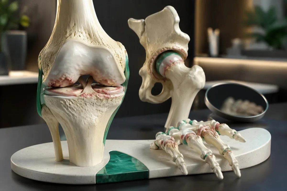
最初的警示信号通常是活动期间或之后的关节疼痛、休息后僵硬(通常持续不到30分钟),以及活动范围逐渐减小。膝盖可能在上下楼梯时疼痛,髋部可能在穿袜子时疼痛,手部则可能在抓握时疼痛。血液检查无法确诊骨关节炎;关节发热且迅速肿胀时,需要紧急评估。

要点
- 与活动相关的疼痛 常见于步行、上下楼梯、抓握或反复负重关节活动时;仅有一种活动会引起疼痛,并不足以确诊。
- 短暂僵硬 典型骨关节炎患者醒来或久坐后出现的僵硬通常不超过30分钟。
- 临床诊断 根据NICE的建议,对于45岁或以上、有关节活动相关疼痛且晨僵缺如或短暂的成年人,这通常已足以作出诊断。
- 膝部症状 包括上下楼梯时疼痛、难以完全伸直关节,以及间歇性肿胀;单独出现无痛弹响不能作为诊断依据。
- 髋部症状 可能包括腹股沟疼痛、难以穿袜子,以及牵涉至膝部的疼痛——即使髋部没有明显痛感。
- 手部症状 常影响拇指根部以及靠近指甲或位于手指中部的指关节;麻木提示可能还存在其他问题或不同的问题。
- 血液检查的局限 意味着没有任何常规 CRP、ESR、类风湿因子或代谢面板检查结果能够确诊或排除骨关节炎。
- 紧急警示信号 包括关节突然发热、肿胀,受伤后无法负重,或腿部肿胀并伴有胸痛或呼吸急促;不要等待 AI 解读。
骨关节炎的早期征兆有哪些?
骨关节炎的早期征兆 包括与活动相关的关节疼痛、休息后短暂僵硬,以及舒适活动范围逐渐减小。症状模式比单一症状更重要:NICE 允许对年龄为 45 岁或以上 、出现与活动相关的疼痛且没有晨僵,或僵硬持续时间不超过 30分钟内避免咖啡因、尼古丁和运动,并进行.
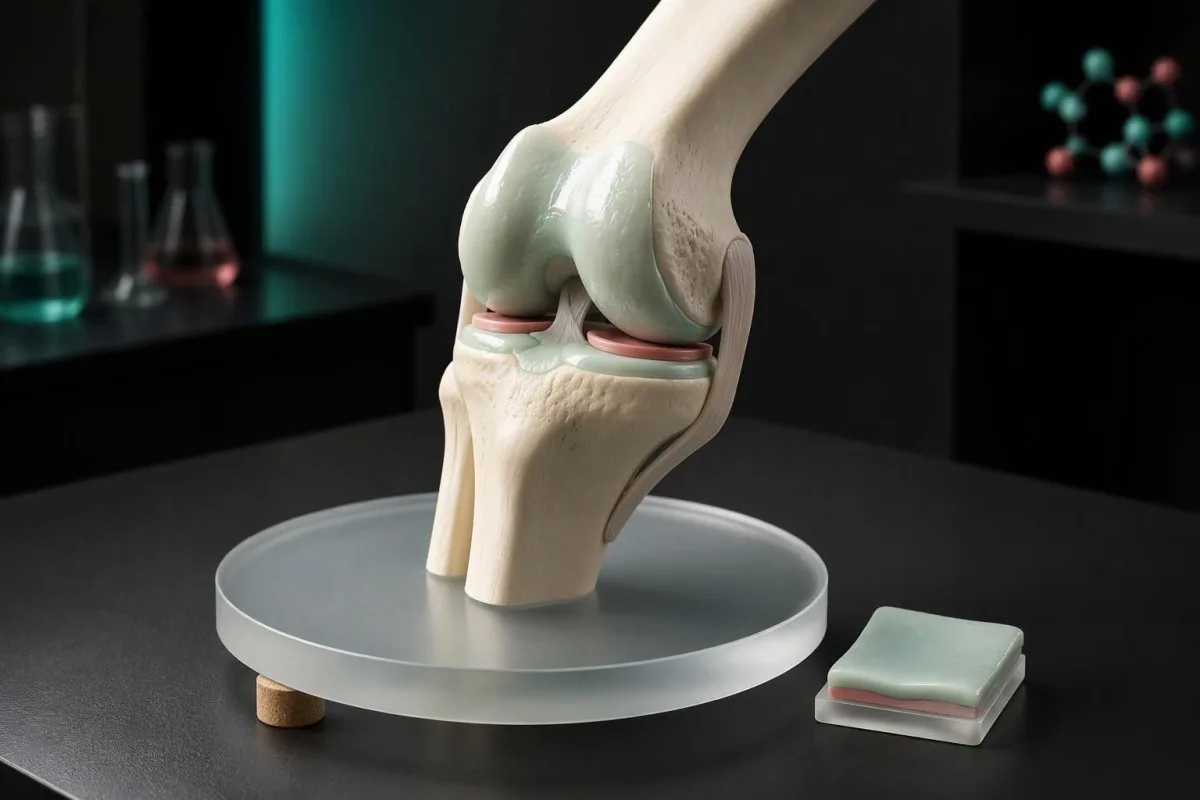
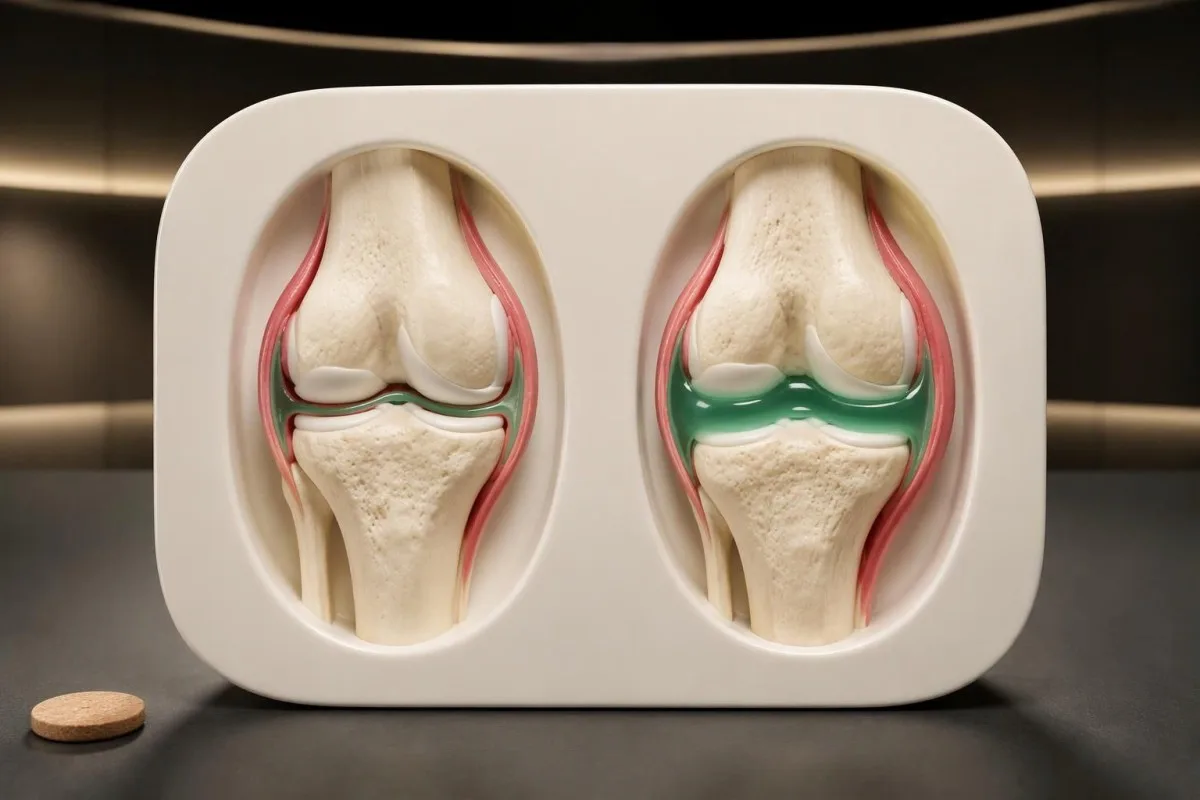
骨关节炎影响整个关节,而不只是覆盖骨骼的软骨。关节内膜、下方骨骼、支撑肌肉和韧带的变化都可能导致症状;NICE 规定的 45 岁 年龄门槛描述的是一种实用的诊断途径,并非排除年轻成人的生物学规则(NICE,2022 年)。
例如,假设有一名 一位 56 岁的人 发现长距离步行后膝部酸痛,早餐后僵硬持续 8分钟 ,并且难以跪下,而几个月前还没有这种情况。综合来看,这些表现比单独出现弹响更支持进行骨关节炎评估,但检查仍需排查肌腱问题、损伤和炎性关节病。
Kantesti 是一个 人工智能血液检测分析仪 用于解释实验室检查结果,但不会将其视为骨关节炎的证据。我们的 组织和临床范围 与面对面关节检查相互独立:记录 3 个细节——诱发症状的活动、僵硬持续时间以及功能丧失——通常比安排一组广泛的实验室检查更能为临床医生提供有用的评估起点。
与活动相关的关节疼痛通常有什么表现?
骨关节炎疼痛 通常会在患病关节负重时或负重后加重,减少负重后会缓解,尤其是在病情早期。一个有用的区分是:休息后最初 几步 时出现的僵硬,与步行 20分钟 后逐渐加重的酸痛;两者可能同时发生在同一个关节。
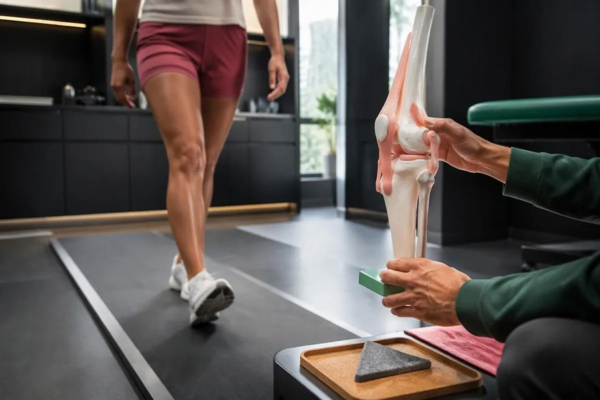
一个可重复的诱因有助于定位问题,但本身并不能确定病因。在假设的日记中,膝痛从 2/10升至5/10 在爬楼梯时加重提示对负荷敏感;肌腱、既往损伤或髌股关节引起的疼痛也可能呈现类似模式,并可能与骨关节炎并存。
静息时疼痛并不能自动排除骨关节炎,因为病情较明确时可能会影响睡眠,或在忙碌一天后出现酸痛。不过,新的持续性静息痛、在 连续数天内, 内迅速恶化,或伴有发热或不明原因体重减轻的疼痛,都值得接受评估,而不应归因于已有的X线检查所见。
小腿疼痛若总是在相似的步行距离后出现,并在停止后 几分钟 内缓解,可能反映的是血液循环问题,而非膝关节问题。我们的 下肢动脉性疼痛 指南解释了这种区别;请留意,仅仅停下来是否能缓解,还是改变关节位置才会带来缓解。
骨关节炎最先出现哪些膝部症状?
膝骨关节炎症状 往往从上下楼梯、从椅子上起身、蹲下或比平时走得更远时疼痛开始。早期不适可能出现在髌骨周围或关节线两侧;一位假设的 60岁 患者若连一层楼梯都难以爬上去,需要检查疼痛模式,而不能仅凭疼痛部位作出诊断。
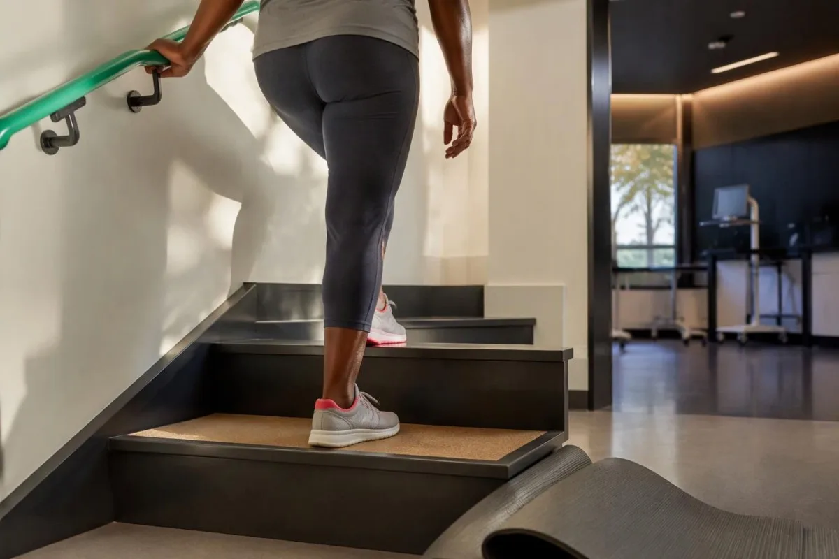
膝骨关节炎可能伴有磨擦感或咔嗒声,但仅凭响声不能说明软骨丢失,也不能确定疼痛程度。应改为比较 2个特征 响声是否伴有疼痛,以及活动是否发生变化;膝关节功能仍正常时的无痛弹响,不如新出现的、无法舒适伸直膝关节那么有参考价值。
间歇性肿胀可能反映关节内有积液,而有无力感可能是疼痛相关的肌肉抑制所致,并非韧带撕裂。膝关节若出现物理性卡住、无法伸直的情况——即 真正的膝关节锁定,哪怕只发生过一次,也需要尽快评估,因为机械性梗阻可能需要采用不同的处理方案。
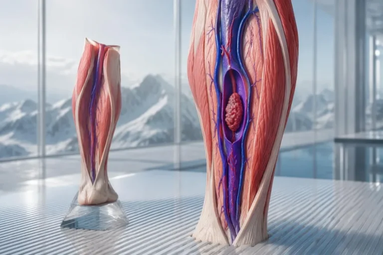
肿胀蔓延至小腿时,不应自动归咎于膝关节炎或膝后充液囊肿。 单侧腿部, 新发肿胀,尤其伴有小腿压痛或存在血栓风险因素时,需要紧急评估;我们的 腿部肿胀紧急处理指南 解释了为何在假定病因无害之前,需要进行检查和适当的检测。
髋关节骨关节炎会引起腹股沟或膝部疼痛吗?
髋骨关节炎 通常会导致腹股沟疼痛和髋部旋转困难,但疼痛也可能延伸至大腿、臀部或膝盖。如果 1 个膝盖 疼痛却找不到令人信服的膝部原因,检查髋部可能会发现疼痛来源;穿袜子或上车时可能会显露出活动受限。
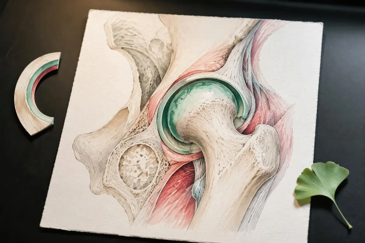
髋部向内旋转时感到不适,可能是检查线索,但没有任何单一动作能够确诊。在一个假设的 58 岁患者, 中,行走后腹股沟酸痛且难以够到脚,比单纯髋部外侧压痛更提示髋关节问题;后者通常指向周围肌腱。
疼痛呈灼烧感并向膝盖以下放射、刺痛或无力,提示可能存在神经因素,而非单纯骨关节炎。我们对 坐骨神经痛和神经体征 的说明区分了这些表现;一个人可能同时有 2 个问题,因此,X 光片显示髋部关节炎,也不应让评估忽视神经系统症状。
髋部功能突然发生变化,应比逐渐加重的僵硬受到更多重视。例如,一个假设的 67岁的人 患者在轻微跌倒后出现明显腹股沟疼痛且无法负重,需要紧急评估是否骨折,即使此前的影像检查显示有骨关节炎;有些骨折在首次普通 X 光片上无法显示。
骨关节炎的手部症状是什么感觉?
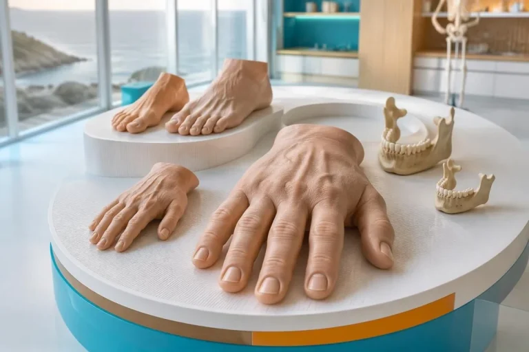
手部骨关节炎症状 通常包括捏握时酸痛、静止一段时间后僵硬,以及灵活性下降。拇指根部关节和 2 组指关节——靠近指甲的关节和手指中部的关节——是常见部位;坚硬的骨性增大可能在之后才出现,而非最初的警示信号。
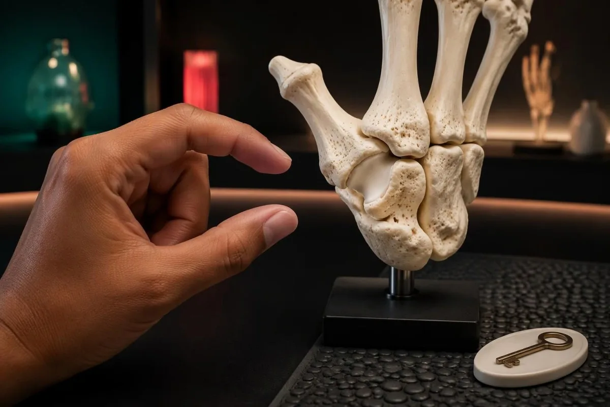
拇指根部骨关节炎可能最初表现为转钥匙、开罐子或长时间握手机时感到不适。一个假设的 62 岁患者 仍可能全手握力很强,却难以精确捏取物品,因为这些动作的受力分布不同;功能下降并不总等同于全身性无力。
麻木并非指关节骨关节炎的典型直接影响。拇指、食指、中指和无名指部分区域出现刺痛,可能提示正中神经受压;我们的指南介绍了 腕管综合征手指分布模式 解释了为什么大约 3½根手指 出现症状时,需要进行与疼痛性指骨关节不同的检查。
肿胀的分布会改变鉴别诊断:指关节处持续的软性肿胀,不如指尖附近的坚实增大常见。如果手部增大伴有 另外2种变化,例如鞋码变大和面部特征改变,我们的 手部增大警示信号 会说明为什么需要考虑内分泌病因,而不是直接认定为关节炎。
骨关节炎导致的僵硬会持续多久?
骨关节炎的僵硬 通常持续不超过 30分钟内避免咖啡因、尼古丁和运动,并进行 起床后,并且坐下后往往持续时间更短。这种短暂的“胶着感”不同于持续的炎症性僵硬,但这一界限只是临床参考,并非精确计时测试:必须综合考虑持续时间、肿胀、受累关节和全身症状。
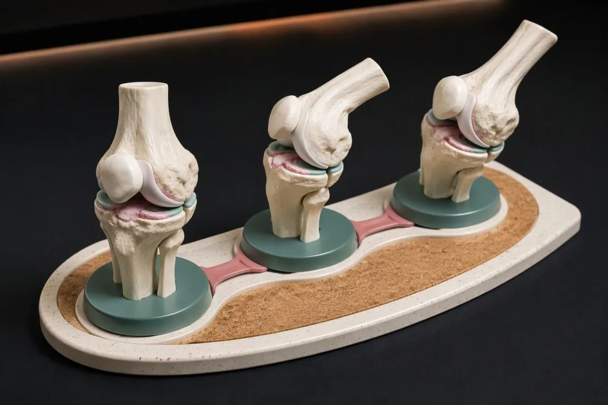
测量僵硬的时间,直到关节活动基本自如,而不是直到疼痛完全消失。在一个假设的 7天 日记中,僵硬持续 5–12分钟 ,随后出现与活动相关的酸痛,这与每天早晨有一小时难以活动的情况属于不同模式;将疼痛与僵硬区分开,可避免高估时间。
年龄超过 50岁以上的成人 的人出现肩部和髋带持续僵硬时,应考虑风湿性多肌痛,尤其是晨僵超过 45分钟. 我们的指南 风湿性多肌痛检测限值 说明了为什么炎症指标正常并不总能排除这一可能;其表现模式比单侧髋关节活动受限更广泛。
之前开始的背痛 45 岁, 持续超过 超过,且活动后改善而非休息后改善,可能提示炎性脊柱疾病。 HLA-B27的临床背景 比单独的基因检测结果更重要;阳性检测结果或僵硬持续时间都不能单独确立诊断。
哪些活动能力变化值得记录?
活动受限 可能表现为膝盖无法完全伸直、髋部旋转困难,或无法舒适地握拢手掌。比较相同的 2 或 3 项日常活动 随时间的变化,而不要强迫关节超出其活动范围;功能的变化往往比在家估算活动角度更能清楚地反映问题。
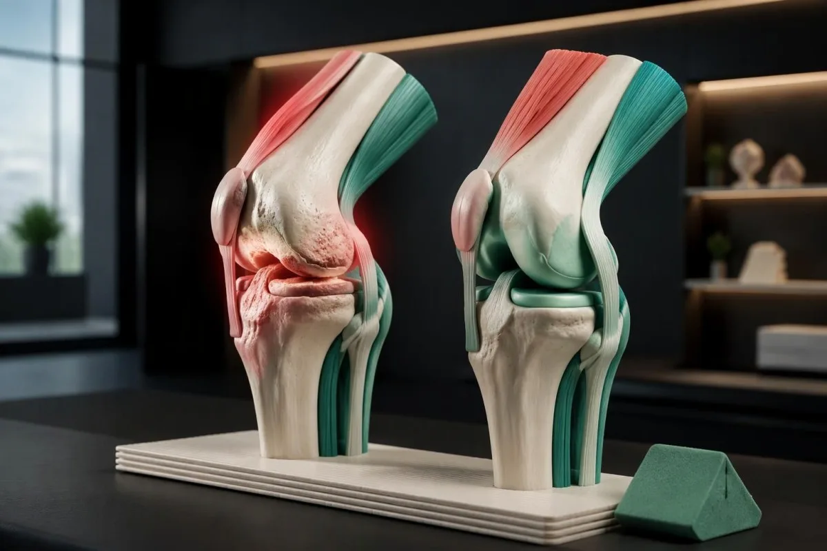
站立时膝盖仍微微弯曲,可能会改变步态,并增加周围肌肉所需的发力。临床医生可能会以大约 0 度, 的伸直位置为基准测量伸展情况,但个体差异、既往损伤和检查方法都很重要;测量到的轻微差异本身不能作为诊断,也不构成强行拉伸的理由。
选择能反映受影响关节情况的活动: 1 段楼梯 膝关节可选择上下楼,髋关节可选择穿袜子,拇指可选择转动熟悉的钥匙。在Kantesti,我们会区分功能观察与实验室结果解读,因为稳定的 CRP 结果无法显示一个人是否已失去完成这些动作的能力。
骨质疏松症和骨关节炎虽然名称相似,但描述的是不同的问题。骨质疏松症通常在发生骨折前没有明显症状,而骨关节炎通常会引起关节相关症状;我们的 骨量减少检测指南 说明了为何即使发生 1 次低创伤性骨折 也足以成为进行骨骼健康评估的理由,但这并不意味着僵硬的手指关节是由骨质疏松症造成的。
骨关节炎会导致肿胀或发热吗?
骨关节炎可导致关节间歇性肿胀和轻微发热,因为关节局部内衬的变化是该病的一部分。突然出现明显发热、严重肿胀的 单个关节 则是另一种情况:即使患者已确诊骨关节炎,也需要紧急评估是否存在感染、晶体性关节炎或其他急性病因。
坚实的骨性增大与软性的液体相关肿胀并非同一种体征。在一个假设的 64 岁患者, 中,长期存在的手指关节硬性凸起可能反映骨关节炎,而逐渐增大的膝盖 12小时 则值得重新评估;变化速度可能比关节此前是否被诊断为关节炎更有参考价值。
Normal blood results cannot safely dismiss an acutely hot joint. Our explanation of WBC 和 CRP 的差异 shows why timing and clinical findings matter; a result collected early on day 1 may not reflect the full response, and joint infection can occur without striking systemic laboratory abnormalities.
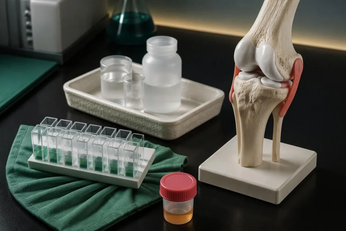
Joint-fluid testing is sometimes needed because examination and blood markers cannot reliably separate all acute causes. Fluid may be checked for crystals, cellular counts, and culture; identifying 1 crystal type does not automatically exclude simultaneous infection, so a clinician must interpret the sample alongside symptoms, risk factors, and the course of the illness.
为什么血液检查无法确诊骨关节炎?
No routine blood test confirms or excludes osteoarthritis. Diagnosis depends on symptoms, examination, and sometimes imaging; CRP, ESR, and antibody tests can investigate alternative explanations but cannot measure whether a particular knee or thumb joint has osteoarthritis. There are 0 standard laboratory confirmation tests for routine clinical osteoarthritis diagnosis.
CRP is a nonspecific marker of systemic inflammation, not a cartilage-loss measurement. If a hypothetical laboratory uses a reference interval below 5 mg/L;而高于, a CRP of 18 mg/L calls for clinical context rather than proving inflammatory arthritis; local osteoarthritis changes, another illness, and other contributing factors can coexist, and reference intervals differ between laboratories.
Rheumatoid factor and anti-CCP testing are most useful when the history and examination suggest inflammatory arthritis, especially persistent synovitis. Our guide to 类风湿因子解读 如何看血液检查报告 解释了为什么 1 positive antibody result cannot replace the examination; negative antibodies also do not exclude rheumatoid arthritis or turn an uncertain joint problem into confirmed osteoarthritis.
Kantesti 是一个 AI 血液检查解读平台 that helps explain reported markers without claiming to identify osteoarthritis from them. Our 生物标志物解读指南 supports checking 3 report details—the units, laboratory reference interval, and collection date—before discussing an abnormal result with a clinician; broad screening panels are not automatically useful for a typical osteoarthritis presentation.
哪些症状模式提示可能是其他诊断?
Persistent soft joint swelling, prolonged morning stiffness, acute redness, nerve symptoms, or illness outside the joints can suggest something other than osteoarthritis. Pattern recognition helps select testing: swelling across several knuckles points toward a different assessment from isolated thumb-base pain, while abrupt swelling in 1 joint raises crystal or infectious causes.
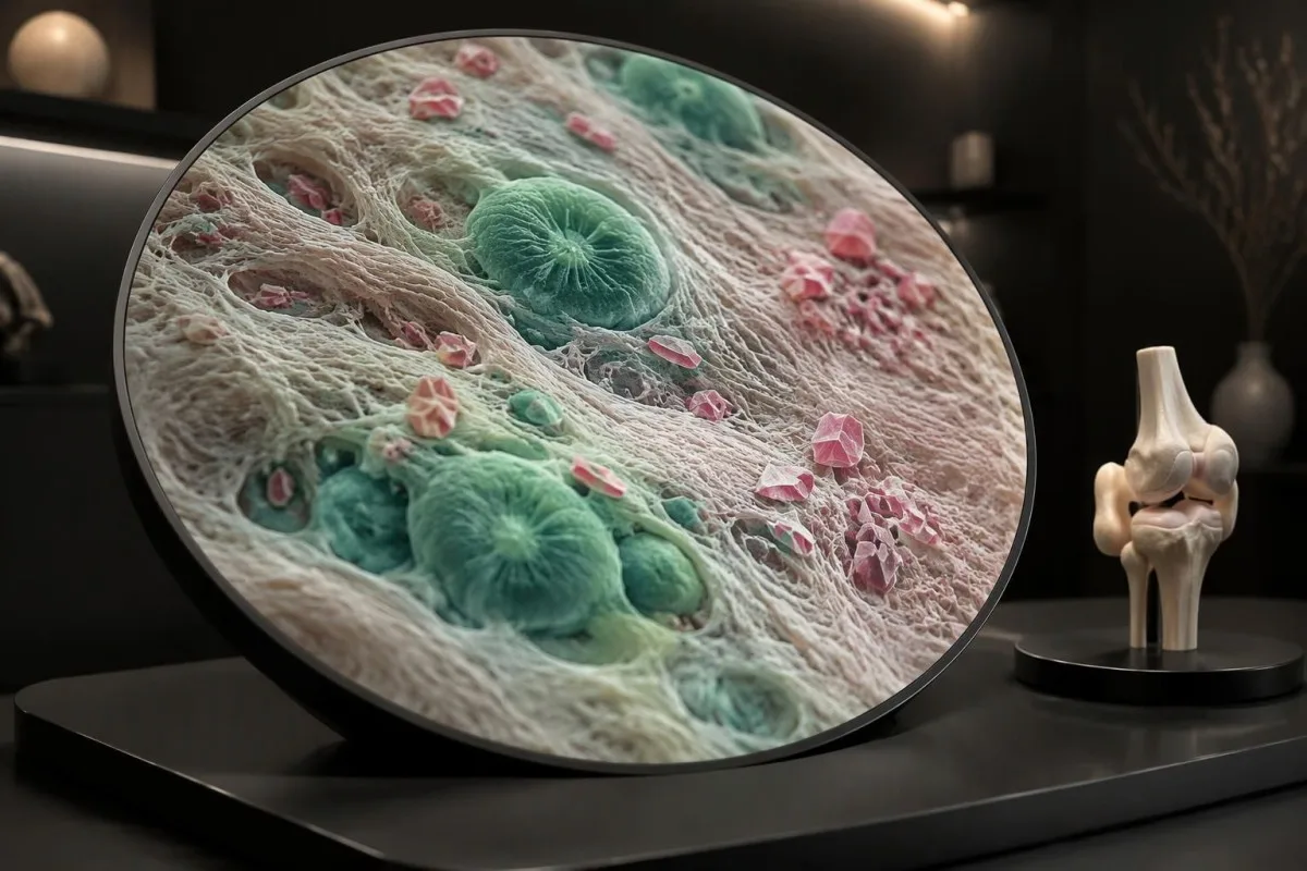
Symmetrical knuckle swelling with prolonged stiffness can suggest rheumatoid arthritis, while Raynaud symptoms, rashes, or muscle weakness may broaden the autoimmune assessment. Our connective tissue testing guide 如何看血液检查报告 解释了为什么 1 antibody, such as anti-RNP, is insufficient by itself; ordering many antibodies without a compatible symptom pattern can create misleading incidental positives.
Gout and calcium pyrophosphate crystal arthritis can cause sudden attacks, and calcium pyrophosphate deposition may also coexist with osteoarthritis. A serum urate below 6 mg/dL during an acute episode does not reliably rule out gout; when the diagnosis or infection risk is uncertain, examining joint fluid can be more informative than repeating a routine blood panel.
Iron overload can produce an osteoarthritis-like pattern, sometimes involving the second and third knuckles. Persistent transferrin saturation around 45% or higher can prompt iron-overload assessment, but does not establish its cause; Kantesti does not recommend this test for every aching joint, and our complete iron study explanation distinguishes saturation from ferritin, which can also rise with inflammation or liver disease.
什么时候需要做X光或MRI扫描?
Imaging helps when symptoms are atypical, the diagnosis is uncertain, or the result could change treatment. NICE does not recommend routine diagnostic imaging for a typical clinical presentation; EULAR likewise advises that imaging is unnecessary in typical osteoarthritis and identifies plain radiography as the first imaging choice when imaging is needed.
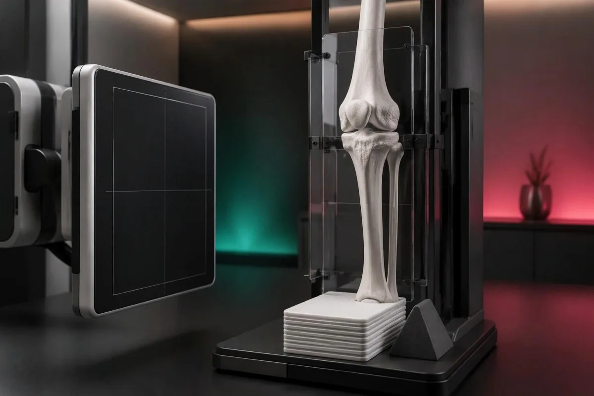
For the knee, weight-bearing radiographs can make joint-space narrowing more apparent than a non-weight-bearing view. EULAR recommends appropriate views and techniques rather than indiscriminate scanning (Sakellariou et al., 2017); importantly, 1 X-ray cannot directly measure pain, and prominent structural changes may coexist with modest symptoms while early symptoms can precede obvious radiographic changes.
MRI is not a routine second step simply because an osteoarthritis X-ray looks mild. It can be useful for a suspected occult fracture, another structural disorder, or selected mechanical symptoms; in a hypothetical 66-year-old with abrupt severe pain and difficulty bearing weight despite an unrevealing X-ray, the clinical concern—not the desire to grade arthritis—determines further imaging.
Repeat scans are usually less useful than reassessing symptoms and function when management remains unchanged. NICE advises against routine follow-up imaging for nonsurgical management (NICE, 2022); Kantesti's 方法学和临床局限 should be read with the same distinction in mind: a technical interpretation benchmark is not proof that an AI can diagnose joint disease from 1 laboratory report.
哪些关节症状需要紧急医疗评估?
A suddenly hot, swollen joint, severe pain with inability to bear weight, or rapid loss of function needs urgent medical evaluation. Seek same-day care for an unexplained acute hot joint, even without fever; a temperature below 38°C does not safely exclude joint infection, particularly in older or immunosuppressed adults.
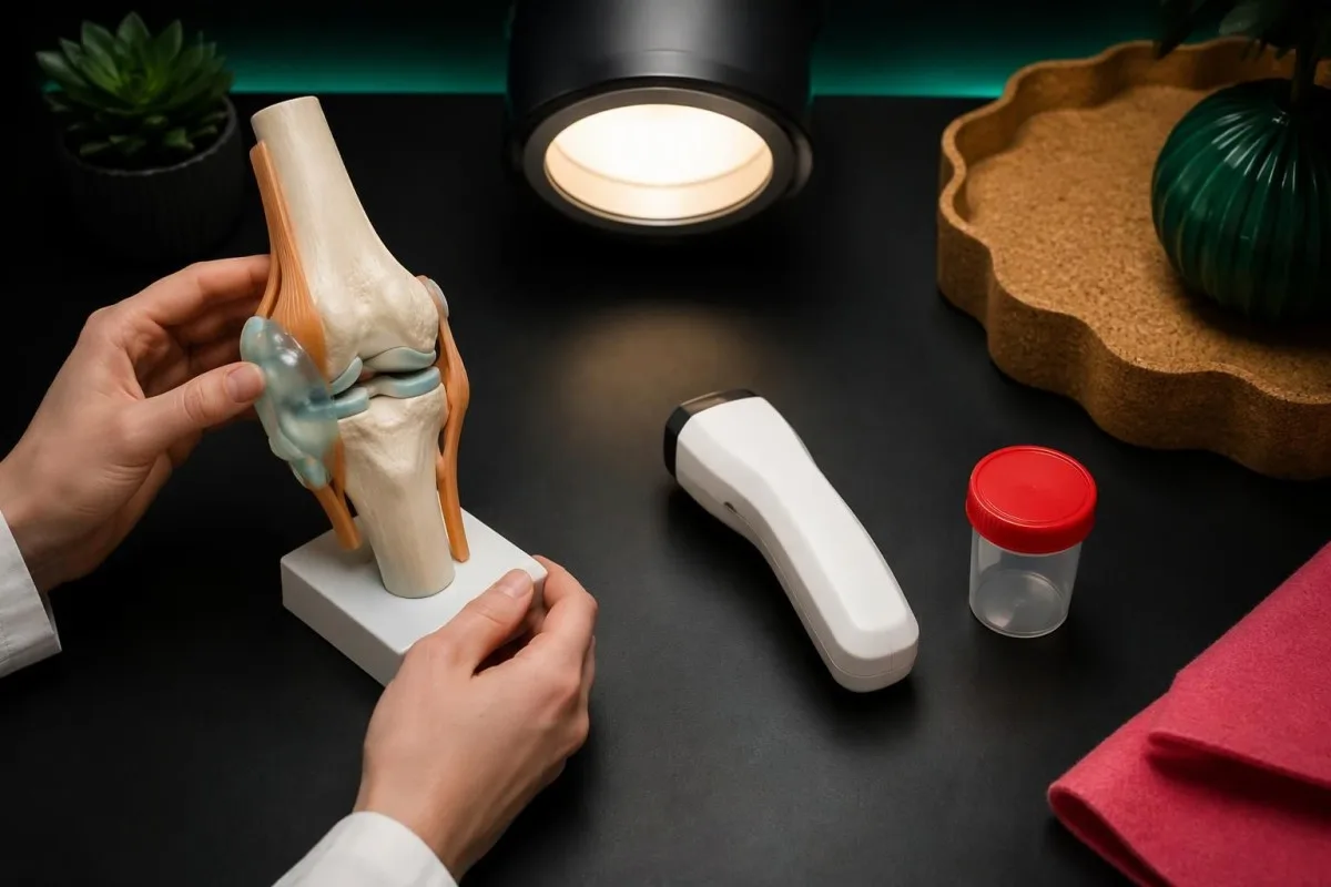
After an injury, inability to bear weight, a visible change in joint shape, or rapidly increasing swelling requires prompt assessment for fracture or another significant injury. Even a low-height fall can matter in someone with fragile bones; an existing diagnosis of osteoarthritis should not delay evaluation of a new event or justify walking through severe pain.
Emergency symptoms can occur near an arthritic joint without being caused by it. Leg swelling with chest pain or breathlessness warrants emergency care; new bladder retention, saddle-area numbness, or rapidly worsening leg weakness also requires immediate assessment, and 1 new neurological red flag is enough to change the plan from a routine arthritis appointment.
A swollen knee developing over 天到数周 after plausible tick exposure may require an infection-focused assessment rather than an osteoarthritis label. Our guide to Lyme test interpretation explains why exposure history and validated testing matter; a hot acute joint still needs urgent assessment, while a random positive antibody result does not establish the cause of swelling.
在安排常规评估期间可以做些什么?
For nonurgent, suspected osteoarthritis, keep moving within tolerable limits and arrange an assessment if symptoms persist or restrict daily life. Exercise is a core treatment once the diagnosis is established; a hypothetical starting plan might use 5–10 分钟 of comfortable walking rather than alternating complete rest with a suddenly much longer session.
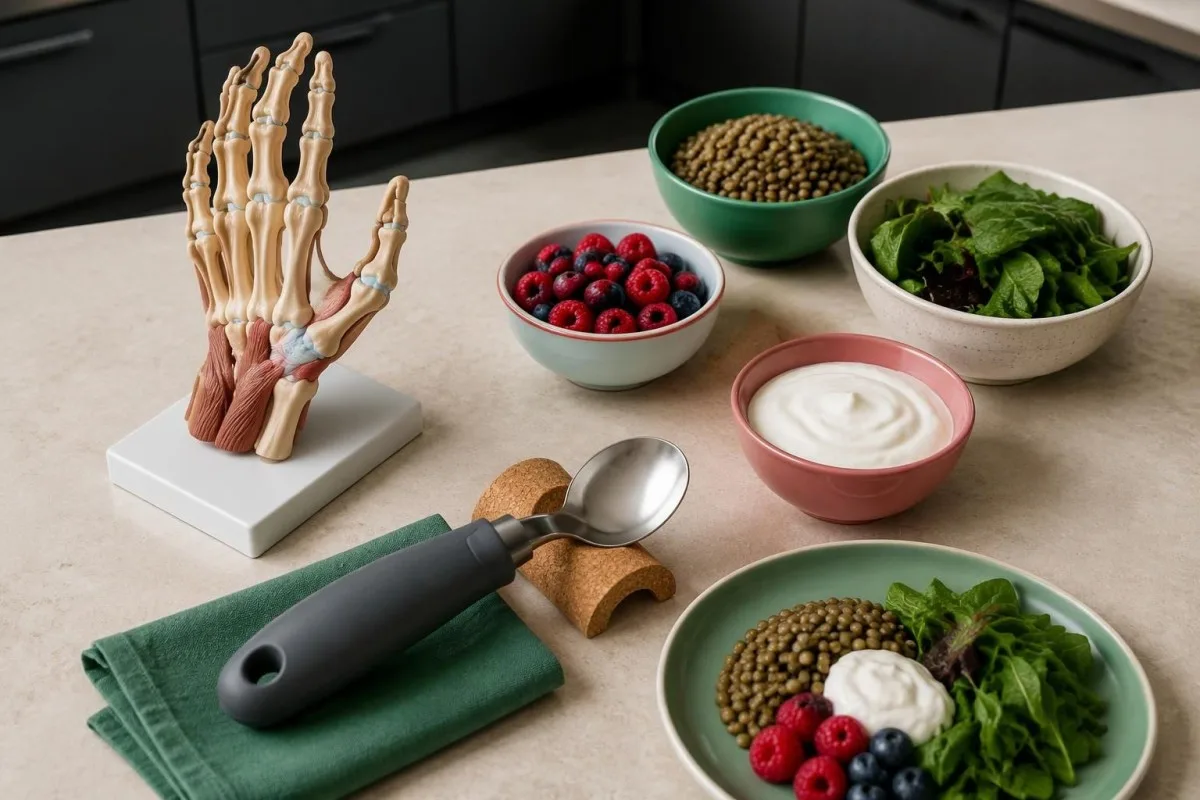
Exercise should be adapted to the affected joint, current capacity, and any warning signs rather than prescribed as one universal routine. The ACR/Arthritis Foundation guideline strongly recommends exercise for hand, hip, and knee osteoarthritis (Kolasinski et al., 2020); try changing 1 variable at a time, such as duration, so a clinician or physiotherapist can identify what is tolerable.
Weight loss can reduce symptoms for people with knee or hip osteoarthritis who are overweight, but it is not a prerequisite for receiving care. NICE states that losing 10% of body weight is likely to help more than 5%; ;我们的 基于证据的食物选择 can support balanced eating, although no specific food has been shown to rebuild an osteoarthritic joint.
Pain treatment needs a safety review, particularly before regularly taking an oral anti-inflammatory medicine. NICE recommends topical NSAIDs for knee osteoarthritis, while oral NSAIDs require consideration of gastrointestinal, kidney, and cardiovascular risks; at Kantesti, we encourage recording the exact mg dose and frequency, and our medication-related laboratory guide explains why accompanying medicines and kidney results matter.
应如何使用实验室检查说明和相关研究?
Use laboratory explanations to prepare better questions, not to self-confirm osteoarthritis or postpone a joint examination. Kantesti 是一个 基于 AI 的血液检测分析工具 that supports understanding of reported results; it cannot determine the cause of a painful knee from 1 CRP value, assess joint warmth remotely, or replace urgent care.

Bring a concise symptom record to a routine appointment: the joint involved, trigger, stiffness duration, swelling, injury history, and lost function. A 7 天日记 is a practical example, not a requirement to wait; our 负责任的 AI 指南 explains why an automated result should not override a clinician's examination or an urgent change in symptoms.
Laboratory reports sometimes contain unrelated findings that still need their own context. For example, 1 urine dipstick abnormality cannot explain knee pain without a relevant history; our 尿液分析解读指南 addresses urinary findings separately, preventing an incidental result from being presented as evidence for osteoarthritis or used to justify unnecessary treatment.
这 2 DOI-linked publications below are supplemental laboratory education resources on urinalysis and iron studies, not osteoarthritis diagnostic studies. The directly relevant clinical references are NICE NG226, EULAR's imaging recommendations, and the ACR/Arthritis Foundation management guideline; a DOI makes a document identifiable, but does not itself establish peer review, clinical validation, or relevance to a joint diagnosis.
常见问题解答
骨关节炎通常最先出现的症状是什么?
骨关节炎最早出现的明显症状通常是活动期间或活动后关节疼痛,休息后伴有短暂僵硬。典型情况下,晨僵通常不超过 30 分钟。膝关节可能在上下楼梯时疼痛,髋关节可能在穿袜子时疼痛,拇指可能在打开容器时疼痛。这些表现有助于评估,但若不进行检查,不足以诊断所有疼痛关节。
血液检查能否显示我是否患有骨关节炎?
No routine blood test can confirm or exclude osteoarthritis. A CRP within a laboratory's reference interval, often below 5 mg/L, does not prove that a joint is healthy or explain its pain. Blood tests may help investigate rheumatoid arthritis, infection, iron overload, or other conditions when the symptoms suggest them. Diagnosis usually relies on the clinical pattern and examination, with imaging used selectively.
如何区分骨关节炎僵硬和类风湿关节炎僵硬?
骨关节炎的僵硬通常持续不超过30分钟,而类风湿关节炎更常导致长时间的晨僵和持续的柔软关节肿胀。持续时间并非绝对的区分标准,且这两种疾病都可能表现出非典型特征。多个指关节出现对称性肿胀,更有必要评估是否患有炎性关节炎。类风湿因子或抗CCP检测结果阴性并不能排除类风湿关节炎。
如果我出现早期骨关节炎症状,需要拍X光片吗?
早期骨关节炎症状不一定需要做 X 线检查。NICE 支持对 45 岁及以上成人进行临床诊断,前提是其关节疼痛与活动有关,且没有晨僵或晨僵持续时间不超过 30 分钟。当症状表现不典型、诊断仍不确定,或检查结果可能改变治疗方案时,影像学检查会更有帮助。X 线片看起来正常,并不能排除所有早期关节变化或其他疼痛原因。
髋骨关节炎会让人感觉膝盖疼痛吗?
髋骨关节炎可引起牵涉痛,疼痛可放射至膝部,即使髋部不适并非主要主诉也是如此。因此,对1例原因不明的膝痛进行评估时,可能需要检查髋关节活动情况,并确认是否有腹股沟疼痛或穿袜困难。膝以下出现刺痛、灼痛或无力,则提示可能存在神经因素。髋部或腹股沟突发剧烈疼痛且无法负重时,需要紧急评估,而不应常规地假定为关节炎。
关节肿胀时,我应该何时紧急就医?
如果关节突然发热、肿胀且疼痛剧烈,请当天就医评估,尤其是在活动关节或负重变得困难时。体温低于 38°C 且血液指标正常,并不能可靠地排除关节感染。受伤后,如果无法负重或关节形状发生改变,也需要尽快接受评估。如同时出现胸痛、呼吸困难或新发严重神经系统症状,请拨打急救电话。
获取血液检查结果解读
上传您的实验室报告,Kantesti 的 AI 会在几秒内用通俗语言解释每项生物标志物。全球已有超过 200 万人使用。.
参考
- Klein, T., Mitchell, S., & Weber, H. (2026). 尿胆原在尿液测试:完整尿液分析指南 2026。. Kantesti AI 医学研究。. DOI
- Klein, T., Mitchell, S., & Weber, H. (2026). 铁代谢检测指南:总铁结合力、铁饱和度和结合能力。. Kantesti AI 医学研究。. DOI
- National Institute for Health and Care Excellence (2022). Osteoarthritis in over 16s: diagnosis and management. NICE guideline NG226.
- Sakellariou G et al. (2017). EULAR recommendations for the use of imaging in the clinical management of peripheral joint osteoarthritis. 《风湿病学年鉴》。. DOI
- Kolasinski SL et al. (2020). 2019 American College of Rheumatology/Arthritis Foundation Guideline for the Management of Osteoarthritis of the Hand, Hip, and Knee. Arthritis Care & Research. DOI
- 200万+分析的检测项目
- 127+国家
- 100+语言
关于医疗团队


E-E-A-T信任信号
经验
由医生主导的临床审阅:实验室解读工作流程。.
专业知识
实验室医学重点:生物标志物在临床情境中的表现。.
权威
经 Thomas Klein 医生医学审核,并由 Sarah Mitchell 医生和 Hans Weber 教授参与撰写。.
可信度
基于循证的解读,并提供清晰的后续路径以减少警报。.
医疗免责声明
本文仅用于教育目的,不构成医疗建议。诊断和治疗决策请始终咨询合格的医疗专业人员。.
发表 · 联系编辑团队
由 Kantesti Ltd 发布,在英格兰和威尔士注册,注册公司编号:. 17090423, ,英国伦敦。.
