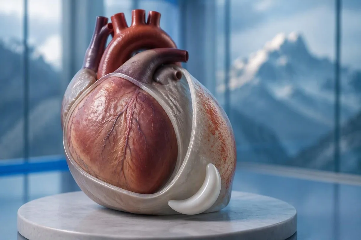
ہاں - pericarditis کا سینے کا درد اکثر لیٹ جانے سے بڑھ جاتا ہے اور بیٹھنے یا آگے جھکنے سے بہتر ہو جاتا ہے۔ لیکن وہ پیٹرن دل کا دورہ خارج نہیں کر سکتا؛ سینے کے نئے غیر واضح درد کو فوری طبی تشخیص کی ضرورت ہے۔.


- پوزیشنل درد اکثر لیٹ جانے سے بڑھ جاتا ہے اور آگے جھکنے سے بہتر ہو جاتا ہے، لیکن کوئی بھی ردعمل pericarditis کی تصدیق نہیں کرتا یا دل کا دورہ خارج نہیں کرتا۔.
- سانس لینے سے متعلق درد گہری سانس لینے یا کھانسنے سے بڑھ سکتا ہے؛ پلمونری ایمبولزم اور پھیپھڑوں کی دیگر حالتیں ایک ہی پیٹرن پیدا کر سکتی ہیں۔.
- ایمرجنسی علامات شدید یا مسلسل سینے میں درد، سانس کی قلت، بے ہوشی، سرد پسینہ، یا بازو، جبڑے، یا کمر تک پھیلنے والا درد شامل ہے۔ ہر علامت کا انتظار کیے بغیر اپنے مقامی ایمرجنسی نمبر پر کال کریں۔.
- ECG تبدیلیاں 2015 کے ESC گائیڈ لائن میں 60% تک معاملات میں شدید pericarditis کی خصوصیت بتائی گئی تھی؛ ایک عام ECG حالت کو خارج نہیں کرتا ہے۔.
- Echocardiography دل کے ارد گرد سیال اور اس کے بھرنے پر اس کے اثر کا اندازہ لگاتا ہے۔ 20 ملی میٹر سے زیادہ کا بہاؤ ایک اعلی خطرے کی خصوصیت ہے، نہ کہ وہ حد جس کے نیچے تمام سیال بے ضرر ہوتا ہے۔.
- ٹروپونن assay-specific 99th percentile سے اوپر دل کے پٹھے کی چوٹ کی نشاندہی کرتا ہے؛ نتیجہ اکیلے دل کا دورہ اور myocarditis کے درمیان فرق نہیں بتا سکتا۔.
- سوزش کے ٹیسٹ جیسے CRP اور ESR، تشخیص اور فالو اپ میں مدد کرتے ہیں، لیکن معمول کے نتائج خود بخود پیری کارڈائٹس کو خارج نہیں کر سکتے۔.
- فالو اپ مناسب طریقے سے منتخب آؤٹ پیشنٹس کے لیے عام طور پر تقریباً 1 ہفتے کے اندر ہوتا ہے؛ بگڑتے ہوئے علامات کے لیے جلد دوبارہ جانچ کی ضرورت ہوتی ہے۔.
pericarditis کا سینے کا درد لیٹ جانے سے کیوں بڑھ جاتا ہے؟
پیری کارڈائٹس کا سینے کا درد اکثر لیٹنے سے بڑھ جاتا ہے اور بیٹھنے یا آگے جھکنے سے کم شدید ہو جاتا ہے۔. پوز میں تبدیلیاں جلن والے پیری کارڈیئم اور قریبی ڈھانچوں کے درمیان مکینیکل تعلق کو متاثر کرتی ہیں، لیکن ایک پوزیشن میں راحت تشخیصی اشارہ ہے—یہ ثبوت نہیں کہ دل کا دورہ ناممکن ہے۔.
دی پیری کارڈیئم دل کے گرد دو تہہ والا تھیلا ہے، اور اس کی بیرونی پرت میں درد کے حساس کنکشن ہوتے ہیں جنہیں دل کے پٹھے خود اسی طرح شریک نہیں کرتے۔ یہ عام وضاحت کہ تہیں صرف “ایک دوسرے پر رگڑتی ہیں” نامکمل ہے: حرکت، گردونواح کے ٹشوز کی جلن، اور انفرادی حسی ردعمل بھی درد کو متاثر کرتے ہیں۔.
ایک فرضی 42 سالہ شخص جسے سانس کی بیماری کے بعد تیز درد ہوتا ہے اور وہ آرام سے سیدھا نہیں سو سکتا، پیری کارڈائٹس کے مطابق پیٹرن رکھتا ہے۔ تشخیص کرنے سے پہلے میں پھر بھی صحیح آغاز، ورزش، سانس کی قلت، اور قلبی تاریخ کے بارے میں پوچھوں گا؛ عمر اور حال ہی میں نزلہ کورونری بیماری یا پلمونری ایمبولزم کو ختم نہیں کرتے۔.
کنٹیسٹی ایک ہے۔ اے آئی بلڈ ٹیسٹ تجزیہ کار جو کہ طبی تشخیص کے بعد رپورٹ کردہ CRP اور ٹراپونن کے نتائج کی وضاحت کرنے میں مدد کر سکتا ہے؛ یہ فیصلہ نہیں کر سکتا کہ پوزیشنل سینے کا درد محفوظ ہے یا نہیں۔ تھامس کلائن کے طور پر، میں اس فرق کو واضح طور پر بیان کرتا ہوں: 1 اطمینان بخش علامت کو کبھی بھی غیر واضح شدید سینے کی شکایت پر غالب نہیں آنا چاہئے؛ کے بارے میں معلومات ادارے میں ہمارے تعلیمی کردار کی وضاحت کرتا ہے۔.
کیا pericarditis کا درد گہری سانس لینے سے بڑھ جاتا ہے؟
پیری کارڈائٹس کا درد عام طور پر گہری سانس لینے، کھانسی، یا کبھی کبھار نگلنے سے بڑھ جاتا ہے۔. ڈاکٹر اسے سانس سے متعلق درد کو پلورائٹک کہتے ہیں، لیکن وہ تفصیل ایک خاص بیماری کے بجائے درد کے پیٹرن کی نشاندہی کرتی ہے؛ پھیپھڑے، سینے کی دیوار، اور پیری کارڈیئم سب اس کو پیدا کر سکتے ہیں۔.
گہری سانس لینے سے ڈایافرام حرکت کرتا ہے اور سینے کے اندر کے تعلقات بدل جاتے ہیں، جو جلن والے پیری کارڈیئل استر کو بڑھا سکتا ہے۔ فرینک اعصاب سے منسلک، جو بنیادی طور پر C3–C5, سے نکلتے ہیں، یہ سمجھانے میں مدد کرتے ہیں کہ کیوں کچھ مریضوں کو صرف سینے کے پیچھے کے بجائے کندھے یا ٹریپیزیس کے کنارے کے قریب منتقل ہونے والی تکلیف محسوس ہوتی ہے۔.
ہر سانس کے ساتھ پکڑنے والا درد جب اچانک شروع ہو تو اس کی وسیع تر تشخیص کی ضرورت ہوتی ہے، خاص طور پر سانس کی قلت یا ٹانگ کے سوجن کے ساتھ۔ پلمونری ایمبولزم کلاسیکی دباؤ کے احساس کے بغیر وہی شکایت پیدا کر سکتا ہے۔ یہاں تک کہ آکسیجن کی سنترپتی 98% بھی اسے خود سے خارج نہیں کر سکتی۔.
جب کوئی معالج سینے کے ایک حصے پر دباتا ہے تو درد سینے کی دیوار کے ماخذ کو زیادہ قابلِ اعتبار بناتا ہے، لیکن یہ کورونری کی مکمل نفی فراہم نہیں کرتا۔ کی ہماری وضاحت غیر واضح درد کے لیے ٹیسٹ معاون لیبارٹری کی اشارے کو ان حالات سے ممتاز کرتی ہے جن کے لیے جانچ اور امیجنگ کی ضرورت ہوتی ہے۔.
pericarditis کی کون سی دیگر علامات اہم ہیں؟
پیری کارڈائٹس کی علامات میں بخار، تھکاوٹ، سانس کی قلت، اور تیز دھڑکن کے ساتھ سینے کا درد شامل ہو سکتا ہے۔. کوئی بھی لازمی نہیں ہے، اور ان کا مجموعہ کسی 1 علامت سے زیادہ اہم ہے؛ شدید کمزوری، بے ہوشی، یا سانس لینے میں تبدیلی سے جانچ کی فوری ضرورت بدل جاتی ہے۔.
ایک درجہ حرارت جو اس سے زیادہ ہو 38°C سے زیادہ ہو is a higher-risk feature in the 2015 ESC pericardial guideline, particularly when the cause is uncertain. A patient with 37.4°C and considerable pain may still need urgent assessment, however; temperature thresholds help clinicians organize risk rather than define a safe home-care boundary.
A fast pulse can reflect pain, fever, dehydration, an arrhythmia, or impaired cardiac filling. I would interpret a resting pulse of 110 beats/min differently in someone comfortable and well hydrated than in someone pale and faint; our دل کی دھڑکن کی جانچ کا گائیڈ explains why laboratory results are only part of that assessment.
Breathlessness when lying flat is not automatically the same thing as chest pain when lying flat. Someone who needs 3 pillows because they cannot breathe comfortably may have congestion, an effusion, or another cardiopulmonary problem; the distinction is covered in our سانس پھولنے کے نتائج گائیڈ کرتے ہیں, but active breathing difficulty should not wait for online interpretation.
پوزیشنل سینے کے درد کو کب ایمرجنسی کی دیکھ بھال کی ضرورت ہوتی ہے؟
Call emergency services for severe, persistent, or rapidly worsening chest pain, especially with breathlessness, fainting, cold sweating, or pain spreading to the arm, jaw, or back. In the UK call 999 or 112; elsewhere use your local emergency number, and do not drive yourself.
Sudden maximal pain, collapse, new confusion, or coughing up blood also warrants emergency assessment because the differential includes pulmonary embolism and aortic emergencies. Do not wait 5 or 15 minutes to see whether leaning forward fixes the problem; concerning accompanying symptoms make a waiting experiment unsafe.
New unexplained chest pain that is not severe still deserves prompt, usually same-day, medical assessment rather than a routine appointment several weeks away. The 2021 AHA/ACC chest pain guideline emphasizes structured assessment and discourages using supposedly “atypical” symptoms to dismiss acute coronary syndrome (Gulati et al., 2021).
Stop exertion while arranging help, and follow the emergency dispatcher’s instructions about any medication rather than starting treatment for presumed pericarditis. Our فوری سینے کے درد کے خون کے ٹیسٹ article explains why 1 laboratory result cannot replace an emergency ECG, examination, and appropriately timed testing.
ڈاکٹر مشتبہ pericarditis کا اندازہ کیسے لگاتے ہیں؟
Clinicians assess suspected pericarditis by combining the history, examination, ECG, echocardiography, and selected blood tests. The widely taught 2015 ESC framework requires at least 2 of 4 findings: typical pain, a pericardial rub, characteristic ECG changes, or a new or worsening pericardial effusion (Adler et al., 2015).
Sharp pain, pain worse lying down, and relief leaning forward are not 3 independent diagnostic criteria; they are different features of the same symptom. This distinction prevents a persuasive story from being mistaken for confirmation, especially when the ECG and imaging have not yet been assessed.
A pericardial friction rub may be transient, so a clinician may listen again after an initial examination rather than treating 1 quiet examination as decisive. Vital signs, signs of poor circulation, heart failure, and the history of recent procedures or systemic illness can matter more urgently than whether the rub is audible.
دی 2025 ESC myocarditis and pericarditis guideline gives contemporary imaging and overlapping myocardial involvement a central place in assessment, so the older checklist is not a complete care pathway (Schulz-Menger et al., 2025). Our clinical standards information should be read as documentation about laboratory interpretation—not evidence that software can reproduce this bedside evaluation.
ECG pericarditis میں کیا دکھاتا ہے؟
An ECG may show widespread ST-segment elevation and PR-segment depression in acute pericarditis, but these changes are not always present. The 2015 ESC guideline reports characteristic ECG changes in up to 60% of cases, so a normal tracing does not exclude pericarditis (Adler et al., 2015).
A 12-لیڈ ECG records electrical activity from several perspectives; clinicians assess which leads are affected, reciprocal changes, rhythm, and comparison with previous tracings. “The ST segment looks elevated” is not enough to separate pericarditis, early repolarization, myocardial infarction, and other causes without the clinical setting.
A heart attack more often produces changes corresponding to a coronary territory, whereas pericarditis may produce a broader pattern—but neither rule is absolute. ECG abnormalities can evolve over گھنٹوں, and repeating the tracing during recurrent or changing symptoms may reveal information absent from the first recording.
Electrolyte disturbances can add another layer of confusion, particularly when kidney disease or medication effects are present. A potassium concentration around 6.0 mmol/L سے اوپر ہو can be clinically urgent depending on context, and our potassium and ECG guide explains why ECG interpretation should never be detached from the rest of the assessment.
pericarditis کے لیے echocardiography کیوں استعمال کیا جاتا ہے؟
Echocardiography checks for fluid around the heart, ventricular function, and signs that fluid is impairing cardiac filling. A normal echocardiogram does not exclude uncomplicated pericarditis; an effusion greater than 20 mm is a recognized high-risk feature, not the definition of every dangerous effusion.
The 2015 ESC guideline describes effusions as small below 10 mm, moderate at 10–20 mm, and large above 20 mm, using the echo-free space on imaging. These measurements help communicate size, but the distribution of fluid and its hemodynamic effects still require interpretation rather than a number-only decision.
Cardiac tamponade occurs when pericardial pressure compromises filling; how quickly fluid accumulates can matter more than its absolute volume. A collection that develops over گھنٹوں may be less well tolerated than a larger, slowly developing collection, and the classic low-pressure, raised-neck-vein, quiet-heart-sound triad is not reliably present in every patient.
دی 2025 ESC guideline supports integrating echocardiography with other imaging when the diagnosis or extent of involvement remains uncertain (Schulz-Menger et al., 2025). An elevated NT-proBNP may add information about cardiac strain, but 1 value cannot diagnose tamponade; our NT-proBNP interpretation guide explains several alternative reasons for elevation.
کیا troponin pericarditis کو دل کے دورے سے ممتاز کر سکتا ہے؟
Troponin identifies heart muscle injury, but an elevated result alone cannot distinguish a heart attack from myocarditis or overlapping myopericarditis. The relevant threshold is the assay-specific 99ویں فیصد (99th percentile) کی بالائی ریفرنس حد سے اوپر کوئی بھی قدر, interpreted alongside symptoms, ECG findings, serial measurements, and sometimes imaging.
There is no universal normal troponin number for every laboratory: troponin I اور troponin T use different assays, and some laboratories apply sex-specific reference limits. If a hypothetical report lists an upper limit of 14 ng/L, a result of 20 ng/L exceeds that particular limit; it does not independently identify the cause, as our troponin I versus T explanation details.
An early normal troponin may precede a detectable rise, which is why emergency departments use validated serial pathways, often with repeat sampling at 1 or 2 hours for suitable high-sensitivity assays. Selected patients can meet validated single-sample rule-out criteria, but that decision depends on assay, symptom timing, ECG, and risk—not a home interpretation of “normal.”
Pericardial symptoms plus raised troponin prompt assessment for myocardial involvement, especially with arrhythmia, breathlessness, or reduced ventricular function. A 30-year-old with a recent viral illness still needs a proper differential; our myocarditis troponin guide explains why neither youth nor the size of the rise settles the diagnosis.
CRP، ESR، اور خون کے گنتی کیا بڑھاتے ہیں؟
CRP, ESR, and a full blood count can support evidence of inflammation, but they do not confirm pericarditis or exclude a heart attack. Many laboratories use a standard CRP upper reference limit around تقریباً, while others use different limits; the printed laboratory range takes precedence.
CRP کی قدر 45 mg/L indicates a substantial inflammatory response in a laboratory with an upper limit of 5 mg/L, but it cannot say whether the source is pericardial, pulmonary, autoimmune, or elsewhere. High-sensitivity CRP measures the same protein at low concentrations; acute illness values should not be interpreted using stable cardiovascular-risk categories.
کنٹیسٹی ایک ہے۔ AI خون کے ٹیسٹ کی رپورٹ کی تشریح that can explain a CRP result against the supplied reference range and help organize its trend alongside other reported results. If CRP changes from 45 to 12 mg/L, the direction may support improvement, but persistent symptoms still matter; our WBC and CRP comparison discusses why these markers can disagree.
ESR often changes more slowly than CRP and is influenced by age, anemia, pregnancy, and protein concentrations, so comparing 2 different markers as though they were interchangeable can mislead follow-up. The 2015 ESC guideline treats inflammatory tests as supportive evidence rather than standalone diagnostic criteria (Adler et al., 2015); our causes of elevated ESR adds context for an unexpectedly persistent elevation.
کون سی وجوہات اور خطرے کی خصوصیات دیکھ بھال کے منصوبے کو تبدیل کرتی ہیں؟
Pericarditis may follow a viral illness, occur after cardiac injury, or accompany autoimmune disease, severe kidney disease, tuberculosis, or other conditions. بخار جو 38°C سے زیادہ ہو, a large effusion, tamponade, or failure to improve with initial treatment are among the features that can justify hospital assessment and a broader search for causes.
دی 2015 ESC guideline lists lack of response to anti-inflammatory treatment within 7 دن as a major adverse prognostic feature, alongside subacute onset and the imaging findings above (Adler et al., 2015). This is a clinician’s reassessment trigger—not permission to wait a week through worsening pain, fever, or breathlessness.
Autoimmune testing is most useful when the history supplies a reason: recurrent episodes, joint symptoms, rashes, mouth ulcers, or known systemic disease. 1 positive antibody does not prove autoimmune pericarditis; our anti‑dsDNA نتیجہ گائیڈ explains why antibody findings need compatible clinical evidence.
Severe kidney dysfunction can change both the cause of pericarditis and medication safety; an eGFR below 15 mL/min/1.73 m² identifies kidney failure-range function but does not itself diagnose uremic pericarditis. Our urgent low-eGFR symptoms guide is relevant to that background, while chest pain in this setting requires direct medical assessment.
کیا دوا سے بہتری pericarditis کی تصدیق کرتی ہے؟
Improvement with ibuprofen, aspirin, or a change in posture does not confirm pericarditis. For appropriately diagnosed uncomplicated cases, clinicians commonly use an anti-inflammatory medicine with colchicine, often for approximately 3 ماہ after a first episode, but the regimen must account for kidney function, interactions, and the underlying cause.
A reduction in pain from 8 out of 10 to 3 out of 10 is useful clinical information, but several chest-pain causes can respond to analgesia. Do not use medication response as a reason to cancel assessment, and do not start high-dose anti-inflammatory treatment for undiagnosed chest pain without professional advice.
Colchicine requires particular care with kidney or liver impairment and certain interacting medicines, including some antibiotics; taking 2 medicines together can alter exposure even when each prescription is familiar. Corticosteroids are not routine first-line treatment for every uncomplicated case, and selected bacterial or systemic causes need a different strategy rather than simply more pain relief.
Kantesti can help organize laboratory follow-up for a clinician through our میڈیکیشن سیفٹی ٹرینڈ گائیڈ, but it should not select the treatment or tapering schedule. A plan that includes 1 named prescriber, clear interaction checks, and instructions for worsening symptoms is more useful than a generic dosing chart.
pericarditis کی علامات بہتر ہونے کے بعد کب ورزش محفوظ ہے؟
Exercise should be restricted during active pericarditis, and return should follow clinical reassessment rather than pain relief alone. The decision changes substantially if troponin, rhythm testing, or imaging suggests myocardial involvement; 1 symptom-free day is not evidence that strenuous activity is safe.
The older 2015 ESC guideline used a minimum 3 ماہ restriction for athletes with isolated pericarditis, while contemporary guidance places greater emphasis on individualized remission and risk assessment. Patients should not borrow an athlete’s timetable—or a myocarditis timetable—without knowing which diagnosis their clinician has established.
For a hypothetical 28-year-old runner, the difference between isolated pericarditis and myocardial involvement can change follow-up testing and return-to-training decisions. I would ask whether symptoms, inflammatory markers, ventricular function, and any relevant rhythm concerns have resolved; a normal resting pulse of 65 beats/min is only one small part of that picture.
Daily activity, physical work, and competitive sport are 3 different workloads, so a written return plan should specify what the patient can actually do. Our خون کے بایومارکرز کی گائیڈ can explain reported measurements, but no biomarker page, wearable reading, or AI result constitutes exercise clearance.
اگر سینے کا درد واپس آجائے یا ٹیسٹ نارمل نظر آئیں تو کیا ہوگا؟
Persistent or recurrent chest pain needs reassessment even when an earlier ECG, echocardiogram, or CRP result was normal. Appropriately selected low-risk outpatients are commonly reviewed within about 1 ہفتے کے اندر۔, but recurrence with faintness, breathlessness, or worsening pain warrants earlier—and sometimes emergency—assessment.
Residual discomfort, recurrent inflammation, chest-wall pain, and a new unrelated problem can feel similar to the patient, so 2 episodes should not automatically receive the same explanation. Cardiac MRI can sometimes identify pericardial or myocardial involvement when first-line tests leave uncertainty; it is selected for a clinical question, not ordered for every ache.
A follow-up note should record symptom timing, temperature, activity tolerance, medication changes, and the actual laboratory reference ranges. CRP values of 8 and 6 mg/L from different laboratories may not represent a meaningful improvement without knowing the methods and context; our post-discharge result changes guide explains why small numerical changes deserve restraint.
کنٹیسٹی ایک ہے۔ اے آئی سے چلنے والا خون کے ٹیسٹ کا تجزیہ کا آلہ that can help organize reported results for discussion, but it cannot verify the cause of renewed chest pain from a PDF. My advice here, as Thomas Klein, is to agree on 3 things before leaving follow-up: the next review date, permitted activity, and the symptoms that require immediate help.
کلینیکل حدود اور متعلقہ تحقیقی اشاعتیں
Symptoms and laboratory interpretation cannot safely replace an acute chest-pain assessment. کے مطابق اکتوبر 3، 2026, the clinical references below support the distinction between pericardial clues, myocardial injury, and emergency risk; the 2 Zenodo resources are background laboratory guides, not trials validating a pericarditis diagnosis.
Kantesti’s medical advisory information describes the professional expertise behind our educational work, but readers should not equate a medical title with an examination of their own chest pain. A personal care plan still needs 1 accountable treating clinician or team who can examine the patient, review the ECG, and act on imaging or troponin findings.
دی اے پی ٹی ٹی نارمل رینج: ڈی ڈائمر، پروٹین سی بلڈ کلاٹنگ گائیڈ is available as a Zenodo educational publication with DOI 10.5281/zenodo.18262555. Its associated clotting-test interpretation guide is relevant when clinicians consider coagulation questions, but a normal aPTT cannot exclude pulmonary embolism and D-dimer must be used within an appropriate clinical probability pathway.
دی سیرم پروٹین گائیڈ: گلوبولنز، البومن اور اے/جی تناسب خون کا ٹیسٹ is a second Zenodo educational publication with DOI 10.5281/zenodo.18316300. The associated serum protein interpretation guide supplies background on systemic laboratory findings; neither albumin nor the A/G ratio independently diagnoses pericarditis, and the discovery links below are searches rather than verified copies of these publications.
اکثر پوچھے گئے سوالات
کیا پیرا کارڈائٹس سینے کا درد لیٹنے سے بدتر ہوتا ہے؟
دل کی جھلی کی سوزش میں سینے کا درد اکثر لیٹنے پر بڑھ جاتا ہے اور بیٹھنے یا آگے جھکنے پر بہتر ہو جاتا ہے۔ یہ نمونہ مشکوک ہے، لیکن یہ طبی تشخیص کا صرف 1 حصہ ہے اور دل کا دورہ پڑنے سے خارج نہیں کیا جا سکتا۔ ڈاکٹر تاریخ کو جسمانی معائنے، ای سی جی، ایکو کارڈیو گرافی، اور منتخب خون کے ٹیسٹوں کے ساتھ جوڑتے ہیں۔ نئی غیر واضح سینے کے درد کے لیے فوری تشخیص کی ضرورت ہے، شدید درد، سانس کی قلت، بے ہوشی، یا ٹھنڈے پسینے کے لیے ایمرجنسی مدد کی ضرورت ہے۔.
کیا آگے جھکنے سے سینے کا درد دل کا دورہ خارج کرتا ہے؟
آگے جھکنے سے سینے کا درد کم ہونا دل کا دورہ خارج از امکان نہیں ہے۔ 2021 AHA/ACC چیسٹ پین کے رہنما خطوط غیر روایتی آواز والے علامات کو نظر انداز کرنے کے بجائے منظم تشخیص کی حمایت کرتے ہیں۔ ECG اور مناسب وقت پر ہائی سینسٹیویٹی ٹراپونن ٹیسٹنگ کی ضرورت ہو سکتی ہے، اور ابتدائی عام نتیجہ خود بخود حتمی نہیں ہوتا ہے۔ شدید یا مسلسل درد یا سانس کی قلت، بے ہوشی، یا ٹھنڈے پسینے سے منسلک ہونے پر ہنگامی خدمات کو کال کریں۔.
کیا آپ کو نارمل ای سی جی کے ساتھ پیریکارڈائٹس ہو سکتا ہے؟
جی ہاں، پیریکارڈائٹس کا سامنا نارمل ای سی جی کے ساتھ بھی ہو سکتا ہے۔ 2015 کے ای ایس سی پیریکارڈیل گائیڈ لائن نے بتایا کہ 60% تک کے شدید کیسز میں ای سی جی کی مخصوص تبدیلیاں ہوتی ہیں، جس کا مطلب ہے کہ ایک قابل ذکر حصہ میں کلاسک پیٹرن نظر نہیں آتا۔ ای سی جی کی تبدیلیاں وقت کے ساتھ ساتھ بدل بھی سکتی ہیں، لہذا جب علامات میں تبدیلی آئے تو ڈاکٹر 12-لیڈ ٹریسنگ کو دہرانے کا کہہ سکتے ہیں۔ صرف نارمل ای سی جی پیریکارڈائٹس یا ہر قسم کے ہارٹ اٹیک کو رد نہیں کر سکتا۔.
کیا ایکوکارڈیوگرام سے ہمیشہ پیری کارڈیائٹس کا پتہ چل جائے گا؟
ایکوکارڈیوگرام ہمیشہ غیر پیچیدہ پیریکارڈائٹس کو ظاہر نہیں کرتا ہے کیونکہ سوزش بصری سیال جمع ہونے کے بغیر بھی ہوسکتی ہے۔ ایکوکارڈیوگرافی خاص طور پر پیرکارڈیل سیال کی پیمائش، وینٹریکولر فنکشن کا جائزہ لینے، اور بھری ہوئی رکاوٹ کو تلاش کرنے کے لیے مفید ہے۔ 2015 ESC درجہ بندی میں 20 ملی میٹر سے زیادہ کے بہاؤ کو بڑا سمجھا جاتا ہے، لیکن چھوٹی تیزی سے جمع ہونے والی جمع بھی خطرناک ہوسکتی ہے۔ جب طبی سوال حل نہ ہو تو مزید امیجنگ پر غور کیا جا سکتا ہے۔.
کیا نارمل ٹراپونن کا مطلب ہے کہ سینے میں درد سنجیدہ نہیں ہے؟
ایک نارمل ٹروپونن نتیجہ خود بخود یہ معنی نہیں رکھتا کہ سینے کا درد بے ضرر ہے۔ وقت، ایسے کی مخصوص 99ویں فیصد شرح، ای سی جی کے نتائج، اور مجموعی خطرے کی تشخیص یہ متعین کرتی ہے کہ نتیجہ کیا خارج کر سکتا ہے۔ توثیق شدہ ہائی سینسیٹیویٹی ٹروپونن راستے اکثر 1 یا 2 گھنٹے میں دوبارہ پیمائش کا استعمال کرتے ہیں، حالانکہ منتخب مریض سنگل سیمپل رول آؤٹ راستے کے اہل ہو سکتے ہیں۔ ٹروپونن خود سے پلمونری ایمبولزم، ایورٹک ایمرجنسی، یا پیریکارڈائٹس کو بھی خارج نہیں کرتا ہے۔.
Pericarditis کی علامات کتنی دیر تک رہتی ہیں؟
مناسب علاج کے ساتھ پیریکارڈائٹس کی علامات دنوں سے ہفتوں میں بہتر ہو سکتی ہیں، لیکن وقت کا تعین اس کی وجہ، دوبارہ ہونے، اور مایوکارڈیل شمولیت کے ساتھ مختلف ہوتا ہے۔ کولچیسن عام طور پر پہلی غیر پیچیدہ قسط کے بعد تقریباً 3 ماہ تک تجویز کی جاتی ہے، جس کا مطلب یہ نہیں ہے کہ اس مدت کے دوران درد باقی رہنا چاہیے۔ 2015 ESC گائیڈ لائن میں سوزش مخالف علاج کے 7 دنوں کے اندر بہتری نہ آنا دوبارہ تشخیص کا ایک تسلیم شدہ محرک ہے۔ شدید درد، سانس لینے میں دشواری، یا بے ہوشی کو مقررہ جائزہ کے منتظر رہنے کے بجائے پہلے ہی جانچنے کی ضرورت ہے۔.
آج ہی اے آئی سے طاقتور خون کے ٹیسٹ کا تجزیہ حاصل کریں
دنیا بھر میں 2M+ صارفین میں شامل ہوں جو فوری اور درست لیب ٹیسٹ تجزیے کے لیے Kantesti پر بھروسہ کرتے ہیں۔ اپنے خون کے ٹیسٹ کے نتائج اپلوڈ کریں اور چند سیکنڈ میں 15,000+ بایومارکرز کی جامع تشریح حاصل کریں۔.
📚 حوالہ دی گئی تحقیقی اشاعتیں
کلائن، ٹی.، مچل، ایس.، اور ویبر، ایچ. (2026). اے پی ٹی ٹی نارمل رینج: ڈی ڈائمر، پروٹین سی بلڈ کلاٹنگ گائیڈ.۔ Kantesti اے آئی میڈیکل ریسرچ۔.
کلائن، ٹی.، مچل، ایس.، اور ویبر، ایچ. (2026). سیرم پروٹین گائیڈ: گلوبولنز، البومن اور اے/جی تناسب خون کا ٹیسٹ.۔ Kantesti اے آئی میڈیکل ریسرچ۔.
📖 بیرونی طبی حوالہ جات
گلتی ایم وغیرہ۔ (2021)۔. 2021 AHA/ACC/ASE/CHEST/SAEM/SCCT/SCMR گائیڈ لائن برائے سینے کے درد کی جانچ اور تشخیص.۔ Circulation۔.
Schulz-Menger J et al. (2025). 2025 ESC Guidelines for the management of myocarditis and pericarditis.۔ European Heart Journal۔.
⚕️ میڈیکل ڈس کلیمر
یہ مضمون صرف تعلیمی مقاصد کے لیے ہے اور طبی مشورہ نہیں ہے۔ تشخیص اور علاج کے فیصلوں کے لیے ہمیشہ کسی مستند صحت کے ماہر سے رجوع کریں۔.
E-E-A-T اعتماد کے اشارے
تجربہ
معالج کی قیادت میں لیب تشریح کے ورک فلو کا کلینیکل جائزہ۔.
مہارت
لیبارٹری میڈیسن کا فوکس یہ کہ بایومارکرز کلینیکل سیاق میں کیسے برتاؤ کرتے ہیں۔.
مستندیت
ڈاکٹر تھامس کلائن نے لکھا، ڈاکٹر سارہ مچل اور پروف. ڈاکٹر ہانس ویبر نے نظرثانی کی۔.
امانت داری
شواہد پر مبنی تشریح واضح فالو اپ راستوں کے ساتھ تاکہ گھبراہٹ کم ہو۔.