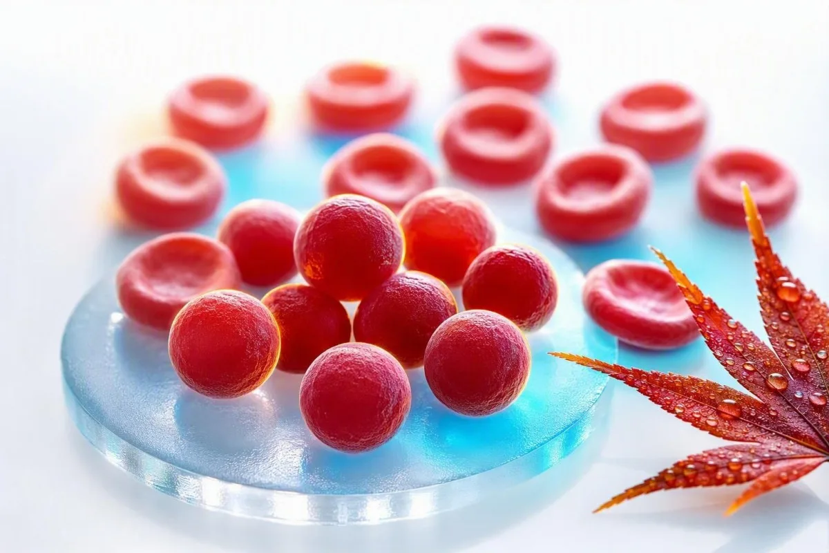
MCHC ya juu ni adimu na mara nyingi ni muhimu zaidi kama dalili kuliko utambuzi. Swali la vitendo ni ikiwa seli nyekundu ni nene na mviringo kweli, au ikiwa sampuli ilipotoka hesabu.


- MCH kwa kawaida ni 32-36 g/dL (320-360 g/L) kwa watu wazima, ingawa kiwango cha maabara yenyewe huchukua kipaumbele.
- MCHC ya juu juu ya 36 g/dL ni ya ajabu na inapaswa kuchochea kuangalia spherocytes, hemolysis, kuungua, au kuingiliwa kwa uchambuzi.
- Spherocytes ni seli ndogo, nyekundu zenye mnene bila rangi ya kati na zinaweza kutokea katika spherocytosis ya kurithi au anemia ya autoimmune hemolytic.
- Agglutinins baridi inaweza kuongeza kwa uwongo MCHC kwa kuganda seli nyekundu, kupunguza idadi ya RBC iliyopimwa, na kupotosha vipimo vilivyohesabiwa.
- Lipemia inaweza kupima hemoglobin kupita kiasi kwenye vichambuzi vingine, na kuunda MCHC iliyoinuliwa hata wakati umbo la seli nyekundu ni la kawaida.
- Kuungua kwa jeraha kunaweza kusababisha MCHC halisi ya juu kupitia upotezaji wa utando wa seli nyekundu za damu, kawaida pamoja na upungufu wa damu na ushahidi wa maabara wa uharibifu wa damu.
- Viashiria vya uharibifu wa damu ni pamoja na reticulocytes zilizoinuliwa, bilirubini isiyo ya moja kwa moja na LDH, na haptoglobin ya chini wakati seli nyekundu za damu zinavunjika mwilini.
- Kurudia vipimo na sampuli ya EDTA iliyokusanywa hivi karibuni, iliyoshughulikiwa ipasavyo mara nyingi ni hatua ya kwanza ya busara wakati MCHC imetengwa na ya juu kidogo tu.
MCHC ya juu kwenye CBC: jibu la moja kwa moja
MCHC ya juu kawaida inamaanisha seli nyekundu za damu ni mnene sana, lakini pia inaweza kumaanisha kuwa uchambuzi ulidanganywa na sampuli. Kwa watu wazima, MCHC zaidi ya takriban 36 g/dL inahitaji muktadha: mwinuko wa kweli mara nyingi huonyesha spherocytes au jeraha kubwa la joto, wakati wataalam wa baridi, lipemia, na uharibifu wa sampuli ni maelezo ya kawaida ya maabara. Kufikia Agosti 23, 2026, sitatoa utambuzi wa shida ya damu kutoka kwa kiashiria hiki pekee.
MCHC huhesabiwa kama hemoglobin kugawanywa na hematocrit, kuzidishwa na 100, kwa hivyo ni mkusanyiko badala ya kipimo cha moja kwa moja cha seli. Hemoglobin ya 15.0 g/dL na hematocrit ya 40% hutoa MCHC ya 37.5 g/dL; idadi inaweza kuongezeka kwa sababu hemoglobin inasomwa sana, hematocrit inasomwa kidogo, au zote mbili. Yetu Mwongozo wa alama za biolojia za CBC ni muhimu wakati viashiria kadhaa vinatambulishwa kwa wakati mmoja.
Ninapokagua paneli yenye MCHC 36.5-37.5 g/dL lakini hemoglobin ya kawaida, bilirubini, reticulocytes, na RDW, udanganyifu mara nyingi ni uwezekano zaidi kuliko ugonjwa. MCHC ya 39-42 g/dL ni ngumu sana kisaikolojia kudumishwa katika seli nyekundu za damu zinazozunguka kwa utimilifu, kwa hivyo maabara inapaswa kufikiria kwa uangalifu uingiliaji kabla ya mtu yeyote kukasirika.
Kantesti ni Mchambuzi wa mtihani wa damu wa AI ambayo inasoma MCHC pamoja na hemoglobin, hematocrit, MCV, RDW, bilirubini, LDH, na matokeo ya awali badala ya kutibu ishara moja ya hatari kama utambuzi. Kanuni ya kliniki ya Dk. Thomas Klein ni rahisi: thibitisha hesabu na sampuli kabla ya kuagiza uchunguzi wa kina; maelezo yetu ya kina RDW na mwongozo wa index ya seli nyekundu yanaelezea kwa nini mbinu hii inayotegemea ruwaza ni muhimu.
MCHC si MCH au MCV
MCHC inaelezea mkusanyiko wa hemoglobin ndani ya kiasi cha vitu vya seli nyekundu za damu zilizopakiwa, wakati MCH inaelezea kiwango cha hemoglobin kwa seli na MCV inaelezea saizi ya seli. Kutaniana vigezo hivi vitatu ni mojawapo ya sababu kwa nini bendera ya kawaida ya CBC inaweza kusikika ya kutisha zaidi kuliko ilivyo.
MCH kwa kawaida ni 27-33 pg kwa kila seli, wakati MCV kwa kawaida ni 80-100 fL kwa watu wazima; hakuna safu inayofafanua MCHC. Seli nyekundu kubwa zinaweza kubeba hemoglobin zaidi na kwa hivyo huongeza MCH, lakini MCHC yao inaweza kubaki kawaida kwa sababu kiasi chao kimeongezeka kwa uwiano. Tazama ulinganisho wetu wa vitendo wa MCV na MCH ikiwa maadili yote mawili yameandikwa.
MCHC ya juu ya kweli mara nyingi huungana na MCV ya kawaida au ya chini-kawaida kwa sababu spherocytes zimepoteza eneo la uso wa utando na kuwa mviringo zaidi, sio kwa sababu zina hemoglobin isiyo na kikomo. Jiometri hiyo ni muhimu: duara hushikilia eneo la uso mdogo kwa kiasi chake, kwa hivyo haina katikati ya rangi kwenye slaidi ya sampuli ya seli iliyoandaliwa vizuri.
Jambo ni kwamba, kipimo cha juu cha MCHC cha damu kinaweza kuwa kweli kihisabati huku kikidanganya kibiolojia. Hematokriti iliyokadiriwa kidogo kupita kiasi baada ya seli nyekundu kuganda itaongeza uwiano hata kama seli nyekundu za mgonjwa hazina msongamano hata kidogo; ndiyo maana a ukaguzi kamili wa hesabu ya damu unapaswa kutangulia hitimisho.
Spherocytes: dalili ya kweli ya MCHC ya juu
Spherocytes ndio sababu inayotambulika zaidi ya MCHC ya juu ya kweli kwa sababu upotezaji wa utando hufanya seli nyekundu kuwa ngumu na zenye msongamano wa hemoglobin. Huonekana katika spherocytosis ya kurithi na upungufu wa damu wa kihemoliti wa kiotomatiki, lakini ugunduzi wa smidi lazima utafsiriwe na dalili na viashiria vya upungufu wa damu.
Hereditary spherocytosis mara nyingi hutoa maadili ya MCHC zaidi ya 36 g/dL, reticulocytosis, jaundice ya vipindi, mawe ya nyongo, splenomegaly, au historia ya familia ya upungufu wa damu. Mwongozo wa 2011 wa British Society for Haematology unabainisha kuwa MCHC inasaidia utambuzi lakini haitoshi kuwa nyeti au maalum kuthibitisha kwa yenyewe (Bolton-Maggs et al., 2012).
Katika upungufu wa damu wa kihemoliti wa kiotomatiki, kingamwili huweka alama kwenye seli nyekundu kwa kuondolewa kwa sehemu ya utando kwenye wengu, na kuacha spherocytes nyuma. Kipimo cha moja kwa moja cha antiglobulin, hesabu ya reticulocyte, bilirubini, LDH, na haptoglobin ni taarifa zaidi pamoja kuliko kielelezo chochote cha pekee; mwongozo wetu wa haptoglobin ya chini zaidi ya upungufu wa damu unashughulikia kizuizi ambacho ni rahisi kukosa.
Hila moja ya kimatibabu: kuhamishwa kwa damu hivi karibuni kunaweza kupunguza au kuficha ruwaza ya asili ya spherocyte ya mgonjwa kwa wiki kadhaa. Christensen et al. wanaelezea kufungwa kwa eosin-5-maleimide kwa mtiririko wa cytometry kama kipimo muhimu kwa tuhuma za spherocytosis ya kurithi, ingawa daktari wa magonjwa ya damu anapaswa kuchagua na kupanga vipimo kwa uangalifu (Christensen et al., 2015).
Jinsi ya kujua ikiwa MCHC ya juu inakuja na hemolysis
MCHC ya juu inakuwa ya maana zaidi kimatibabu inapofuatana na upungufu wa damu, ongezeko la hesabu ya reticulocyte, ongezeko la bilirubini isiyo ya moja kwa moja, ongezeko la LDH, na haptoglobin ya chini. Kundi hilo linaonyesha ongezeko la ubadilishanaji wa seli nyekundu, ingawa hakuna kiashirio kimoja kinachokamilika.
Asilimia ya reticulocyte zaidi ya 2% bado inaweza kuwa haitoshi katika upungufu wa damu mkubwa, kwa hivyo madaktari mara nyingi huhesabu hesabu kamili ya reticulocyte au kiashirio cha uzalishaji wa reticulocyte badala yake. Kwa mfano, hesabu ya reticulocyte 4% yenye hematokriti 24% inaweza kuwakilisha tu mwitikio wa wastani wa marongo baada ya marekebisho; makala yetu kuhusu dalili za reticulocyte za juu inatembea kupitia tofauti.
Bilirubini isiyoandikishwa huongezeka wakati heme inavyochakatwa kwa kasi kuliko ini liwezalo kukiunganisha, huku LDH ikiongezeka kwa sababu chembe nyekundu za damu zina LDH nyingi. Haptoglobin chini ya kikomo cha chini cha maabara inasaidia uharibifu wa ndani wa mishipa, lakini inaweza kuonekana kuwa kawaida wakati wa kuvimba kwa sababu haptoglobin ni protini ya awamu ya papo hapo. Barcellini na Fattizzo wanajadili mtego huu wa uchunguzi katika uhakiki wao wa anemia ya autoimmune hemolytic (Barcellini & Fattizzo, 2015).
Mkojo mweusi, uchovu unaoongezeka haraka, kukosa pumzi wakati wa kupumzika, maumivu ya kifua, kizunguzungu, au macho ya njano na homa vinastahili tathmini ya kimatibabu ya siku hiyo hiyo. Chembe zilizovunjika ni dalili tofauti na ya haraka zaidi kwenye sehemu ya smear kuliko spherocytes; maelezo yetu ya wakati schistocytes zinahitaji mapitio ya haraka husaidia kufafanua kwa nini.
Kwa nini kuungua kunaweza kusababisha MCHC iliyoinuliwa kweli
Jeraha kubwa la joto linaweza kusababisha MCHC ya juu sana kwa sababu joto huharibu utando wa chembe nyekundu za damu, na kusababisha upotezaji wa utando, chembe zinazofanana na spherocytes, na uharibifu. Hii kwa ujumla ni ugunduzi wa hospitalini baada ya kuungua kwa dhahiri kwa kimatibabu, sio maelezo ya matokeo ya mtihani wa nasibu wa CBC kwa wagonjwa wa nje.
Uharibifu wa joto unaweza kuunda microspherocytes ndani ya masaa ya kuungua sana, na MCHC inaweza kuongezeka kadiri uwiano wa utando kwa kiasi unavyoshuka. Hemoglobin pia inaweza kushuka kwa siku zinazofuata kutokana na mabadiliko ya maji, taratibu, upimaji damu, na uharibifu unaoendelea, kwa hivyo MCHC moja haiwezi kupima kiwango cha uharibifu.
Mara kwa mara nimeona wagonjwa wakilenga MCHC ya 37.1 g/dL baada ya kulazwa kwa dharura huku wakipuuza uharibifu mwingi zaidi unaoweza kuchukuliwa hatua: kupungua kwa hemoglobin, kuongezeka kwa kreatini, mabadiliko ya potasiamu, au laktati. Katika hali hiyo, wataalamu wa matibabu wanashughulikia mtu na fiziolojia ya kuungua, sio kiashirio; chapisho letu la ratiba ya vipimo baada ya hospitali linaeleza kwa nini maadili yanaweza kusonga haraka.
Kantesti AI ni jukwaa la tafsiri ya viashiria vya AI ambayo inaweza kuashiria MCHC ya juu pamoja na hemoglobin yenye nguvu na matokeo ya figo, lakini michomo ya papo hapo inahitaji tathmini ya moja kwa moja hospitalini na haiwezi kushughulikiwa kwa usalama kutoka kwa tafsiri ya ripoti. Matokeo baada ya kuungua kali yanapaswa kulinganishwa na masaa 24-72 yaliyopita, sio dhidi ya wastani wa idadi ya watu wa jumla.
Madawa baridi: artifact ya MCHC ya juu ambayo madaktari huangalia
Vigandamizi vya baridi vinaweza kutoa MCHC ya juu bandia kwa kusababisha chembe nyekundu za damu kuganda kwenye joto la kawaida kabla ya uchambuzi. Kifaa cha uchanganuzi kinaweza kuhesabu chembe kadhaa zilizounganishwa kama chembe moja, na kusababisha hesabu ya chini ya RBC bandia, MCV ya juu bandia, na MCHC iliyokadiriwa iliyojaa kupita kiasi.
Muundo wa kawaida wa vigandamizi vya baridi ni hesabu ya chini ya RBC yenye MCV ya juu isiyo na uwiano, MCH ya juu, na MCHC ya juu, mara nyingi bila hadithi inayolingana ya kimatibabu. Kuchomesha mfumo wa EDTA hadi 37°C na kuendesha upya CBC kwa kawaida huboresha maadili, ambayo ni muhimu zaidi kuliko kurudia mtihani wa nasibu wiki baadaye.
Vigandamizi vya baridi vinaweza kutokea kwa muda mfupi baada ya maambukizo fulani ya mfumo wa upumuaji au kuendelea katika ugonjwa wa vigandamizi vya baridi na baadhi ya magonjwa ya limfu. Dalili kama vile mabadiliko ya rangi au usumbufu katika vidole na mfiduo wa baridi huongeza uzito wa kimatibabu, lakini muundo wa maabara wenyewe unaweza kuwa dalili ya kwanza; mtihani wa marudio baada ya uharibifu hutoa maswali ya vitendo ya kuchukua sampuli tena.
Usijipate kwa joto kali au jaribu kubadilisha matokeo nyumbani. Uliza ikiwa maabara iliona kuganda kwa chembe nyekundu za damu, ikiwa sampuli ilichomwa na kuendeshwa upya, na ikiwa hematocrit ya mwongozo ilifanywa; majibu hayo matatu mara nyingi hufafanua suala hilo.
Lipemia na hemolysis ya sampuli inaweza kupotosha MCHC
Lipemia na uharibifu wa sampuli ndani ya mwili unaweza kuongeza MCHC bandia kwa sababu vifaa vingi vya uchanganuzi hupima hemoglobin kwa kutumia photometri baada ya chembe nyekundu za damu kufutwa. Lipidi zenye mawingu au hemoglobin huru kwenye sampuli zinaweza kuongeza kunyonya kwa macho na kufanya hemoglobin ionekane kuwa ya juu kuliko ilivyo.
Lipemia huonekana mara nyingi baada ya triglycerides za juu sana, kawaida juu ya karibu 400-500 mg/dL, ingawa usumbufu hutegemea ala na urefu wa wimbi la kipimo. Maabara inaweza kutumia kinyago cha plasma, urefu mbadala wa wimbi, au mbinu ya kubadilisha plasma badala ya kukubali tu matokeo ya haemoglobini ya kiotomatiki.
Hemolysis ya In-vitro hutokea baada ya kukusanywa au kushughulikiwa, si ndani ya mwili, na inaweza kuathiri zaidi ya MCHC. Potasiamu, LDH, AST, na fosfeti huweza kupanda katika sampuli iliyoharibika, ndiyo sababu MCHC ya juu iliyotengwa pamoja na potasiamu ya juu isiyotarajiwa inahitaji ukaguzi wa ubora wa sampuli; ona mwongozo wetu wa makosa ya potasiamu yanayohusiana na hemolysis.
Safu ya plasma ya waridi au nyekundu inayoonekana ni kidokezo cha maabara, si uthibitisho wa upungufu wa damu wa hemolytic kwa mgonjwa. Hatua inayofuata ya vitendo ni mkusanyiko mpya na utunzaji mtulivu na uchakataji wa haraka, hasa ikiwa ripoti ya asili inajumuisha fahirisi ya uharibifu au maoni kwamba matokeo yanaweza kuathiriwa.
Matatizo ya ukusanyaji, uhifadhi, na mirija ambayo hubadilisha vipimo vya CBC
Mchanganyiko duni wa sampuli, kutojaza, kuganda, na uchambuzi uliocheleweshwa unaweza kubadilisha fahirisi za CBC, ingawa uhifadhi uliocheleweshwa mara nyingi hupunguza badala ya kuongeza MCHC kwa kuruhusu seli kuvimba. Kwa hivyo, MCHC ya juu inapaswa kusomwa na maoni ya sampuli ya maabara, si tu nambari kwenye lango la mgonjwa.
Sampuli za CBC kwa ujumla hukusanywa katika mirija ya EDTA, na mirija iliyojaa kidogo inaweza kuwa na uwiano mwingi wa kiantikoagulant kwa sampuli ambao hubadilisha vipimo vya seli. Mabonge madogo yanaweza kuondoa seli kutoka kwa uchambuzi na kufanya hesabu ya kiotomatiki kuwa haikubaliani ndani; muhtasari wetu wa viongezeo vya mirija ya damu unaeleza kwa nini aina ya mirija si maelezo madogo.
Uhifadhi kwenye joto la kawaida kwa vipindi virefu husababisha seli nyekundu kuchukua maji, na kuongeza MCV na huwa unapunguza MCHC. Mwelekeo huo wa mabadiliko ni muhimu: MCHC ya juu sana baada ya sampuli iliyocheleweshwa haielezwi kiotomatiki na umri pekee, kwa hivyo baridi agglutination au usumbufu wa macho bado unaweza kuwepo.
Sampuli ya kapilari inaweza kufaa katika mazingira fulani lakini inakabiliwa zaidi na kutofautiana kwa mkusanyiko kuliko sampuli ya mishipa ya EDTA iliyokusanywa vizuri. Ikiwa MCHC ya juu haina dalili zinazofanana au uharibifu unaohusiana, mimi huuliza upya wa mishipa safi kabla ya kuichukulia kama utambuzi mpya sugu.
Tumia CBC za awali kuamua ikiwa mabadiliko ni halisi
Kupanda kwa ghafla kwa MCHC kutoka msingi wa 33-35 g/dL hadi 38 g/dL kunapendekeza zaidi suala la sampuli au kichambuzi kuliko spherocytosis ya urithi inayoendelea polepole. Mitindo haibadilishi doa, lakini ni kidokezo chenye nguvu cha udhibiti wa ubora.
Watu wengi wana kiwango chao cha MCHC cha kibinafsi kinachobadilika kwa muda, mara nyingi hutofautiana chini ya 1 g/dL wakati hali za upimaji zinafanana. Hundi ya delta ya maabara hulinganisha matokeo mapya na ya awali ili kutambua mabadiliko yasiyo ya kweli kabla ya matokeo kutolewa; makala yetu kuhusu ukaguzi wa mabadiliko (delta checks) inaonyesha jinsi mtandao huu wa usalama unavyofanya kazi.
Kantesti ni huduma ya kutafsiri vipimo vya maabara ya AI ambayo inalinganisha maadili ya sasa na ya awali ya CBC, ikisaidia kutofautisha ishara ya kudumu kutoka kwa mtoaji wa mara moja. Katika mtiririko wetu wa kazi wa ukaguzi, mabadiliko ya MCHC yana umuhimu zaidi wakati yanasafiri na hemoglobin, MCV, bilirubini, na reticulocytes katika mwelekeo unaoeleweka kibiolojia.
Kuna vighairi. Uharibifu mpya wa autoimmune, kuchomwa moto sana, majibu ya kuhamisha damu, au baridi kali sana inaweza kubadilisha matokeo haraka, kwa hivyo thamani ya kihistoria iliyo thabiti inatia moyo lakini kamwe si sababu ya kupuuza jaundice mpya, mkojo mweusi, au upungufu wa damu.
Hali zinazohusishwa na MCHC lakini mara chache husababishwa
Upungufu wa maji mwilini, upungufu wa chuma, upungufu wa vitamini B12, na mafadhaiko ya kawaida kwa kawaida havisababishi MCHC ya juu sana. Zinaweza kubadilisha maadili mengine ya CBC, lakini hazipaswi kutumiwa kama maelezo rahisi kwa MCHC ya 38 g/dL bila kuthibitisha sampuli.
Upungufu wa chuma kwa kawaida hupunguza MCH na MCHC na mara nyingi hupunguza MCV mara tu upungufu unapojitokeza. Upungufu wa mapema wa chuma unaweza kuwa na MCV ya kawaida, lakini hauelezei viashiria vya hyperchromic; maelezo yetu ya transferrin saturation kuwa chini husaidia kutenganisha upatikanaji wa chuma kutoka kwa mkusanyiko wa CBC.
Upungufu wa vitamini B12 au folate mara nyingi husababisha macrocytosis, na MCV juu ya 100 fL, badala ya MCHC ya juu. Ikiwa MCV na MCH zote mbili ni za juu lakini MCHC ni ya kawaida, hiyo ni mwelekeo tofauti wa utambuzi; angalia dalili za ufuatiliaji wa MCV ya juu.
Upungufu wa G6PD husababisha hemolysis ya vipindi baada ya vichocheo maalum vya oxidative kwa watu walioathirika, lakini MCHC sio mtihani wa kutegemewa wa uchunguzi kwa hilo. Wakati wa kipindi kikali, smear na alama za ubadilishaji zinaweza kufichua zaidi, wakati upimaji wa enzyme unaweza kuhitaji kurudiwa baada ya kupona kwa sababu seli nyekundu za damu vijana zina G6PD zaidi.
Vipimo vya ufuatiliaji vinavyofaa zaidi baada ya MCHC iliyoinuliwa
Hatua inayofuata bora kwa MCHC ya juu iliyotengwa kwa kawaida ni kurudia CBC na ukaguzi wa maabara, ikifuatiwa na smear ya pembeni na jopo la hemolysis ikiwa tu ongezeko litaendelea au dalili zingine zipo. Kuagiza kila jaribio la upungufu wa damu mara moja kunaweza kusababisha kelele zaidi kuliko majibu.
Ukaguzi wa mstari wa mbele uliozingatiwa unajumuisha kurudia CBC, ukaguzi wa slaidi ya sampuli ya seli, hesabu ya reticulocyte, jumla na isiyo ya moja kwa moja ya bilirubini, LDH, haptoglobin, na upimaji wa moja kwa moja wa antiglobulin wakati hemolysis ya autoimmune inawezekana. Smear inaweza kutambua spherocytes, agglutination, fragments, polychromasia, au morphology ya kawaida ambayo hufanya artifact kuwa na uwezekano mkubwa zaidi.
Ikiwa spherocytosis ya urithi inabaki kuwa na uwezekano, daktari wa damu anaweza kuzingatia kumfunga eosin-5-maleimide, upimaji wa uvumilivu wa osmotic, au upimaji wa maumbile. Vipimo hivi vina hatari tofauti za hasi bandia baada ya kuongezewa damu na wakati wa hemolysis ya papo hapo, ndio sababu mpangilio wa jaribio unapaswa kufuata historia ya kliniki badala ya orodha ya vitufe; yetu mwongozo wa upimaji wa G6PD huonyesha shida ya jumla ya wakati.
Kantesti AI hutafsiri MCHC ya juu kwa kuangalia hesabu dhidi ya alama za karibu za CBC na hemolysis, kisha kutambua maswali ya kumpeleka kwa daktari. Haibadilishi ukaguzi wa morphology ya seli, hasa wakati maabara imetoa bendera ya kichanganuzi au wakati upungufu wa damu unazidi kuwa mbaya.
Wakati MCHC ya juu inahitaji tathmini ya haraka ya matibabu
MCHC ya juu yenyewe sio kizingiti cha dharura, lakini MCHC ya juu yenye dalili za hemolysis ya papo hapo, upungufu mkubwa wa damu, au kuchoma kali kunaweza kuhitaji huduma ya siku hiyo hiyo. Dalili na kiwango cha mabadiliko huamua uharaka kwa ufanisi zaidi kuliko ikiwa bendera imewekwa H.
Tafuta tathmini ya haraka kwa upungufu mpya wa kupumua wakati wa kupumzika, maumivu ya kifua, kufa ganzi, kuchanganyikiwa, udhaifu unaozidi kuwa mbaya haraka, mkojo wenye rangi ya chai, au njano ya macho yenye homa. Dalili hizi zinaweza kuambatana na upungufu mkubwa wa damu au hemolysis, hasa wakati hemoglobin imeshuka kwa zaidi ya 2 g/dL kutoka kwa msingi unaojulikana.
Wasiliana na daktari aliyeagiza kipimo ndani ya siku ikiwa MCHC inabaki juu ya 36 g/dL kwenye sampuli ya kurudia, upungufu wa damu upo, au ripoti inataja agglutination au spherocytes. Kwa mtu aliye imara, mwenye afya njema na MCHC 36.2 g/dL na matokeo mengine ya kawaida, kuchora tena bila uharaka mara nyingi ni busara; yetu wa muundo wa bilirubini inaweza kusaidia kuunda majadiliano yajayo.
Usianze chuma, folate, steroidi, au virutubisho kwa ajili tu ya kusahihisha MCHC. Hakuna kinachotibu sababu kuu za kweli, na steroidi hasa zinaweza kuficha tathmini ya hemolysis ya autoimmune ikiwa zitaanza bila usimamizi wa kliniki.
Jinsi Kantesti huweka MCHC katika muktadha wa kimatibabu
Kantesti husoma MCHC ya juu kama shida ya kutambua muundo: magonjwa halisi ya seli nene na vitu bandia vya sampuli hutoa mchanganyiko tofauti wa CBC, kemia, bendera, na mitindo ya zamani. Lengo ni kufanya swali la kliniki lijalo liwe wazi zaidi, si kuelezea ugonjwa adimu kutoka kwa kipimo kimoja kilichohesabiwa.
Kantesti ni Zana ya uchambuzi wa vipimo vya damu inayotumia AI hutumiwa na watu zaidi ya milioni 2 katika nchi 127 kupanga matokeo ya maabara kwa muktadha. Kwa MCHC ya juu, akili bandia yetu huangalia kama hemoglobin na hematocrit vinaunga mkono matokeo kihisabati, kama MCV iko juu kuliko ilivyotarajiwa, na kama bilirubini, LDH, reticulocytes, au haptoglobin zinaelekeza kwenye mzunguko wa chembe nyekundu za damu.
Jukwaa letu pia hutafuta kutokubaliana kwa ndani: MCHC 40 g/dL na maoni ya kawaida ya smear na hakuna upungufu wa damu kunaweza kuhitaji kidokezo cha bandia, wakati MCHC 37 g/dL na spherocytes, reticulocytosis, na kuongezeka kwa bilirubini isiyo ya moja kwa moja kunastahili ufuatiliaji wa kimatibabu. Mbinu ya kiufundi na mipaka yake imeelezwa katika yetu mwongozo wa teknolojia ya AI.
Ninawaonya wagonjwa kuhifadhi PDF ya maabara, tarehe ya ukusanyaji, hali ya kufunga, ugonjwa wa hivi karibuni, historia ya kuongezewa damu, na maoni yoyote ya maabara. Rekodi hiyo ndogo mara nyingi hueleza tofauti inayoonekana haraka kuliko kurudia upimaji mpana, hasa wakati matokeo yakilinganishwa miezi baadaye.
Maswali ambayo hufanya miadi ya MCHC ya juu kuwa na tija zaidi
Uliza kama MCHC ilirudiwa, kama maabara iliona spherocytes au agglutination, na kama kuna dalili za upungufu wa damu au hemolysis. Maswali haya hubadilisha mazungumzo kutoka “kwa nini nambari moja iko juu?” hadi njia dhahiri ya kliniki inayoweza kupimwa.
Maneno muhimu ni pamoja na: “Je, sampuli ilikuwa na hemolysis au lipemic?”, “Je, kinachochambua kiliashiria agglutination ya chembe nyekundu za damu?”, na “Je, tunaweza kulinganisha matokeo haya na MCHC yangu ya awali?” Uhakiki wa smear wa mwongozo ni muhimu sana wakati MCHC inarudiwa zaidi ya 36 g/dL au upungufu wa damu haueleweki.
Dk. Thomas Klein anapendekeza kuchukua orodha ya dawa, maambukizi ya hivi karibuni, dalili zinazosababishwa na baridi, historia ya familia ya mawe ya nyongo au upungufu wa damu, na kuongezewa damu ndani ya miezi 3 iliyopita. Historia hiyo inaweza kuelekeza kwenye spherocytosis ya kurithi, hemolysis ya kinga ya mwili, au vipindi vya muda mfupi vya baridi sana haraka kuliko utafutaji wa jumla mtandaoni.
Maudhui ya matibabu ya Kantesti yanapitiwa na usimamizi wa daktari, lakini daktari wako mwenyewe anaweza kukuchunguza, kuomba kurudiwa kwa joto, na kuzungumza moja kwa moja na maabara. Wasomaji wanaotaka kuelewa mfumo wetu wa uhakiki wa kliniki wanaweza kukutana na bodi ya ushauri wa matibabu, wakati usumbufu unaoendelea au wenye dalili unapaswa kujadiliwa kila wakati ana kwa ana.
Utafiti, kutokuwa na uhakika, na mipaka ya MCHC moja
MCHC ni kidokezo muhimu cha uchunguzi, si kipimo cha utambuzi cha pekee cha spherocytosis ya kurithi, hemolysis ya kinga ya mwili, kuungua, au ugonjwa wa baridi agglutinin. Thamani yake huongezeka sana wakati sampuli ya kurudiwa, umbo la chembe, viashiria vya mzunguko, na historia ya kliniki vyote vinasema hadithi moja.
Hakuna mpango wa MCHC wa ulimwengu wote unaotambua spherocytosis ya kurithi kwa sababu mbinu za kinachochambua, umri, reticulocytosis, na upungufu wa chuma unaojitokeza unaweza kubadilisha unyeti. Mwongozo wa BSH unasaidia kuunganisha historia ya familia, matokeo ya smear, reticulocytes, bilirubini, na upimaji thibitishi inapohitajika badala ya kutegemea kizingiti kimoja (Bolton-Maggs et al., 2012).
Kantesti inasaidia ufuatiliaji wenye ufahamu kwa kutambua ruwaza za matokeo na kutokuwa na uhakika, si kwa kutoa utambuzi wa mwisho wa magonjwa ya damu. Yetu mbinu ya uthibitisho wa kimatibabu inaeleza kwa nini maadili ya ajabu, bendera za kinachochambua, na dalili za dharura hubaki sababu za uhakiki wa maabara na daktari wa binadamu.
Kwa muktadha zaidi wa magonjwa ya damu, angalia chapisho la utafiti Aina ya Damu B Negativu, Mwongozo wa Kipimo cha LDH & Hesabu ya Reticulocyte na rasilimali pana ya mawasiliano ya kliniki Kuhara Baada ya Kufunga, Madoa Meusi kwenye Kinyesi na Mwongozo wa GI 2026. Zote mbili ni machapisho ya ziada ya elimu badala ya mbadala wa smear ya pembeni au tathmini ya mtaalamu.
Maswali Yanayoulizwa Mara Kwa Mara
Co nene dhiidhin ka sarreeya oo MCHC ah maxay ka dhigan tahay baaritaanka dhiigga?
Kiwango cha juu cha MCHC kwa kawaida huashiria kuwa mkusanyiko wa hemoglobin ndani ya kiasi cha chembe nyekundu za damu uko juu ya kiwango cha maabara, kwa kawaida zaidi ya 36 g/dL kwa watu wazima. Nyanyuko halisi hutokea na spherocytes kutokana na spherocytosis ya kurithi, anemia ya hemolitiki ya kiotomatiki, au jeraha kubwa la joto. Vipuu baridi, lipemia, na ugomvi wa sampuli vinaweza pia kusababisha matokeo ya juu ya uwongo. CBC rudufu, uchunguzi wa slaidi ya sampuli ya chembe, na viashiria vya ugomvi vinaelezea ni maelezo yapi yanafaa.
Je, MCHC ya juu ni hatari?
MCHC nyingi si hatari yenyewe; ni kielelezo cha kuhesabiwa cha CBC badala ya ugonjwa. MCHC ya 36.2 g/dL yenye hemoglobin ya kawaida na hakuna dalili mara nyingi hutibiwa kwa jaribio la kawaida la marudio, huku MCHC nyingi pamoja na kupungua kwa hemoglobin, jaundice, mkojo mweusi, au kupumua kwa shida zinahitaji tathmini ya haraka. Thamani zilizo juu ya 39 g/dL zina uwezekano mkubwa wa kuonyesha usumbufu wa sampuli au kipimajoto. Hatari ya kimatibabu hutokana na sababu ya msingi, kama vile hemolysis kubwa, sio nambari ya MCHC pekee.
Husababisha upungufu wa maji mwilini MCHC ya juu?
Kupunguka kwa maji kwa kawaida hakusababishi MCHC kuwa juu sana kwa sababu huongeza mkusanyiko wa plasma zaidi ya kubadilisha mkusanyiko wa hemoglobin ndani ya seli nyekundu za damu. Kupunguka kwa maji kunaweza kuongeza hemoglobin na hematocrit pamoja, na kuacha MCHC karibu na kiwango chake cha kawaida cha 32-36 g/dL. Ikiwa MCHC ni 37-40 g/dL, wataalamu wanapaswa kuzingatia spherocytes, mkusanyiko baridi wa chembe, lipemia, au tatizo la upimaji badala yake. CBC iliyokusanywa vyema tena ni muhimu zaidi kuliko kujaribu kurekebisha thamani kwa maji ya ziada.
Je, spherocytes hufanya MCHC kuwa juu kila wakati?
Sferositi spesso aumentano MCHC sopra 36 g/dL, ma non sempre lo fanno e l'alto MCHC non dimostra la presenza di sferociti. L'sferocitosi ereditaria può essere mascherata da carenza di ferro, trasfusione recente o malattia lieve, mentre l'emolisi autoimmune può mostrare reperti variabili a seconda del momento. È necessario un vetrino del campione di cellule esaminato da un professionista esperto di laboratorio per identificare gli sferociti. Il conteggio dei reticolociti, la bilirubina, la LDH, l'aptoglobina e il test diretto all'antiglobulina aggiungono contesto diagnostico.
Kan kalla antikroppar orsaka hög MCHC?
Vidhuruaji baridi wanaweza kuongeza MCHC kwa bandia kwa kuganda seli nyekundu kabla ya uchunguzi wa CBC wa kiotomatiki kupimwa. Muundo huo mara nyingi hujumuisha idadi ndogo ya RBC, MCV ya juu, MCH ya juu, na MCHC juu ya 36 g/dL ambayo haiendani na dalili za mgonjwa. Kuchoma sampuli ya EDTA hadi 37°C na kuirudia tena kunaweza kurudisha kawaida idadi ya vipimo. Dalili za kudumu za vidhuruaji baridi au hemolysis zinapaswa kutathminiwa na daktari au mtaalamu wa magonjwa ya damu.
Kwa vipimo gani ninafaa kuuliza baada ya MCHC kuwa juu?
Baada ya MCHC ya juu inayoendelea, vipimo vya kawaida vinavyofuata ni kurudia CBC, uchunguzi wa sampuli ya seli, hesabu ya reticulocyte, vipande vya bilirubini, LDH, haptoglobin, na wakati mwingine mtihani wa moja kwa moja wa antiglobulin. Daktari anaweza kuomba kufungwa kwa eosin-5-maleimide au upimaji mwingine maalum ikiwa utambuzi wa spherocytosis wa kurithi unashukiwa. MCHC zaidi ya 36 g/dL na kurudia upimaji wa kawaida na hakuna upungufu wa damu inaweza kuhitaji uchunguzi mwingi. Mfuatano bora wa vipimo hutegemea dalili, maadili ya awali ya CBC, historia ya familia, na bendera za maabara.
Pata Uchambuzi wa Vipimo vya Damu kwa AI Leo
Jiunge na zaidi ya watumiaji 2 milioni duniani kote wanaoamini Kantesti kwa uchambuzi wa papo hapo na sahihi wa vipimo vya maabara. Pakia matokeo yako ya vipimo vya damu na upate tafsiri ya kina ya viashiria vya 15,000+ ndani ya sekunde.
📚 Machapisho ya Utafiti Yanayorejelewa
Klein, T., Mitchell, S., & Weber, H. (2026). Mwili wa damu Aina ya B Negativu, Mwongozo wa Kipimo cha Damu cha LDH na Hesabu ya Reticulocyte. Kantesti uchambuzi wa damu kwa AI ya utafiti wa matibabu.
Klein, T., Mitchell, S., & Weber, H. (2026). Kuhara Baada ya Kufunga, Madoa Meusi kwenye Kinyesi na Mwongozo wa GI 2026. Kantesti uchambuzi wa damu kwa AI ya utafiti wa matibabu.
📖 Marejeo ya Nje ya Tiba
⚕️ Kanusho la Kimatibabu
Makala haya ni kwa madhumuni ya elimu tu na si ushauri wa kimatibabu. Wasiliana na mtoa huduma aliyehitimu kila wakati kwa maamuzi ya utambuzi na matibabu.
E-E-A-T Trust Signals
Uzoefu
Mapitio ya kimatibabu inayoongozwa na daktari ya mifumo ya tafsiri ya maabara.
Utaalamu
Kuzingatia dawa za maabara kuhusu jinsi viashiria (biomarkers) vinavyobadilika katika muktadha wa kliniki.
Mamlaka
Imeandikwa na Dk. Thomas Klein kwa mapitio ya Dk. Sarah Mitchell na Prof. Dk. Hans Weber.
Uaminifu
Tafsiri inayotegemea ushahidi yenye njia zilizo wazi za ufuatiliaji ili kupunguza tahadhari za hofu.