viputo vichache baada ya mkondo wa mkojo wa kasi kwa kawaida ni fisikia. Mfumo ambao hurudia kufunika bakuli, hudumu baada ya kumwaga, au huambatana na uvimbe unastahili kupimwa albmini ya mkojo badala ya kukisia.


- Viputo vifupi ambavyo hutoweka ndani ya sekunde kwa kawaida huonyesha kasi ya mkojo, mabaki ya kisafishaji choo, au mkojo wenye mkusanyiko badala ya ugonjwa wa figo.
- Mkojo wenye povu unaoendelea unatia wasiwasi zaidi wakati safu nene nyeupe inabaki kwa dakika 1-2 mara kwa mara.
- ACR ya mkojo chini ya 3 mg/mmol (30 mg/g) ni kawaida hadi kutolewa kwa albmini kidogo kwa watu wazima wengi.
- Albuminuria ya 3-30 mg/mmol (30-300 mg/g) inapaswa kuthibitishwa na sampuli za asubuhi mapema mara kwa mara kwa angalau miezi 3.
- ACR zaidi ya 30 mg/mmol (300 mg/g) ni albimuria iliyoongezeka sana na inahitaji tathmini ya haraka ya kimatibabu.
- Ishara za hatari ni pamoja na uvimbe wa uso au miguu, damu inayoonekana, ugumu wa kupumua, kupungua kwa pato la mkojo, homa, au ujauzito na maumivu ya kichwa na shinikizo la damu.
- Mambo ya ukusanyaji: epuka mazoezi mazito kwa masaa 24, tumia sampuli safi ya asubuhi ya katikati ya kukojoa, na umwambie daktari kuhusu hedhi au UTI.
- Vipimo vya damu husaidia: kreatini, eGFR, alilbumin ya seramu, glukosi, na HbA1c vinafanya iwe wazi ikiwa protini ya mkojo inaonyesha ugonjwa wa figo, kisukari, au mfumo wa mwili.
Jinsi ya kutofautisha viputo visivyo na madhara kutoka kwa mkojo wenye povu unaoendelea
Sababu za mkojo wenye povu huanzia mkondo mzito unaogonga maji hadi alilbumin inayovuja kupitia kichujio cha figo. Mapepe, yasiyo na madhara ni makubwa, ya wazi, na hupotea haraka; povu linalotia wasiwasi ni jembamba, jeupe, linajirudia, na hudumu kwa takriban dakika 1-2 licha ya bakuli safi.
Mvutano wa uso huelezea maputo mengi ya mara moja. Mkondo wa kwanza wa asubuhi wenye nguvu, upungufu wa maji mwilini, vitu vilivyojaa sana, au mabaki ya wakala wa kusafisha vinaweza kutega hewa kwenye uso wa maji bila kupoteza protini yoyote; hii hutokea mara nyingi baada ya safari ndefu, homa, au mazoezi magumu.
Muundo huendana zaidi kuliko uchunguzi mmoja. Katika miaka 15 ya taaluma yangu ya kimatibabu, huuliza wagonjwa kuzingatia povu kwa siku tatu tofauti wakitumia bakuli safi, isiyo na rangi; safu ya povu jembamba inayoweza kurudiwa ni sababu nzuri zaidi ya kupima kuliko kisa kinachoonekana kwa nguvu baada ya mazoezi.
Povu pekee haiwezi kugundua protiniuria kwa sababu fosfati, chumvi za bile, manii, na kuvimba kwa mfumo wa mkojo pia kunaweza kubadilisha mvutano wa uso. Ikiwa mkojo ni mawingu badala ya kuwa na povu, tofauti huhamia kwenye fuwele, seli, au maambukizi; mwongozo wetu wa dalili za mkojo mawingu husaidia kutenganisha mifumo hii.
Kanuni rahisi ya uchunguzi wa nyumbani
Usipige picha bakuli la choo au kujaribu “vipimo vya kutikisa” nyumbani. Badala yake, zingatia ikiwa povu hutokea kwenye bakuli safi kwa angalau siku 3 kati ya 7, ikiwa hudumu zaidi ya dakika 1, na ikiwa uvimbe au mabadiliko ya shinikizo la damu yapo. Maelezo hayo hufanya ombi la maabara kuwa muhimu zaidi.
Kwa nini protini katika mkojo inaweza kuunda povu thabiti
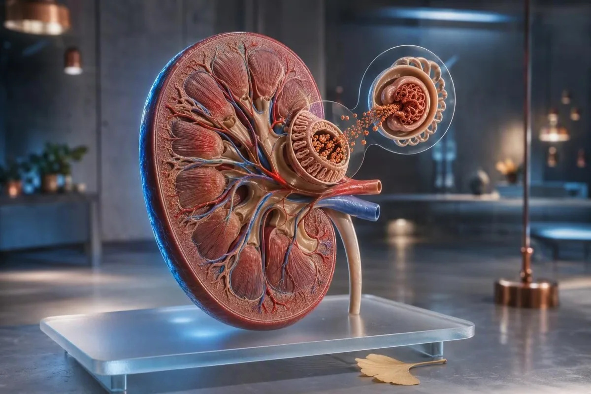
Protini ya povu kwenye mkojo mara nyingi huwa alilbumin, protini yenye chaji hasi ambayo kwa kawaida hukaa katika mfumo wa damu. Wakati kichujio cha glomerulus kinapoanza kuruhusu zaidi, alilbumin hupunguza mvutano wa uso na inaweza kuunda viputo vidogo, imara.
Mtu mzima mwenye afya kwa ujumla hutoa chini ya 30 mg ya alilbumin kwa siku, ingawa uchunguzi wa kawaida wa dipstick unaweza usigundue upotezaji mdogo wa alilbumin. Uwiano wa alilbumin-kreatini wa mkojo, au ACR, unaosikika zaidi hurekebisha alilbumin kwa mkusanyiko wa mkojo na unapendelewa kwa uchunguzi wa awali.
Alilbumin sio protini pekee inayohusika. Minyororo mepesi kutoka kwa magonjwa ya seli za plasma, protini za tubular baada ya dawa fulani, na protini za muda mfupi wakati wa homa zinaweza kutoka kwa dipstick iliyoundwa hasa kwa alilbumin; ndio maana mstari hasi hauwezi kutatua kila kisa cha povu kinachoendelea.
Dk. Thomas Klein hupitia matokeo ya mkojo pamoja na kreatini ya seramu, eGFR, alilbumin, na glukosi badala ya kutibu dalili inayoonekana kama utambuzi. Yetu mwongozo wa protini ya seramu inaeleza kwa nini matokeo ya chini ya alilbumin ya damu na ACR ya mkojo iliyoinuka pamoja huleta uzito zaidi kuliko moja pekee.
Sababu za kawaida za mkojo wenye povu zisizo za figo
Povu nyingi za muda mfupi kwenye mkojo zina sababu isiyo ya figo: mkojo wenye mkusanyiko, kukojoa kwa kasi, shahawa za hivi majuzi, mabaki ya sabuni, homa, au mazoezi ya nguvu. Sababu hizi kwa kawaida hupotea ndani ya masaa 24-48 baada ya unyevu wa kawaida na kupona.
Upungufu wa maji mwilini hufanya mkojo kuwa mweusi na wenye mkusanyiko zaidi, kwa hivyo viputo vinaweza kuonekana kuwa mnene hata wakati usafirishaji wa alilbumin uko kawaida. Lenga mkojo wa rangi ya majani ya mkuyu kwa siku inayofuata badala ya kulazimisha maji mengi; kunywa lita kadhaa haraka kunaweza kupunguza sodiamu kwa watu walioathirika.
Mazoezi yanaweza kuongeza kwa muda alilbumin kwenye mkojo. Mbio za kilomita 10, kikao cha mafunzo ya uvumilivu wa kiwango cha juu, au kukabiliwa na joto kwa muda mrefu kunaweza kutoa protini kwenye sampuli iliyokusanywa mara tu baada ya hapo, ndio maana maabara kwa kawaida hupendekeza kuepuka mazoezi makali kwa masaa 24 kabla ya kupima ACR.
Manii kwenye urethra baada ya kumwaga inaweza kutoa viputo vya muda na mstari wa protini unaoonekana kuwa na athari ndogo. Mkojo unaoonekana kuwa wa waridi au rangi ya chai hubadilisha swali kabisa na unapaswa kutathminiwa kwa kutumia mwongozo huu wa damu kwenye mkojo, hasa baada ya miaka 45 au kwa mtu yeyote aliyeathiriwa na uvutaji sigara.
Lini povu inayoendelea inapendekeza upotezaji wa protini unaohusiana na kimatibabu
Povu la kudumu linafaa kupimwa kwa protini ya mkojo linapotokea mara kwa mara na hasa linapohusishwa na kisukari, shinikizo la damu, uvimbe, au kupungua kwa eGFR. Albuminuria inaweza kuwa ishara ya mapema ya figo hata wakati kreatini bado iko ndani ya kiwango cha maabara.
KDIGO inafafanua ACR chini ya 3 mg/mmol, au 30 mg/g, kama A1; 3-30 mg/mmol kama A2; na zaidi ya 30 mg/mmol kama A3 albuminuria. Makundi haya yanatabiri hatari ya figo na moyo kwa usahihi zaidi yanapounganishwa na eGFR, hayatumiwi peke yake (KDIGO, 2024).
Kisukari na shinikizo la damu ndio vyanzo vya kawaida vya muda mrefu vya albuminuria duniani kote, lakini sio pekee. Glomerulonephritis, glomerulopathy inayohusiana na unene kupita kiasi, lupus, pre-eclampsia, usingizi wa kupumua, na baadhi ya dawa za kuzuia uvimbe pia zinaweza kuchangia; matokeo yanayoendelea yanahitaji daktari kubaini utaratibu.
Kantesti ni Mchambuzi wa mtihani wa damu wa AI ambayo huweka kreatini, eGFR, albmini ya damu, sukari, na HbA1c katika muktadha mmoja wa muda mrefu. Haiwezi kupima ACR ya mkojo kutoka kwa sampuli ya damu, lakini inaweza kusaidia kutambua muundo wa kimetaboliki au figo unaofanya matokeo ya mkojo kuwa ya haraka zaidi.
Ni vipimo gani vya protini ya mkojo vinajibu swali bora zaidi
Kipimo cha ACR cha mkojo wa asubuhi mapema ndicho kipimo bora cha kwanza kwa kupoteza kwa albmini kwa watu wengi wazima. Uwiano wa protini-kreatini, au PCR, ni muhimu wakati protini isiyo ya albmini inashukiwa au wakati protini jumla inahitaji kupimwa.
ACR huripotiwa kama mg/mmol nchini Uingereza na nchi nyingine nyingi, au mg/g nchini Marekani. ACR ya 3 mg/mmol inalingana na takriban 30 mg/g; ubadilishaji huu ni muhimu kwa sababu wagonjwa wakati mwingine huchanganya vitengo tofauti kwa mabadiliko makubwa ya matokeo.
Matokeo ya protini kwenye kipima cha mkono yenye kiwango cha athari kidogo au 1+ ni dalili ya uchunguzi, sio utambuzi. Mkojo wenye alkali, mkojo mkali, na uchafuzi unaweza kusababisha matokeo chanya yanayodanganya, wakati mkojo uliochujwa na protini zisizo za albmini zinaweza kutoa uhakikisho wa uwongo; upimaji wa kiasi huondoa kutokuwa na uhakika huo.
Mkusanyiko wa saa 24 sasa umeachwa kwa ajili ya hali maalum, kama vile kupoteza protini nyingi sana, tathmini ya ujauzito, au sampuli za kando zinazokinzana. Ni rahisi kukusanya kidogo, kwa hivyo soma maelezo yetu ya vitendo ukusanyaji wa mkojo wa saa 24 kabla ya kuanza moja.
Jinsi ya kukusanya vipimo vya protini ya mkojo kwa usahihi
Upimaji sahihi wa protini ya mkojo huanza na sampuli safi ya katikati ya mkojo wa asubuhi mapema iliyokusanywa baada ya saa 24 bila mazoezi mazito. Mkojo wa kwanza baada ya kuamka unapendelewa kwa sababu mkao, unywaji maji, na shughuli za mchana huathiri kidogo utoaji wa albmini.
Osha mikono, tumia chombo kilichotakaswa kilichotolewa, anza kukojoa kwenye choo, kisha chukua sehemu ya katikati bila kugusa ndani ya sufuria au kifuniko. Peleka sampuli mara moja; ikiwa kuchelewa hakuepukiki, fuata maagizo ya maabara ya kuhifadhi kwenye jokofu badala ya kuiacha kwenye gari lenye joto.
Weka kando vipimo visivyo vya lazima wakati wa UTI yenye dalili, homa, kuhara, hedhi, au ndani ya saa 24 baada ya mazoezi makali. Hali hizi zinaweza kuongeza kwa muda utoaji wa albmini, wakati uchafuzi wa hedhi unaweza pia kuleta damu na protini inayoonekana; matokeo ya esterase ya leukositi inaweza kufafanua muundo unaowezekana wa maambukizi.
Orthostatic proteinuria inastahili kutajwa, hasa kwa vijana na vijana wa kiume na wa kike. Protini inayoonekana kwenye mkojo wa mchana lakini haipo kwenye sampuli ya asubuhi mapema mara nyingi huwa haina madhara, ingawa daktari anapaswa kuthibitisha muundo badala ya kudhania.
Jinsi ya kutafsiri matokeo ya ACR na protini-kreatinine
ACR chini ya 3 mg/mmol kwa kawaida huleta uhakikisho, wakati ACR iliyothibitishwa ya 3 mg/mmol au zaidi inahitaji ufuatiliaji unaotegemea hatari. Sampuli moja iliyoinuliwa haithibitishi ugonjwa sugu wa figo kwa sababu mazoezi, maambukizi, na miinuko ya sukari inaweza kusababisha albuminuria ya muda mfupi.
Kwa watu wazima wasio na kisukari, ACR iliyoibuka hivi karibuni mara nyingi hurudiwa kwa kutumia sampuli ya asubuhi mapema ndani ya wiki. Ugonjwa sugu wa figo unahitaji uharibifu ulioonekana kwa angalau miezi 3, isipokuwa kama kuna majeraha ya papo hapo ya figo au protini nyingi sana inayohitaji ukaguzi wa haraka zaidi (KDIGO, 2024).
ACR zaidi ya 70 mg/mmol, takriban 700 mg/g, kwa kawaida huhitaji majadiliano ya haraka na mtaalamu hata kama eGFR imehifadhiwa, hasa ikiwa kuna damu kwenye mkojo. Protini nyingi za aina ya nephrotic kwa ujumla ni PCR zaidi ya 300 mg/mmol au utoaji wa protini zaidi ya 3.5 g/siku, mara nyingi huambatana na uvimbe na kiwango cha chini cha albmini ya damu.
Kantesti ni huduma ya kutafsiri vipimo vya maabara ya AI ambacho kinaweza kupanga vipimo vya damu vya figo kwa tarehe na kuashiria mwelekeo unaoshuka wa eGFR. Tafsiri ya mwelekeo inasaidia, lakini kamwe haibadili, uchunguzi wa kiini wa mkojo wa daktari, uthibitisho wa ACR, na ukaguzi wa dawa.
Vipimo vya damu vinavyofanya matokeo ya mkojo wenye povu kuwa na maana
Kreatini, eGFR, albmini ya damu, sukari, HbA1c, elektroliti, na lipidi husaidia kueleza ikiwa protini ya mkojo inaonyesha uharibifu wa kuchuja kwa figo au shida kubwa zaidi ya kimetaboliki. Kreatini ya kawaida haizuii albuminuria ya mapema.
eGFR huhesabiwa kutoka kwa creatinine na huanguka baadaye kuliko kuvuja kidogo kwa albumin kwa watu wengi wenye kisukari. eGFR chini ya 60 mL/min/1.73 m² kwa miezi 3 au zaidi hukutana na kigezo kimoja cha ugonjwa sugu wa figo, lakini maadili lazima yafasiriwe kulingana na umri, misuli ya mwili, na ugonjwa wa hivi karibuni.
Alabamu ya chini ya seramu chini ya takriban 35 g/L pamoja na protini nyingi kwenye mkojo na uvimbe mpya wa kifundo cha mguu huongeza wasiwasi wa kupoteza protini nyingi kwenye figo. Kinyume chake, alabamu ya juu na mkojo mweusi mara nyingi huonyesha upungufu wa maji mwilini; yetu eGFR baada ya upungufu wa maji mwilini inaeleza kwa nini kupima tena baada ya kupona kunaweza kuwa na maana.
American Diabetes Association inapendekeza angalau UACR ya kila mwaka na upimaji wa eGFR kwa aina ya 2 ya kisukari na kwa aina ya 1 ya kisukari inayodumu kwa miaka 5 au zaidi (ADA Professional Practice Committee, 2024). HbA1c inatuambia kuhusu wastani wa sukari mwilini, lakini ongezeko kali la sukari bado linaweza kuongeza kwa muda mfupi alabamu ya mkojo kabla ya HbA1c kubadilika.
Dawa, virutubisho, na magonjwa ambayo yanaweza kuathiri protini ya mkojo
Dawa za maumivu za kuzuia uvimbe, lithiamu, baadhi ya tiba za kansa, na maambukizi makali yanaweza kubadilisha uchujaji wa figo au usindikaji wa protini kwenye mirija. Usisimamishe dawa iliyoagizwa kwa sababu tu mkojo unaonekana kuwa na povu; omba uchunguzi wa mkojo na damu ya figo.
NSAIDs kama vile ibuprofen na naproxen zinaweza kupunguza mtiririko wa damu kwenye figo, hasa wakati wa upungufu wa maji mwilini, kushindwa kwa moyo, au CKD iliyopo. Hatari huongezeka wakati NSAID inapotumiwa pamoja na diureti na kizuizi cha ACE au ARB, mchanganyiko ambao waganga wakati mwingine huuelezea kama “pigo mara tatu.”
Dawa za SGLT2 zinaweza kusababisha kushuka kidogo kwa eGFR mapema lakini kwa kawaida hupunguza albuminuria kwa wakati kwa wagonjwa waliochaguliwa ipasavyo. Mabadiliko hayo yanayotarajiwa ya hemodynamiki yanatofautiana na uharibifu unaoendelea; angalia mabadiliko ya eGFR na matibabu ya SGLT2 kwa tofauti za vitendo.
Dozi kubwa ya vitamini C inaweza kuingilia baadhi ya vipimo vya mkojo kwa kutumia vipande, wakati creatine inaweza kuleta ugumu katika tafsiri ya creatinine bila lazima kuiathiri vibaya figo. Leta orodha kamili ya dawa zilizoagizwa, dawa za maumivu zinazouzwa bila dawa, poda za protini, na bidhaa za mitishamba kwa miadi—majina na vipimo ni muhimu.
Kisukari na shinikizo la damu: ukaguzi wa hatari wenye mavuno mengi zaidi
Kisukari na shinikizo la damu hufanya povu la kudumu kuwa muhimu zaidi kimatibabu kwa sababu zote mbili zinaweza kusababisha albuminuria miaka mingi kabla ya dalili kuonekana. Shinikizo la damu la 140/90 mmHg au zaidi na ACR ya 3 mg/mmol au zaidi inapaswa kusababisha uchunguzi wa kina katika huduma ya msingi.
Kwa watu wengi wenye CKD na albuminuria, waganga hulenga shinikizo la kawaida la sistoli la damu katika ofisi chini ya 120 mmHg inapovumiliwa, ingawa malengo lazima yapatanishwe kwa umri, hatari ya kuanguka, ujauzito, na njia ya kipimo. Vipimo vya nyumbani vinavyofanywa vibaya havipaswi kusababisha mabadiliko ya dawa pekee.
Albuminuria pia ni kiashiria cha hatari ya mishipa. Sababu tunayoihofia ongezeko la ACR pamoja na shinikizo la damu ni kwamba pamoja huashiria msongo kwenye endotheliamu na glomeruli, wakati wowote kati ya vitu hivyo mara moja wakati wa ugonjwa mkali unaweza kuwa wa muda mfupi; yetu mwongozo wa maabara ya shinikizo la damu unaelezea uchunguzi mpana zaidi.
Kantesti ni jukwaa la tafsiri ya viashiria vya AI hutumiwa katika nchi 127+ kulinganisha mwenendo wa sukari, HbA1c, vigezo vya figo, na lipids. Kwa mkojo wenye povu wa kudumu, kipimo kinachofaa kinachofuata bado ni ACR ya mkojo wa maabara, bora ukaguziwe na timu ile ile ya kimatibabu inayoshughulikia shinikizo la damu au kisukari.
Mkojo wenye povu wakati wa ujauzito na kwa watoto unahitaji sheria tofauti
Mkojo wenye povu wakati wa ujauzito unahitaji tathmini ya siku hiyo hiyo ikiwa utatokea na maumivu ya kichwa, mabadiliko ya maono, maumivu ya tumbo la juu, uvimbe wa ghafla, au shinikizo la damu lililoongezeka. Protiniuria baada ya wiki 20 inaweza kuwa sehemu ya pre-eclampsia, lakini mwonekano pekee hauwezi kugundua.
Katika ujauzito, uwiano wa protini-creatinine wa 30 mg/mmol au zaidi, au 300 mg ya protini katika masaa 24, unasaidia protiniuria kubwa katika mazingira sahihi ya kimatibabu. Sampuli ya mkojo inayoonekana ya kawaida haiwezi kutenga pre-eclampsia ikiwa shinikizo la damu ni 140/90 mmHg au zaidi na dalili za kutisha.
Watoto mara nyingi huwa na protiniuria ya muda mfupi baada ya homa, michezo, au upungufu wa maji mwilini. Protini ya kudumu kwenye sampuli tatu, shinikizo la damu la juu, uvimbe, au damu kwenye mkojo kunahitaji tathmini ya watoto; upimaji wa asubuhi ya kwanza ni muhimu sana kwa kutenga protiniuria ya kimaeneo kwa vijana.
Mabadiliko ya ujauzito huchuja, kwa hivyo maadili ya creatinine yanayoonekana “kawaida” kwa watu wazima wasio wajawazito huenda hayathibitishi. Mwongozo wetu wa maadili ya GFR wakati wa ujauzito unatoa muktadha, lakini dalili za haraka zinapaswa kwenda moja kwa moja kwenye chumba cha dharura cha uzazi badala ya tafsiri ya mtandaoni.
Ishara za hatari: lini mkojo wenye povu unahitaji huduma ya haraka ya kimatibabu
Tafuta tathmini ya haraka ya kimatibabu kwa mkojo wenye povu na upungufu wa pumzi, uvimbe wa miguu au uso unaoongezeka kwa kasi, mkojo uliopungua sana, damu inayoonekana, homa na maumivu ya kiuno, au dalili za onyo za ujauzito. Mchanganyiko huu unaweza kuashiria ugonjwa mkali wa figo, moyo, maambukizi, au unaohusiana na ujauzito.
Kuvimba kwa uso mpya wakati wa kuamka, viatu kukaza jioni, na ongezeko la uzito la zaidi ya kilo 2 katika siku 2-3 kunaweza kuonyesha uhifadhi wa maji. Dalili hizo zikifuatana na povu la kudumu, wahudumu wa afya huangalia protini ya mkojo, kreatini, albmini, shinikizo la damu, na wakati mwingine matokeo ya kifua mara moja.
Mkojo wenye rangi ya chai baada ya mazoezi makali yenye maumivu makali ya misuli sio povu la kawaida la kutishia; unaweza kuashiria kutolewa kwa myoglobin na unahitaji vipimo vya damu na mkojo vya haraka. Hali hiyo hiyo inatumika kwa mkojo wenye rangi ya cola na upungufu wa pato, ambao unaweza kuonyesha kutokwa na damu kwa glomerulus au majeraha ya misuli.
Dk. Thomas Klein anawashauri wagonjwa wasisubiri matokeo ya kipimo cha nyumbani wakati dalili za hatari zipo. Kwa mabadiliko ya rangi yanayohusiana na juhudi, makala yetu kuhusu hemoraji ya mwanariadha inaelezea mwisho wa kawaida wa wigo na dalili ambazo si za kawaida.
Wataalamu wa afya hufanya nini baada ya matokeo chanya ya protini ya mkojo
Baada ya matokeo chanya ya ACR au PCR, wahudumu wa afya kwa kawaida huthibitisha matokeo, hupima shinikizo la damu na eGFR, huchunguza chembechembe za mkojo, na hupitia historia ya kisukari, dawa, dalili za autoimmune, na historia ya familia. Hatua inayofuata inategemea wingi, kudumu, na uwepo wa damu au upungufu wa uchujaji.
Uchunguzi wa darubini ya mkojo unaweza kutambua seli nyekundu za damu, seli nyeupe za damu, na maganda ambayo nambari ya protini pekee haiwezi kueleza. Maganda ya seli nyekundu za damu au seli nyekundu za damu zenye umbo la ajabu zinaonyesha chanzo cha glomerulus na huharakisha rufaa kwa nephrology; maganda rahisi ya hyaline yanaweza kutokea baada ya upungufu wa maji mwilini au mazoezi, kama ilivyoelezwa katika mwongozo wetu wa maganda ya hyaline.
Ikiwa albuminuria imethibitishwa, matibabu kwa kawaida hulenga sababu: udhibiti wa shinikizo la damu, usimamizi wa sukari, msaada wa uzito na uvutaji sigara, na dawa za kulinda figo pale zinapohitajika. Kipimo cha ultrasound cha figo, kipimo cha kinga, uchunguzi wa umeme wa damu, au biopsy ya figo ni vipimo maalum - sio matokeo ya kiotomatiki ya kipimo cha 1+.
Mbinu ya kimatibabu ya Kantesti inachunguzwa dhidi ya viwango vya usalama vilivyopangwa, na wasomaji wanaweza kuchunguza mbinu ya uthibitisho wa kimatibabu. yetu. Jukumu letu ni kufanya matokeo ya vipimo vya damu yaliyopo kuwa rahisi kujadili, sio kutambua ugonjwa wa glomerulus kutoka kwa picha au thamani moja ya maabara.
Mpango wa vitendo wa siku saba kwa povu inayoendelea
Kwa mkojo wenye povu la kudumu bila dalili za hatari, panga kipimo cha ACR cha mkojo cha huduma ya msingi na uchunguzi wa mkojo ndani ya wiki 1-2, ikiwezekana kwa kutumia sampuli ya asubuhi mapema. Rekodi shinikizo la damu, unyevu, mazoezi, dawa, na ikiwa povu hudumu zaidi ya dakika 1 kwa siku saba.
Siku 1-3: kunywa maji kama kawaida, epuka mazoezi makali, na uangalie tu kwenye bakuli safi la choo bila kiboreshaji kilichoongezwa. Usijaribu “kuondoa” tatizo kwa maji mengi; lengo ni sampuli inayowakilisha, sio iliyopunguzwa bandia.
Siku 4-7: omba uchunguzi wa mkojo pamoja na ACR, na uulize ikiwa kreatini, eGFR, potasiamu, albmini ya damu, glukosi, na HbA1c zinafaa kwa hali zako. Leteni matokeo ya awali kwa sababu mabadiliko kutoka ACR 2 hadi 8 mg/mmol yana maana zaidi kuliko nambari yoyote bila muktadha; ufuatiliaji wa matokeo kwa muda mrefu inaweza kusaidia kupanga tarehe.
Kufikia Septemba 4, 2026, ujumbe wa busara unabaki wa kutia moyo lakini imara: matone mengi ya pekee hayadhuru, wakati povu laini la mara kwa mara linastahili kipimo cha mkojo cha kuhesabu. Ikiwa unahitaji usaidizi wa kuandaa maswali kwa daktari, kama rasilimali ya uwazi na kutazama ripoti hiyo kama mwanzo uliopangwa wa huduma, kamwe si kama ruhusa ya kuanza, kusitisha, au kubadilisha matibabu yaliyowekwa. yetu huweka viwango vya kimatibabu vinavyotumiwa na maudhui yetu ya elimu.
Maswali Yanayoulizwa Mara Kwa Mara
Je, mkojo wenye povu huwa ni ishara ya protini kwenye mkojo?
Hapana, mkojo wenye povu mara nyingi si ishara ya kupoteza protini. Mkojo unaotoka kwa kasi, upungufu wa maji mwilini, mabaki ya dawa ya chooni, homa, mazoezi, na shahawa vinaweza kuunda viputo vinavyopotea baada ya sekunde chache. Povu jeupe jepesi linaloendelea kuonekana kwa siku kadhaa linaweza kuashiria uwepo wa protini kwenye mkojo (proteinuria) na linapaswa kuchunguzwa kwa kutumia kipimo cha uwiano wa alb_umin na kreatini kwenye mkojo (urine albumin-creatinine ratio - ACR). ACR ya 3 mg/mmol, sawa na 30 mg/g, au zaidi huchukuliwa kuwa si kawaida na huhitaji uchunguzi wa kimatibabu na mara nyingi vipimo vya kurudiwa.
Muda gani povu katika mkojo hudumu kabla ya kuwa na wasiwasi?
Povu ambalo hubaki kama safu nene kwa takriban dakika 1-2 mara kwa mara ni la wasiwasi zaidi kuliko viputo vikubwa vinavyopotea haraka. Muda si kipimo cha utambuzi kwa sababu kemikali za chooni na mkusanyiko wa mkojo vinaweza kubadilisha utulivu wa viputo. Ikiwa hali hiyo inajirudia kwa siku 3 au zaidi, panga uchunguzi wa mkojo na ACR ya mkojo ndani ya wiki 1-2. Tafuta huduma ya haraka mapema ikiwa povu hutokea pamoja na uvimbe, damu inayoonekana, ugumu wa kupumua, homa, au kupungua kwa kiasi cha mkojo.
Ni kipimo gani cha protini katika mkojo ninapaswa kuuliza?
Kipimo bora cha kwanza kwa watu wazima wengi ni uwiano wa dutu ya albamini na kreatini kwenye mkojo wa asubuhi na mapema, unaoitwa ACR au UACR. ACR chini ya 3 mg/mmol, au 30 mg/g, kwa ujumla huwa kawaida au imeongezeka kidogo, wakati 3-30 mg/mmol huonyesha ongezeko la wastani la albamini kwenye mkojo. Uwiano wa protini na kreatini unaweza kuongezwa wakati upotevu wa jumla wa protini au protini isiyo ya albamini unashukiwa. Kipimo cha kawaida cha kutumia karatasi maalumu (dipstick) ni muhimu kwa uchunguzi lakini si sahihi kama ACR kwa upotevu mdogo wa albamini.
Je, upungufu wa maji mwilini husababisha mkojo kuwa na povu?
Ndiyo, upungufu wa maji mwilini unaweza kufanya mkojo kuwa na mkusanyiko zaidi na unaweza kuleta mapovu au povu linaloonekana zaidi bila kupoteza protini kwenye figo kwa kudumu. Mara nyingi hutokea na mkojo wa njano wenye rangi ya giza, kiu, kukabiliwa na joto, kutapika, kuhara, au kipindi kirefu bila kunywa. Rejea ulaji wa kawaida wa maji kwa saa 24-48 kabla ya kipimo kisicho cha dharura, lakini epuka kunywa kupita kiasi ili tu kupunguza matokeo. Ikiwa povu itaendelea kuwepo baada ya unyweshaji, omba kipimo cha mkojo cha ACR.
Je, mazoezi yanaweza kusababisha protini na povu kwenye mkojo?
Mazoezi makali yanaweza kusababisha albulmuria ya muda na mkojo wenye povu, hasa baada ya kukimbia, mazoezi mazito ya kubeba vitu vizito, mashindano ya uvumilivu, au kufanya mazoezi katika hali ya joto. Athari kawaida hupotea ndani ya saa 24-48, ndiyo sababu sampuli ya ACR inapaswa kukusanywa baada ya kuepuka mazoezi makali kwa saa 24. Proteinuria inayoendelea katika sampuli ya asubuhi baada ya kupona haifikishwi kwa urahisi na mazoezi pekee. Mkojo wa rangi nyeusi au ya cola pamoja na maumivu makali ya misuli unahitaji tathmini ya haraka ya kimatibabu.
ACR hatari ni ipi?
ACR iliyo juu ya 30 mg/mmol, au 300 mg/g, ni albominuria iliyoongezeka sana na inahitaji tathmini ya haraka ya kimatibabu, hasa ikiwa eGFR imepungua au mkojo una damu. ACR iliyo juu ya 70 mg/mmol, kama 700 mg/g, mara nyingi huchochea mjadala wa haraka wa mtaalamu hata kama kreatini ni kawaida. Hatari huamuliwa na mchanganyiko wa ACR, eGFR, shinikizo la damu, hali ya ugonjwa wa kisukari, dalili, na vipimo vya kurudia. ACR moja iliyoinuliwa kwa kawaida inapaswa kuthibitishwa isipokuwa hali ya kimatibabu ni ya haraka.
Pata Uchambuzi wa Vipimo vya Damu kwa AI Leo
Jiunge na zaidi ya watumiaji 2 milioni duniani kote wanaoamini Kantesti kwa uchambuzi wa papo hapo na sahihi wa vipimo vya maabara. Pakia matokeo yako ya vipimo vya damu na upate tafsiri ya kina ya viashiria vya 15,000+ ndani ya sekunde.
📚 Machapisho ya Utafiti Yanayorejelewa
Klein, T., Mitchell, S., & Weber, H. (2026). Mfumo wa Uthibitishaji wa Kitaaluma v2.0 (Ukurasa wa Uthibitishaji wa Tiba). Kantesti uchambuzi wa damu kwa AI ya utafiti wa matibabu.
Klein, T., Mitchell, S., & Weber, H. (2026). Kichambuzi cha Uchambuzi wa Damu kwa AI: Vipimo 2.5M Vilivyofanyiwa Uchambuzi | Ripoti ya Afya ya Kimataifa 2026. Kantesti uchambuzi wa damu kwa AI ya utafiti wa matibabu.
📖 Marejeo ya Nje ya Tiba
⚕️ Kanusho la Kimatibabu
Makala haya ni kwa madhumuni ya elimu tu na si ushauri wa kimatibabu. Wasiliana na mtoa huduma aliyehitimu kila wakati kwa maamuzi ya utambuzi na matibabu.
E-E-A-T Trust Signals
Uzoefu
Mapitio ya kimatibabu inayoongozwa na daktari ya mifumo ya tafsiri ya maabara.
Utaalamu
Kuzingatia dawa za maabara kuhusu jinsi viashiria (biomarkers) vinavyobadilika katika muktadha wa kliniki.
Mamlaka
Imeandikwa na Dk. Thomas Klein kwa mapitio ya Dk. Sarah Mitchell na Prof. Dk. Hans Weber.
Uaminifu
Tafsiri inayotegemea ushahidi yenye njia zilizo wazi za ufuatiliaji ili kupunguza tahadhari za hofu.