High serum viscosity alone does not diagnose hyperviscosity syndrome. New visual changes, persistent mucosal bleeding, or neurological symptoms can make the result an emergency—even below a commonly quoted threshold.

Key takeaways
- Serum viscosity test measures how strongly serum resists flow; it does not directly measure whole-blood thickness or clotting tendency.
- Emergency symptoms include sudden vision loss, new weakness, confusion, seizures, or substantial bleeding; call emergency services rather than waiting for another test.
- Reference intervals depend on the laboratory, method, temperature, and units. An interval around 1.4–1.8 cP is illustrative, not a universal standard.
- Around 4 cP is a commonly discussed higher-risk range, but hyperviscosity syndrome can occur below it and some people remain asymptomatic above it.
- Serum versus plasma matters because plasma retains fibrinogen while serum does not; their results and reference intervals are not interchangeable.
- Immunoglobulin concentration influences viscosity differently by protein type: IgM may increase viscosity around 3,000 mg/dL, whereas IgG usually requires much higher concentrations.
- Plasma exchange can rapidly reduce circulating abnormal proteins when symptomatic hyperviscosity is suspected; urgent treatment need not wait for a send-out viscosity result.
- Follow-up should combine symptoms, eye examination, quantitative immunoglobulins, protein studies, and same-method trends—not an isolated high flag.
When does high serum viscosity need emergency care?
High serum viscosity needs emergency assessment when it accompanies new visual changes, persistent mucosal bleeding, or neurological symptoms. Sudden vision loss, weakness, difficulty speaking, seizures, or marked confusion warrant emergency services immediately; a value below 4 cP does not make those symptoms safe.

An elevated serum viscosity test result alone does not establish hyperviscosity syndrome, because the syndrome requires clinically significant effects on circulation. Around 4 cP is a useful warning context, not a diagnostic boundary; clinicians assess the person, the protein disorder, and the laboratory method together before deciding what treatment is needed.
For a clearly hypothetical comparison, consider 2 people with the same measured viscosity of 3.6 cP: one feels well, while the other has new blurred vision and repeated nosebleeds. The second person needs urgent emergency assessment, whereas the first needs prompt discussion with the ordering clinician rather than an automatic diagnosis or an automatic plasma exchange.
Kantesti is an AI blood test analyzer that can help explain a reported viscosity value, but no 60-second interpretation can replace emergency assessment or examination of the retina. Information about our organization explains our role; if symptoms are happening now, use local emergency services rather than messaging a website or waiting for a repeat result.
What does a serum viscosity test actually measure?
A serum viscosity test measures serum's resistance to flow, usually through a calibrated laboratory viscometer. Serum is the liquid remaining after clotting and separation of cellular material; viscosity reported in cP or mPa·s describes a physical property, not the time needed to form a clot.

The unit conversion is exact: 1 centipoise equals 1 millipascal-second, written 1 cP = 1 mPa·s. Large proteins, their concentrations, and their tendency to associate with one another influence the result; two samples with the same total protein concentration can therefore have different viscosities, especially when their immunoglobulin composition differs.
Serum viscosity is not whole-blood viscosity, which also depends strongly on hematocrit, cellular deformability, and flow conditions. A person with a hematocrit of 60%, for example, could have a cellular cause of increased whole-blood viscosity without a proportionate serum abnormality; our serum protein interpretation guide explains the separate contribution of dissolved proteins.
Osmolality and viscosity answer different questions, even when both tests use serum. Sodium-related confusion at a measured osmolality of 320 mOsm/kg cannot be explained simply by calling serum 'thick'; the serum osmolality guide describes particle concentration, whereas viscosity reflects resistance to movement and interactions between molecules.
How does serum viscosity differ from plasma viscosity?
Serum viscosity and plasma viscosity are different tests because plasma retains fibrinogen, while serum is obtained after clotting. A plasma reference interval cannot be applied to a serum result, even when both reports use mPa·s; 1 mPa·s equals 1 cP, but that conversion does not make the specimens equivalent.

Plasma viscosity can reflect changes in fibrinogen as well as immunoglobulins, so clinicians sometimes use it in assessing inflammatory activity. A hypothetical plasma result of 1.9 mPa·s and serum result of 1.9 cP are numerically equal in absolute units, but they must still be interpreted against two different specimen-specific reference intervals.
Test temperature also changes interpretation: measurements performed at 25°C and 37°C should not be compared as though temperature were irrelevant. Some laboratories, including European services, use plasma viscosity for inflammatory assessment while specialist laboratories offer serum viscosity for paraprotein-related concerns; the laboratory's actual test name matters more than the country or the familiar-looking number.
When a clinician is investigating a monoclonal protein, quantitative IgG, IgA, and IgM help explain which proteins may be contributing to resistance to flow. Those 3 immunoglobulin classes do not behave identically, and our immunoglobulin panel explanation shows why a viscosity result should be read alongside the protein pattern rather than substituted for it.
How should you read serum viscosity test results?
Read serum viscosity test results using the reference interval, units, specimen, and temperature printed by the reporting laboratory. Some laboratories use an interval around 1.4–1.8 cP, while others report a dimensionless relative viscosity; similar-looking numbers do not prove that the methods or thresholds are equivalent.
An absolute viscosity of 2.0 cP is not automatically interchangeable with a relative viscosity of 2.0. Relative viscosity compares the sample with a reference fluid under specified conditions; because the reference fluid's viscosity changes with temperature, converting a ratio requires the laboratory's method rather than an assumed universal multiplier.
The commonly cited 4 cP warning range comes from clinical experience with paraprotein-related hyperviscosity, not from a sharp biological switch at 4.00. Gertz's review in Blood describes substantial individual variation in the relationship between viscosity and symptoms, which is why symptom-based treatment decisions remain central rather than a numerical definition alone (Gertz, 2018).
Kantesti's interpretation of a report should preserve the laboratory's own units and limits, rather than replacing them with a generic range from a different method. Before comparing 2 reports, check all four details—specimen, units, method, and temperature—and use our biomarker reference guide to understand why reference intervals are not interchangeable treatment thresholds.
| Level | Range | What it means |
|---|---|---|
| Within the laboratory interval | Use the report; some intervals are around 1.4–1.8 cP | An in-range serum result does not rule out other causes of urgent visual or neurological symptoms. |
| Above the laboratory limit | No universal mildly elevated band | Review the protein pattern, symptoms, method, and reason for testing; a high flag alone is not a syndrome. |
| Below the commonly quoted warning range | Below 4 cP | Symptomatic hyperviscosity remains possible; symptoms override reassurance from this number. |
| Higher-risk numerical context | Around 4 cP or higher | Arrange prompt clinical assessment, often same-day specialist discussion; compatible symptoms require emergency care. |
What causes high serum viscosity?
High serum viscosity most often raises concern about increased immunoglobulins, particularly monoclonal IgM associated with Waldenström macroglobulinemia or certain other B-cell disorders. IgM may increase viscosity around 3,000 mg/dL; the concentration is a clue to risk, not a stand-alone diagnosis of cancer or hyperviscosity syndrome.

Protein shape matters as much as protein quantity. IgM's large pentameric structure can increase viscosity at approximately 3 g/dL, while IgG-related hyperviscosity often involves concentrations around 10 g/dL or more; IgG subclass and molecular association also affect this relationship, so these approximate values are not interchangeable diagnostic thresholds (Weaver et al., 2020).
A monoclonal disorder is not the only explanation: marked polyclonal immunoglobulin elevation and cryoproteins can also increase viscosity. The 2 principal questions are whether one protein-producing clone dominates and whether that protein behaves unusually; serum electrophoresis patterns help separate a narrow monoclonal band from a broader polyclonal increase.
Total protein and the gamma gap can identify a reason to investigate, but neither replaces the viscosity measurement or identifies its cause. For example, a hypothetical total protein of 10.0 g/dL and albumin of 3.0 g/dL produce a 7.0 g/dL gap; our gamma gap interpretation explains why additional protein studies are still needed.
Why does an elevated result not diagnose the syndrome?
Hyperviscosity syndrome is a clinical complication, not simply an above-range laboratory result. A person with 4.5 cP and no detectable organ effects may require a different plan from someone with 3.2 cP and retinal abnormalities; protein characteristics, examination findings, and the individual's symptom threshold influence the decision.

There is no single viscosity cutoff that reliably diagnoses or excludes symptomatic hyperviscosity in every patient. A previously documented symptom threshold—such as symptoms occurring near 4.2 cP in a hypothetical earlier episode—can inform that person's future care, but a past threshold cannot safely override new visual or neurological findings.
The classic 3 symptom domains are visual disturbance, mucosal bleeding, and neurological dysfunction, but the full triad is not required. Retinal findings may appear before obvious visual complaints, while headache or fatigue alone is nonspecific; this combination of incomplete presentations and common alternative causes explains why clinical examination is more useful than a symptom checklist alone.
Kantesti is an AI blood test interpretation platform that can place a viscosity result alongside immunoglobulins and CBC findings, not certify that the syndrome is present or absent. Rouleaux on a smear may support a protein-related process, but 1 finding of stacked cellular elements does not prove impaired organ circulation or justify emergency treatment by itself.
Which visual changes make the result urgent?
New blurred vision, double vision, or loss of vision with high serum viscosity requires urgent assessment, because retinal circulation may be affected. Sudden loss of vision in 1 or both eyes is an emergency regardless of the viscosity result; do not wait for a complete symptom triad or another laboratory measurement.
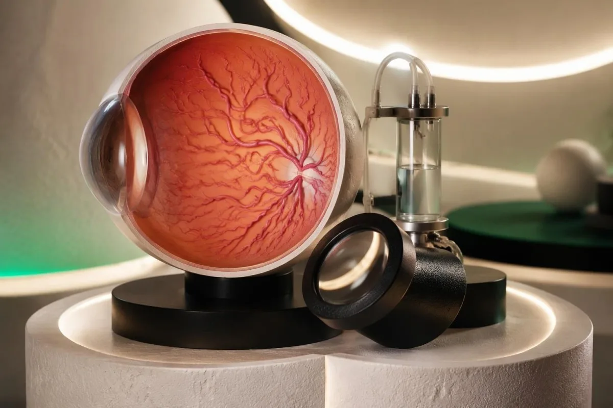
A dilated retinal examination can identify enlarged, tortuous retinal vessels and retinal hemorrhages associated with hyperviscosity. A hypothetical patient reporting no visual difficulty at 4.8 cP could still have relevant retinal findings; conversely, a complaint of blurred vision at 2.2 cP may come from another urgent eye condition rather than protein-related hyperviscosity.
Eye pressure testing is not a substitute for looking at the retina when hyperviscosity is suspected. The glaucoma testing explanation describes different tools and targets; a normal pressure measurement does not rule out retinal circulation abnormalities, and 1 reassuring eye test cannot answer every cause of visual change.
Tell the assessing team whether the change began suddenly or gradually, affects 1 eye or both, and coincides with headache, nosebleeds, or confusion. Bring the actual viscosity report and any IgM result if readily available—but do not delay travel to collect paperwork, and do not drive yourself when vision is impaired.
Why do nosebleeds or gum bleeding change urgency?
New, recurrent, or persistent nose or gum bleeding can be a hyperviscosity warning sign, especially alongside visual changes or an elevated monoclonal protein. Bleeding can occur despite a normal platelet count and an INR near 1.0, because routine coagulation tests do not fully assess platelet function or all acquired bleeding disorders.

Abnormal circulating proteins can interfere with hemostasis, and some monoclonal disorders cause acquired von Willebrand abnormalities as well as increased viscosity. A hypothetical platelet count of 220 × 10^9/L does not exclude these mechanisms; our coagulation panel limitations explain why normal routine clotting results cannot settle the cause of mucosal bleeding.
A small gum bleed after vigorous brushing is not equivalent to repeated spontaneous bleeding plus blurred vision. However, a nosebleed continuing after about 15–20 minutes of uninterrupted pressure, a heavy bleed, faintness, or breathing difficulty needs urgent care; an elevated viscosity result lowers the threshold for immediate assessment rather than encouraging repeated home observation.
Clinicians distinguish a new acquired problem from a lifelong bleeding tendency by reviewing previous procedures, medication exposure, and family history. Those 3 pieces of history help decide whether targeted testing is needed; the von Willebrand bleeding guide describes related patterns, but unexplained active bleeding with suspected hyperviscosity should not wait for outpatient confirmation.
Which neurological symptoms need immediate action?
New confusion, marked drowsiness, trouble walking, seizures, or focal neurological deficits require emergency assessment when hyperviscosity is possible. Sudden weakness on 1 side, speech difficulty, or severe imbalance must be treated as a possible stroke; the viscosity result cannot distinguish stroke from other neurological emergencies.

A viscosity value below 4 cP does not rule out stroke, metabolic disturbance, or another cause of acute neurological dysfunction. Record the time symptoms started or the last time the person was normal, and use the stroke warning signs guide to recognize why emergency services—not a repeat blood test—are the appropriate first step.
Headache and dizziness are less specific than a seizure or new focal weakness, but their context still changes urgency. For example, a hypothetical new headache with repeated nosebleeds and blurred vision at 3.4 cP warrants emergency assessment, whereas an isolated longstanding mild headache needs evaluation without automatically being attributed to the viscosity number.
Slowly progressive numbness in both feet is a different pattern from neurological symptoms developing over 10 minutes. IgM-associated neuropathy, including anti-MAG-related disease, can occur without symptomatic hyperviscosity; our anti-MAG neuropathy explanation helps separate a chronic nerve problem from an acute circulation-related emergency, although rapid progression still requires urgent care.
Which tests explain an elevated viscosity result?
The workup for high serum viscosity usually combines quantitative immunoglobulins, serum protein electrophoresis, immunofixation, CBC, and kidney-related chemistry with clinical examination. These tests answer different questions; the 3 major immunoglobulin classes—IgG, IgA, and IgM—help identify the protein context but do not independently diagnose symptomatic hyperviscosity.

Electrophoresis estimates the amount and distribution of serum proteins, while immunofixation identifies a monoclonal protein's type. A hypothetical M-spike of 2.8 g/dL and IgM of 3,400 mg/dL are related but not identical measurements; the immunofixation results guide explains why a band should be characterized before assumptions are made about its diagnosis.
Serum free light chains can add information about a clonal process, but kidney impairment also influences concentrations and interpretation of the ratio. A hypothetical creatinine of 2.0 mg/dL therefore changes the context of an abnormal light-chain result; our free light chain ratio guide explains why kidney-aware interpretation is needed rather than a simple positive-or-negative label.
Kantesti AI can help organize 2 or more related reports for discussion, but distinguishing Waldenström macroglobulinemia, myeloma, and other protein disorders may require marrow examination and specialist testing. Ask which diagnosis is actually suspected and which result would change management; a high viscosity number alone does not identify the responsible cell population or establish a cancer diagnosis.
How is symptomatic hyperviscosity treated urgently?
Therapeutic plasma exchange is the principal rapid treatment for symptomatic paraprotein-related hyperviscosity, because it removes circulating proteins contributing to resistance to flow. A typical procedure processes approximately 1–1.5 estimated plasma volumes; strongly suspected organ-threatening hyperviscosity can require treatment before a send-out serum viscosity result returns.
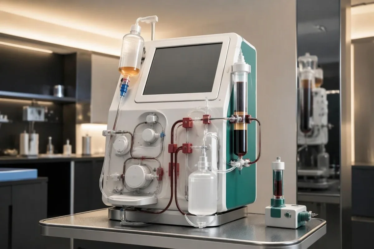
The number of exchanges depends on symptoms, protein distribution, rebound, and repeated measurements rather than a universal schedule. IgM is predominantly intravascular—approximately 75% in the review by Weaver and colleagues—so its removal can produce rapid benefit; IgG-related disease may require a different exchange pattern because more protein resides outside the circulation (Weaver et al., 2020).
Transfusion decisions need coordination because adding cellular material can increase whole-blood viscosity in an already compromised circulation. A hypothetical hemoglobin of 7.0 g/dL does not justify a blanket instruction to transfuse immediately or never transfuse; lifesaving treatment must not be withheld, and the crossmatch testing explanation addresses compatibility rather than the separate decision about timing.
Plasma exchange also affects proteins involved in clotting, especially across repeated procedures, and replacement fluid is selected for the clinical situation. Specialists may monitor fibrinogen and other hemostatic results after 1 or more sessions; our clotting test interpretation guide explains those measurements, but aspirin, anticoagulants, or supplements are not substitutes for removal of viscosity-producing proteins.
Why can viscosity rise again after treatment?
Viscosity can rise again because plasma exchange removes circulating proteins without stopping the cells that produce them. Disease-directed treatment is therefore needed when a protein-producing disorder is responsible; 1 successful exchange does not establish durable control, and some therapies can temporarily increase IgM before longer-term improvement occurs.
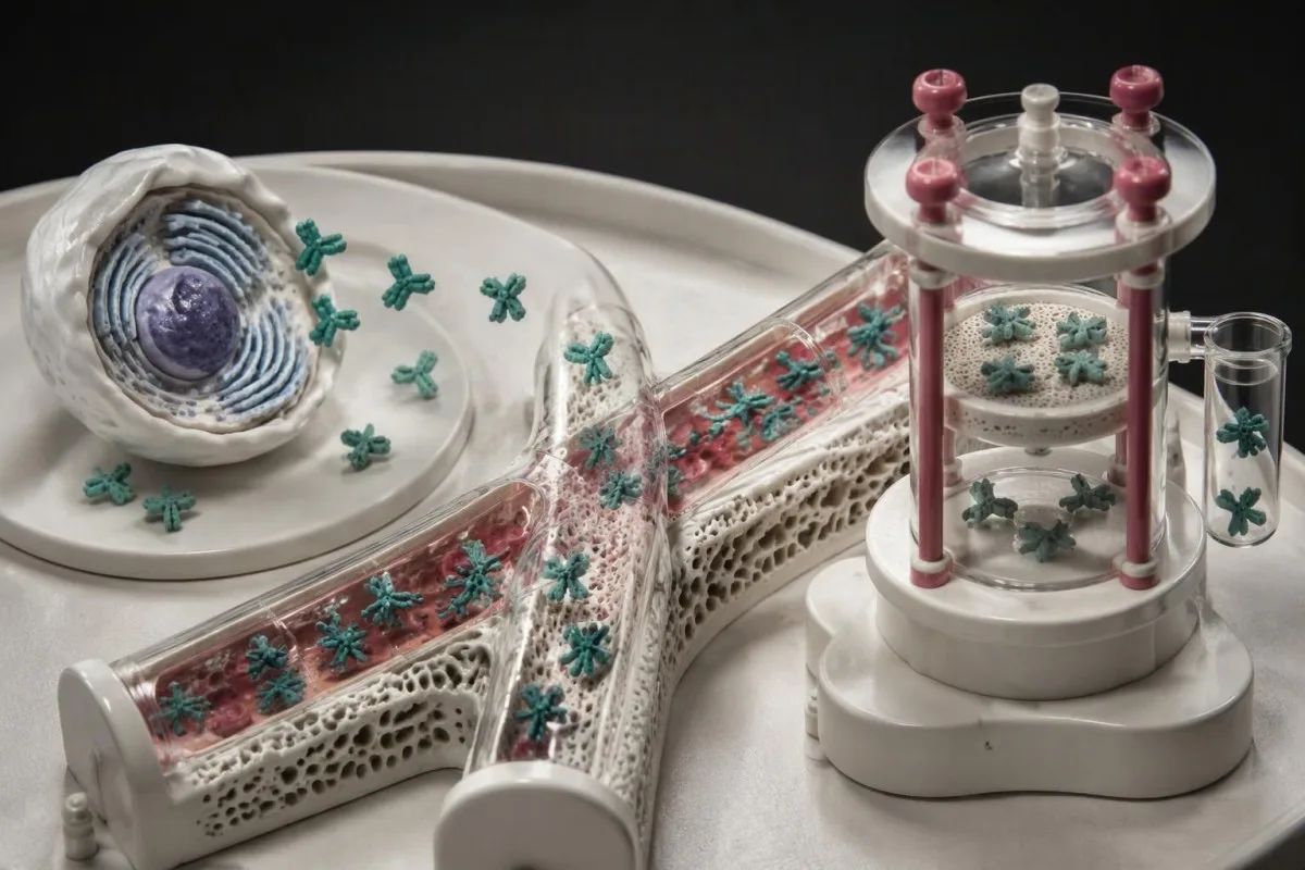
Rituximab-associated IgM flare is a recognized concern in Waldenström macroglobulinemia, but its clinical importance depends on the starting IgM level, viscosity, symptoms, and regimen. Some specialist protocols consider preventive exchange when IgM is at least 4,000 mg/dL or viscosity exceeds 3.5 cP; these are treatment-specific prevention thresholds, not universal diagnostic definitions of hyperviscosity syndrome (Weaver et al., 2020).
A rising protein level immediately after treatment does not always mean the therapy has failed. For example, a hypothetical rise from 3,800 to 4,600 mg/dL needs interpretation against symptom changes and the treatment timeline, not just percentage change; blood test timing and trends explains why the sequence of illness, treatment, and sampling matters.
Ask the hematology team which 3 monitoring signals matter most for the prescribed regimen: symptoms, immunoglobulin concentration, and viscosity are often central, but schedules are individualized. Do not stop anticancer treatment or attempt to compensate with extra fluids on your own; new visual, bleeding, or neurological symptoms still require urgent assessment even if a temporary flare was discussed beforehand.
How should an asymptomatic high result be followed up?
An asymptomatic elevated result needs clinician-led follow-up matched to its magnitude, cause, and trend, not automatic emergency treatment or a standard repeat interval. A result around 4 cP or higher, a rapid rise, or an established high-risk protein disorder often warrants same-day specialist discussion even when the person currently feels well.
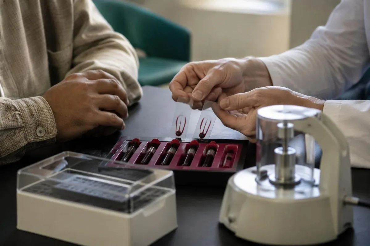
Use the same laboratory method when possible, because a change from 2.1 to 2.4 cP cannot be interpreted confidently without knowing whether the specimen and temperature matched. Our between-visit result comparison explains why analytical variation and biological change are different; there is no universal percentage increase that diagnoses hyperviscosity or determines a repeat date.
Fasting is usually not required specifically for serum viscosity, although accompanying tests may have different instructions. Suspected cryoproteins can require specimen handling near 37°C before separation according to the laboratory's protocol; tell the collecting service about that concern in advance, and do not warm, refrigerate, or otherwise handle your own specimen based on an internet instruction.
Kantesti is an AI-powered blood test analysis tool that can help organize serial results, but even 2 carefully compared reports cannot replace a symptom check or retinal examination. The service is provided by Kantesti LTD, UK Company No. 17090423; review privacy and your data before sharing reports, and give your clinician both the original values and a clear timeline of any symptoms.
Research publications and the limits of AI interpretation
Clinical reviews support symptom-led assessment of hyperviscosity, while AI technical benchmarks evaluate a different question: interpretation performance under specified test conditions. A benchmark using 100,000 synthetic cases is not evidence that an AI system can examine the retina, diagnose an emergency, or determine whether an individual patient needs plasma exchange.

Gertz's 2018 Blood review and Weaver and colleagues' 2020 Frontiers in Oncology review describe clinical hyperviscosity assessment and management, including why individual symptom thresholds matter. These 2 sources support the distinction between an abnormal measurement and a clinical syndrome; neither provides a universal serum reference interval that should override the laboratory's own method or the person's current condition.
The 2 repository publications listed below describe a synthetic technical benchmark and a clinical validation framework associated with Kantesti. Their titles and repository identifiers should not be treated as proof of independent clinical efficacy, peer-reviewed emergency-triage performance, or validated sensitivity for hyperviscosity syndrome; those conclusions would require examination of the methods and appropriate real-patient evidence.
Our technical and clinical standards explain the intended interpretation context, but the practical boundary remains clear: 1 laboratory flag is not an emergency diagnosis, and 1 reassuring AI explanation is not clearance to ignore symptoms. Bring the report to the responsible clinician; new visual change, mucosal bleeding, or neurological dysfunction should move the decision from online interpretation to direct medical assessment.
Frequently asked questions
What is the normal range for a serum viscosity test?
The normal range for a serum viscosity test is the reference interval supplied by the reporting laboratory. Some laboratories use an interval around 1.4–1.8 cP, but others use a dimensionless relative viscosity or a different absolute interval. One cP equals 1 mPa·s, but a relative viscosity of 1.8 is not automatically the same as 1.8 cP. Check the specimen, units, method, and temperature before comparing results.
Is high serum viscosity always an emergency?
High serum viscosity alone is not always an emergency and does not independently diagnose hyperviscosity syndrome. Around 4 cP or higher is a commonly discussed higher-risk context that often merits same-day specialist discussion, but urgency depends on symptoms and the underlying protein disorder. New visual changes, recurrent or persistent mucosal bleeding, or neurological symptoms require urgent direct assessment. Sudden vision loss, weakness, seizures, or substantial bleeding warrant emergency services regardless of the number.
Can hyperviscosity syndrome happen below 4 cP?
Hyperviscosity syndrome can occur below 4 cP because symptom thresholds vary between individuals and protein types. A hypothetical result of 3.3 cP with retinal abnormalities and new nosebleeds is more concerning than the same result without organ effects. The 4 cP value is a warning context, not a reliable exclusion threshold. Compatible symptoms should not be dismissed while waiting for a higher result.
What is the difference between serum and plasma viscosity?
Serum viscosity measures resistance to flow in the liquid remaining after clotting, while plasma viscosity measures a specimen that retains fibrinogen. Both tests may use cP or mPa·s, and 1 cP equals 1 mPa·s, but their reference intervals are not interchangeable. Plasma viscosity may reflect fibrinogen-related changes that a serum measurement does not capture in the same way. The report's specimen type and measurement temperature must be checked before interpretation.
Does a high serum viscosity result mean cancer?
A high serum viscosity result does not by itself diagnose cancer. Monoclonal IgM disorders, including Waldenström macroglobulinemia, are important causes, but marked polyclonal immunoglobulin increases and cryoproteins can also contribute. IgM around 3,000 mg/dL can increase viscosity, although that concentration alone proves neither cancer nor symptomatic hyperviscosity. Protein electrophoresis, immunofixation, clinical examination, and sometimes marrow testing identify the underlying explanation.
Can drinking water or taking aspirin lower serum viscosity?
Drinking water and taking aspirin are not established treatments for paraprotein-related hyperviscosity syndrome. Correcting dehydration may help general circulation, but it does not remove the abnormal immunoglobulins responsible for a result such as 4.5 cP. Aspirin affects platelet function rather than removing those proteins and may worsen mucosal bleeding. Symptomatic hyperviscosity usually requires urgent specialist treatment, often plasma exchange, followed by treatment of the underlying disorder.
Do I need to fast before a serum viscosity test?
Fasting is usually not required for a serum viscosity test itself, although other tests collected at the same appointment may require it. Follow the collecting laboratory's instructions rather than fasting or stopping medicines on your own. If cryoproteins are suspected, the laboratory may require handling near 37°C before specimen separation. Tell the collecting service about the suspected condition in advance; patients should not attempt to manage specimen temperature themselves.
Get your blood test results explained
Upload your lab report and Kantesti's AI explains each biomarker in plain language within seconds. Used by more than 2 million people worldwide.
References
- Klein, T., Mitchell, S., & Weber, H. (2026). A Pre-Registered, Rubric-Based Automated Technical Benchmark of the Kantesti Blood-Test Interpretation Engine on 100,000 Synthetic Test Cases. Kantesti AI Medical Research. DOI
- Klein, T., Mitchell, S., & Weber, H. (2026). Clinical Validation Framework v2.0 (Medical Validation Page). Kantesti AI Medical Research. DOI
- Gertz MA (2018). Acute hyperviscosity: syndromes and management. Blood. DOI
- Weaver A et al. (2020). Hyperviscosity Syndrome in Paraprotein Secreting Conditions Including Waldenstrom Macroglobulinemia. Frontiers in Oncology. DOI
- 2M+Tests analysed
- 127+Countries
- 100+Languages
About the medical team


E-E-A-T Trust Signals
Experience
Physician-led clinical review of lab interpretation workflows.
Expertise
Laboratory medicine focus on how biomarkers behave in clinical context.
Authoritativeness
Medically reviewed by Dr. Thomas Klein, with contributions from Dr. Sarah Mitchell and Prof. Dr. Hans Weber.
Trustworthiness
Evidence-based interpretation with clear follow-up pathways to reduce alarm.





Medical disclaimer
This article is for educational purposes only and does not constitute medical advice. Always consult a qualified healthcare provider for diagnosis and treatment decisions.
Published · Contact the editorial team
Published by Kantesti Ltd, registered in England & Wales, company no. 17090423, London, United Kingdom.
