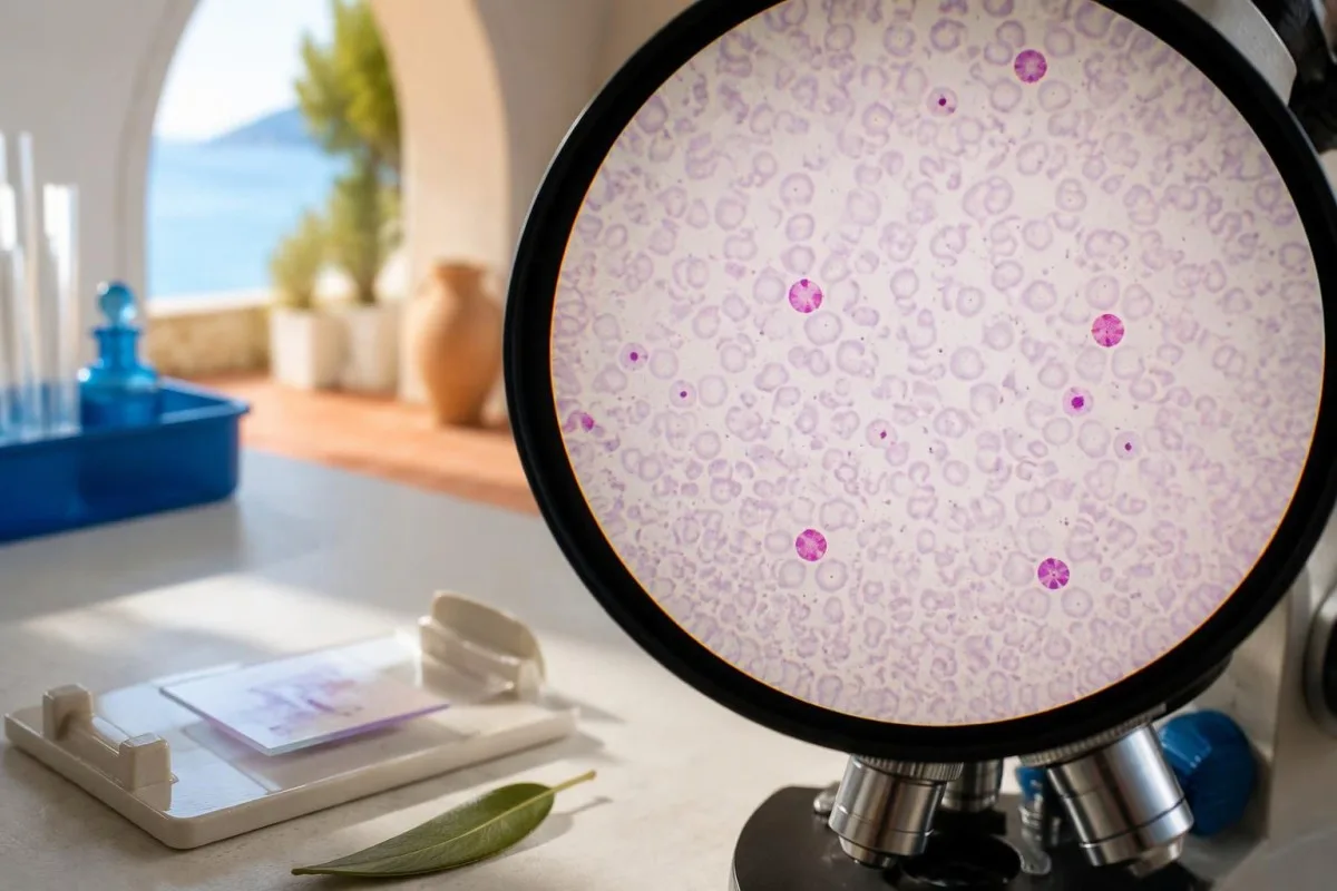
ຜົນບວກໂດຍທົ່ວໄປຊີ້ວ່າເມັດເລືອດແດງຂອງທາລົກໄດ້ເຂົ້າສູ່ການໄຫຼວຽນຂອງເລືອດມານດາ. ປະລິມານທີ່ຄາດຄະເນຊ່ວຍໃຫ້ແພດຄຳນວນປະລິມານ Rh immune globulin ເພີ່ມເຕີມເມື່ອມີຂໍ້ບົ່ງຊີ້, ແຕ່ເຮໂມໂກລບິນຂອງທາລົກໃນມານດາອາດເຮັດໃຫ້ຜົນຄາດເຄື່ອນ.

ຂໍ້ສະຫຼຸບສຳຄັນ
- លទ្ធផលវិជ្ជមាន ໝາຍຄວາມວ່າການຍ້ອມສີກວດພົບເຊວທີ່ມີເຮໂມໂກລບິນ ແລະທົນຕໍ່ກົດ; ໂດຍປົກກະຕິເຊວເຫຼົ່ານີ້ແມ່ນເມັດເລືອດແດງຂອງທາລົກ, ແຕ່ເຊວຂອງມານດາທີ່ມີ HbF ກໍສາມາດຕິດສີໄດ້ເຊັ່ນກັນ.
- ຄຳຖາມສອງຂໍ້ທີ່ແຕກຕ່າງກັນ ຕ້ອງປະເມີນແຍກກັນ: ມີເລືອດທາລົກເຂົ້າສູ່ກະແສເລືອດຂອງແມ່ຫຼາຍປານໃດ, ແລະທາລົກເກີດພາວະເລືອດຈາງຫຼືບໍ່.
- ການຄາດຄະເນປະລິມານ ມັກໃຊ້ອັດຕາສ່ວນເຊວຂອງທາລົກໃນຄັນ × 50 ເພື່ອປະເມີນປະລິມານເລືອດລວມຂອງທາລົກໃນຄັນເປັນ mL, ໂດຍສົມມຸດວ່າປະລິມານເລືອດຂອງແມ່ແມ່ນ 5,000 mL.
- ອິມມູໂນໂກລບູລິນ Rh ໃນຂະໜາດ 300 micrograms, ເທົ່າກັບ 1,500 IU, ໂດຍປົກກະຕິຄຸ້ມຄອງເລືອດລວມຂອງທາລົກໃນຄັນໄດ້ເຖິງ 30 mL ຫຼືເມັດເລືອດແດງຂອງທາລົກໃນຄັນ 15 mL.
- ຜົນ 0.8% ເທົ່າກັບເລືອດລວມຂອງທາລົກໃນຄັນປະມານ 40 mL ພາຍໃຕ້ສົມມຸດຖານ 5,000 mL; ແມ່ທີ່ມີ RhD ເປັນລົບແລະມີສິດຮັບການປ້ອງກັນ ອາດຕ້ອງໄດ້ຮັບຢາປ້ອງກັນເພີ່ມ.
- HbF ຂອງມານດາ ອາດສູງຂຶ້ນໃນໄລຍະຖືພາ, ເມື່ອມີພະຍາດຮີໂມໂກລບິນ, ຫຼືໄດ້ຮັບການຮັກສາທີ່ເພີ່ມ HbF; ຄ່າເປີເຊັນ HbF ບໍ່ແມ່ນການວັດຄ່າດຽວກັນກັບຄ່າເປີເຊັນຂອງເຊວທາລົກ.
- ການກວດການໄຫຼຂອງຈຸລັງ (flow cytometry) ອາດຊ່ວຍໃຫ້ແຈ້ງຊັດຂຶ້ນ ເມື່ອຜົນສູງກວ່າຄາດ ຫຼືບໍ່ແນ່ນອນ, ໂດຍສະເພາະເມື່ອໃຊ້ວິທີທີ່ຜ່ານການຮັບຮອງ ແລະໃຊ້ຕົວຊີ້ວັດສອງຊະນິດເພື່ອແຍກເຊວທາລົກອອກຈາກ F cells ຂອງແມ່.
- ການປະເມີນດ່ວນ ຈຳເປັນເມື່ອທາລົກເຄື່ອນໄຫວໜ້ອຍລົງ, ໄດ້ຮັບບາດເຈັບບໍລິເວນທ້ອງຢ່າງສຳຄັນ, ມີເລືອດອອກທາງຊ່ອງຄອດ ຫຼືເຈັບທ້ອງຮຸນແຮງ; ຢ່າລໍຖ້າຜົນການກວດ ຫຼືໄລຍະເວລາປ້ອງກັນຕາມປົກກະຕິ 72 ຊົ່ວໂມງ.
ຜົນບວກຂອງການກວດ Kleihauer-Betke ກວດພົບຫຍັງແທ້?
A ຜົນກວດ Kleihauer-Betke ເປັນບວກ ຜົນກວດມັກຊີ້ວ່າມີເມັດເລືອດແດງຂອງທາລົກໃນຄັນຢູ່ໃນເລືອດຂອງແມ່, ຊຶ່ງຊີ້ບອກວ່າອາດມີ ການຕົກເລືອດລະຫວ່າງທາລົກກັບມານດາ. ແຕ່ການຍ້ອມສີກວດພົບເຊວທີ່ຍັງມີເຮໂມໂກລບິນຂອງທາລົກຢູ່ ບໍ່ແມ່ນຕົວຕົນຂອງທາລົກໂດຍກົງ; ແພດຈະພິຈາລະນາເປີເຊັນ, ສະພາບທາງຄລີນິກ ແລະປະຫວັດ HbF ຂອງມານດາປະກອບກັນ ກ່ອນນຳຜົນໄປໃຊ້ກຳນົດການໃຫ້ Rh immune globulin.
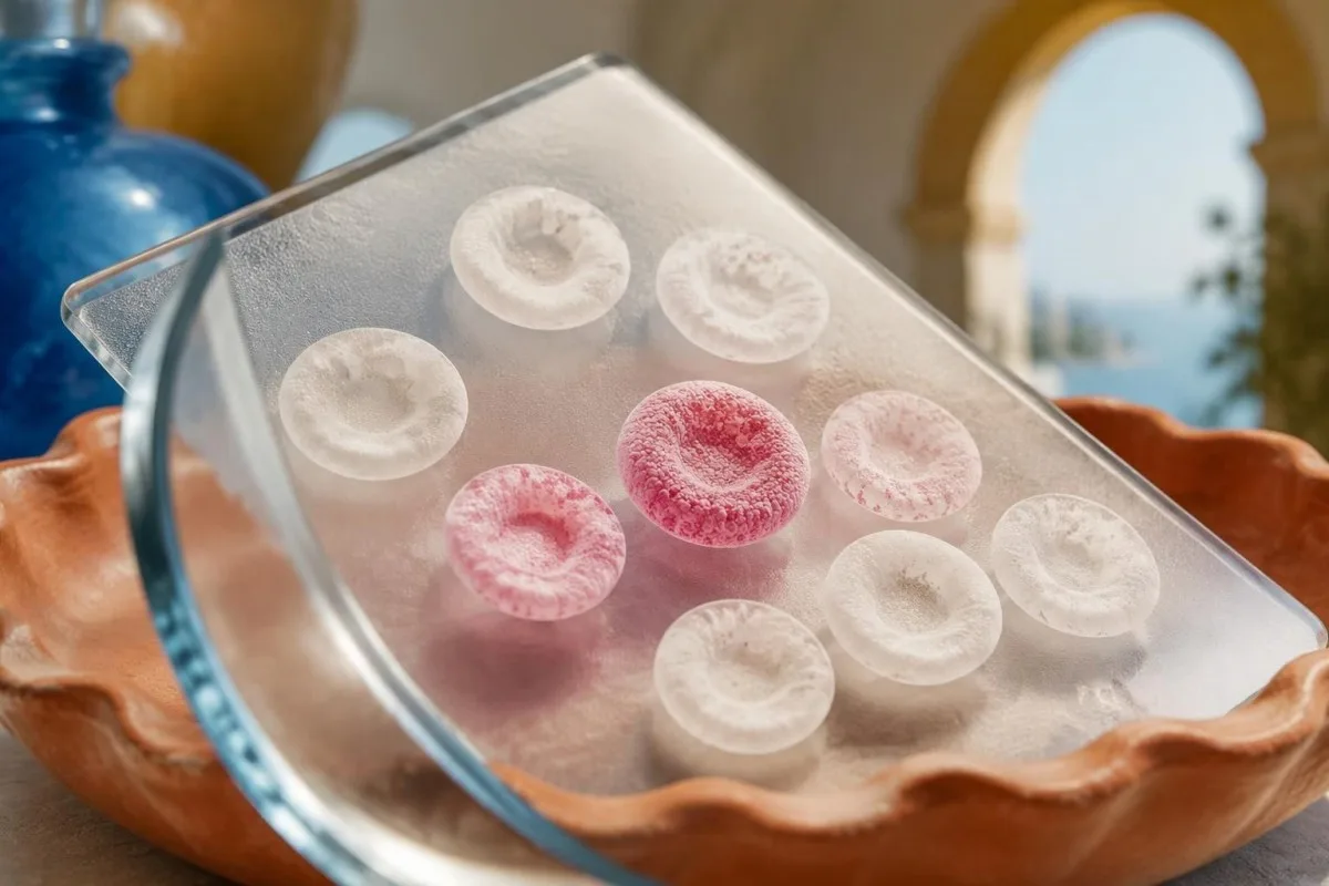
ການກວດ Kleihauer-Betke ໃຊ້ການຊະລະລ້າງດ້ວຍກົດ ເພື່ອກຳຈັດເຮໂມໂກລບິນຂອງຜູ້ໃຫຍ່ສ່ວນໃຫຍ່ອອກຈາກຟິມເລືອດມານດາທີ່ກຽມໄວ້. ເຊວທີ່ມີ ຮີໂມໂກລບິນ F ຫຼື HbF, ຈະກັກສີຍ້ອມໄວ້ຫຼາຍກວ່າ, ໃນຂະນະທີ່ເຊວສ່ວນໃຫຍ່ຂອງຜູ້ໃຫຍ່ຈະກາຍເປັນຮູບເງົາຈາງໆ; ຜົນ 0.2% ບອກສັດສ່ວນຂອງເຊວທີ່ນັບວ່າເປັນເຊວຊະນິດທາລົກ, ບໍ່ແມ່ນຄວາມເຂັ້ມຂຸ້ນ HbF ໂດຍລວມຂອງແມ່.
ຜົນກວດ Kleihauer-Betke ເປັນບວກ ບໍ່ໄດ້ຢືນຢັນວ່າແມ່ເກີດມີແອນຕິບໍດີ anti-D. ການຖ່າຍໂອນຂອງເຊວ ແລະການກະຕຸ້ນພູມຄຸ້ມກັນໃຫ້ໄວຕໍ່ແອນຕິເຈນ ແມ່ນ 2 ຂະບວນການທີ່ແຕກຕ່າງກັນ, ດັ່ງນັ້ນແພດຈຶ່ງກວດທົບທວນກຸ່ມເລືອດ ແລະການກວດຫາແອນຕິບໍດີດ້ວຍ; ຄຳອະທິບາຍຂອງພວກເຮົາກ່ຽວກັບ a ການກວດກອງແອນຕິບໍດີໃນການຖືພາຕາມປົກກະຕິ ອະທິບາຍການປະເມີນອີກດ້ານໜຶ່ງນັ້ນ.
ຄຳວ່າ ການຕົກເລືອດ ໝາຍເຖິງເລືອດທີ່ເຄື່ອນຈາກການໄຫຼວຽນຂອງເລືອດທາລົກໄປສູ່ການໄຫຼວຽນຂອງເລືອດມານດາ ໂດຍບໍ່ຈຳເປັນຕ້ອງເຫັນເລືອດອອກທາງຊ່ອງຄອດ. ການຖ່າຍໂອນເລືອດ 5 mL ຕາມສົມມຸດຖານອາດເກີດຂຶ້ນໂດຍບໍ່ມີອາການພາຍນອກ, ໃນຂະນະທີ່ການຖ່າຍໂອນແບບສະເພາະໜ້າທີ່ມີປະລິມານຫຼາຍເມື່ອທຽບກັບການໄຫຼວຽນຂອງເລືອດທາລົກ ອາດເຮັດໃຫ້ທາລົກເລືອດຈາງໄດ້ ແມ່ນແຕ່ເມື່ອມານດາຮູ້ສຶກສະບາຍດີ.
ແຄນເທສຕີ ເປັນ AI ເຄື່ອງວິເຄາະເລືອດ ທີ່ອາດຊ່ວຍອະທິບາຍຄ່າເປີເຊັນຂອງເຊວທາລົກທີ່ລາຍງານໄວ້ ເຊັ່ນ 0.2%, ແຕ່ບໍ່ສາມາດຢືນຢັນຕົ້ນກຳເນີດຂອງເຊວ ຫຼືກຳນົດຂະໜາດຢາຕ້ານ-D ໄດ້. ຂໍ້ມູນຊີ້ຂາດແມ່ນມາຈາກວິທີການຂອງຫ້ອງປະລິກ, ທີມງານການໃຫ້ເລືອດ, ແລະການປະເມີນທາງສູຕິກຳ—ບໍ່ແມ່ນອາໄສພຽງສັນຍານຜົນບວກ.
ເຊວຂອງທາລົກເຂົ້າສູ່ເລືອດມານດາໄດ້ແນວໃດ?
ເຊວຂອງທາລົກເຂົ້າສູ່ເລືອດມານດາ ເມື່ອພວກມັນຂ້າມຜ່ານຮອຍຕໍ່ຂອງຮົກ, ບໍ່ວ່າຈະເປັນການຖ່າຍໂອນປະລິມານໜ້ອຍໃນຊ່ວງຖືພາແລະການຄອດ ຫຼືຫຼັງຈາກການຂັດຂວາງທີ່ກ່ຽວຂ້ອງກັບການບາດເຈັບ, ຫັດຖະການ ຫຼືພາວະແຊກຊ້ອນຂອງຮົກ. ການຖ່າຍໂອນທີ່ກວດພົບໄດ້ ບໍ່ໄດ້ໝາຍຄວາມວ່າເປັນການຕົກເລືອດປະລິມານຫຼາຍ ຫຼືອັນຕະລາຍສະເໝີໄປ; ຜົນ 1 ຄັ້ງບໍ່ສາມາດລະບຸສາເຫດຂອງມັນໄດ້ຢ່າງໜ້າເຊື່ອຖື.
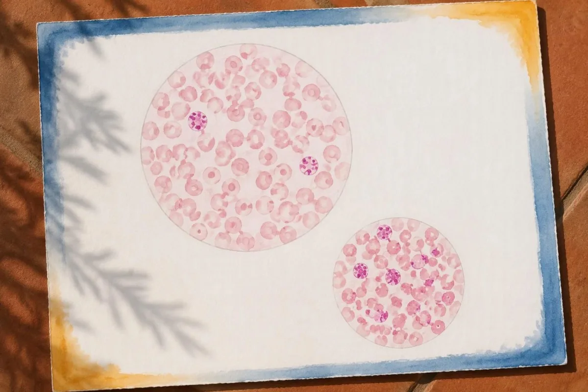
ການຕົກເລືອດຈາກທາລົກສູ່ແມ່ອາດເກີດຕາມຫຼັງການຄອດ, ການບາດເຈັບບໍລິເວນທ້ອງ, ການໝູນທ່າທາລົກຈາກພາຍນອກ, ຫຼືຫັດຖະການແບບຮຸກລ້ຳໃນໄລຍະຖືພາ. ຫຼັງ 20 ອາທິດ, ພິທີການຫຼາຍສະບັບໃນອັງກິດເພີ່ມການກວດການຕົກເລືອດຈາກທາລົກສູ່ແມ່ເຂົ້າໃນການຈັດການ anti-D ຫຼັງເຫດການທີ່ອາດກະຕຸ້ນການສ້າງພູມໄວຕໍ່ເມັດເລືອດ, ຂະນະທີ່ການຕັດສິນໃຈໃຫ້ຢາປ້ອງກັນໃນຊ່ວງຕົ້ນຂອງການຖືພາ ໃຊ້ກົດເກນທີ່ແຕກຕ່າງກັນຕາມແຕ່ລະເຫດການ.
ການຕົກເລືອດຈາກທາລົກສູ່ແມ່ທີ່ມີຄວາມສຳຄັນທາງຄລີນິກບາງກໍລະນີ ບໍ່ມີເຫດການກະຕຸ້ນທີ່ເຫັນໄດ້ຊັດ. ດັ່ງນັ້ນ ຜົນບວກສົມມຸດຖານໃນອາທິດທີ 32 ໂດຍບໍ່ມີການບາດເຈັບ ກໍຍັງຄວນໄດ້ຮັບການແປຜົນ; ການບໍ່ມີອຸບັດເຫດບໍ່ໄດ້ຫັກລ້າງວ່າມີການຖ່າຍໂອນເຊວ, ແລະການກວດບໍ່ໄດ້ພິສູດວ່າຮົກມີບັນຫາໂຄງສ້າງສະເພາະ.
ການກວດ fetal fibronectin ແລະການກວດເມັດເລືອດແດງຂອງທາລົກໃນຄັນຕອບຄຳຖາມທີ່ແຕກຕ່າງກັນ ເຖິງແມ່ນວ່າຈະມີຄຳວ່າ fetal ຄືກັນກໍຕາມ. ຢ່າງທຳອິດໃຊ້ປະເມີນຄວາມສ່ຽງຕໍ່ການຄອດກ່ອນກຳນົດໃນບາງສະຖານະການ, ສ່ວນຢ່າງຫຼັງໃຊ້ປະເມີນການຖ່າຍໂອນຂອງເຊວ; ຄູ່ມືຂອງພວກເຮົາກ່ຽວກັບ a ຜົນກວດ fetal fibronectin ເປັນບວກ ອະທິບາຍວ່າເຫດໃດການກວດທັງສອງຈຶ່ງໃຊ້ແທນກັນບໍ່ໄດ້.
ຜົນກະທົບຈາກການສູນເສຍເລືອດຂອງທາລົກໃນຄັນ ຂຶ້ນກັບທັງປະລິມານແລະຄວາມໄວ. ການສູນເສຍ 20 mL ໃນໄລຍະຫຼາຍມື້ຕາມສົມມຸດຖານ ເປີດໂອກາດໃຫ້ຮ່າງກາຍຕອບສະໜອງທາງສະລີລະວິທະຍາແຕກຕ່າງຈາກການສູນເສຍ 20 mL ຢ່າງວ່ອງໄວ, ດັ່ງນັ້ນແພດຈຶ່ງປະເມີນການເຄື່ອນໄຫວຂອງທາລົກ, ຮູບແບບອັດຕາການເຕັ້ນຂອງຫົວໃຈ ແລະພາວະເລືອດຈາງ ແທນການກຳນົດຄວາມຮຸນແຮງຈາກອັດຕາສ່ວນໃນເລືອດແມ່ພຽງຢ່າງດຽວ.
ມີການສັ່ງກວດການຕົກເລືອດລະຫວ່າງທາລົກກັບມານດາເມື່ອໃດ?
ການກວດຫາພາວະເລືອດອອກຈາກເລືອດຂອງແມ່ໄປສູ່ທາລົກ is ordered to estimate fetal-cell exposure for anti-D prophylaxis or to investigate suspected fetal blood loss. Common settings include delivery, significant pregnancy trauma, and unexplained fetal anemia; after birth, many protocols collect the maternal sample at least 30 minutes later to allow mixing.
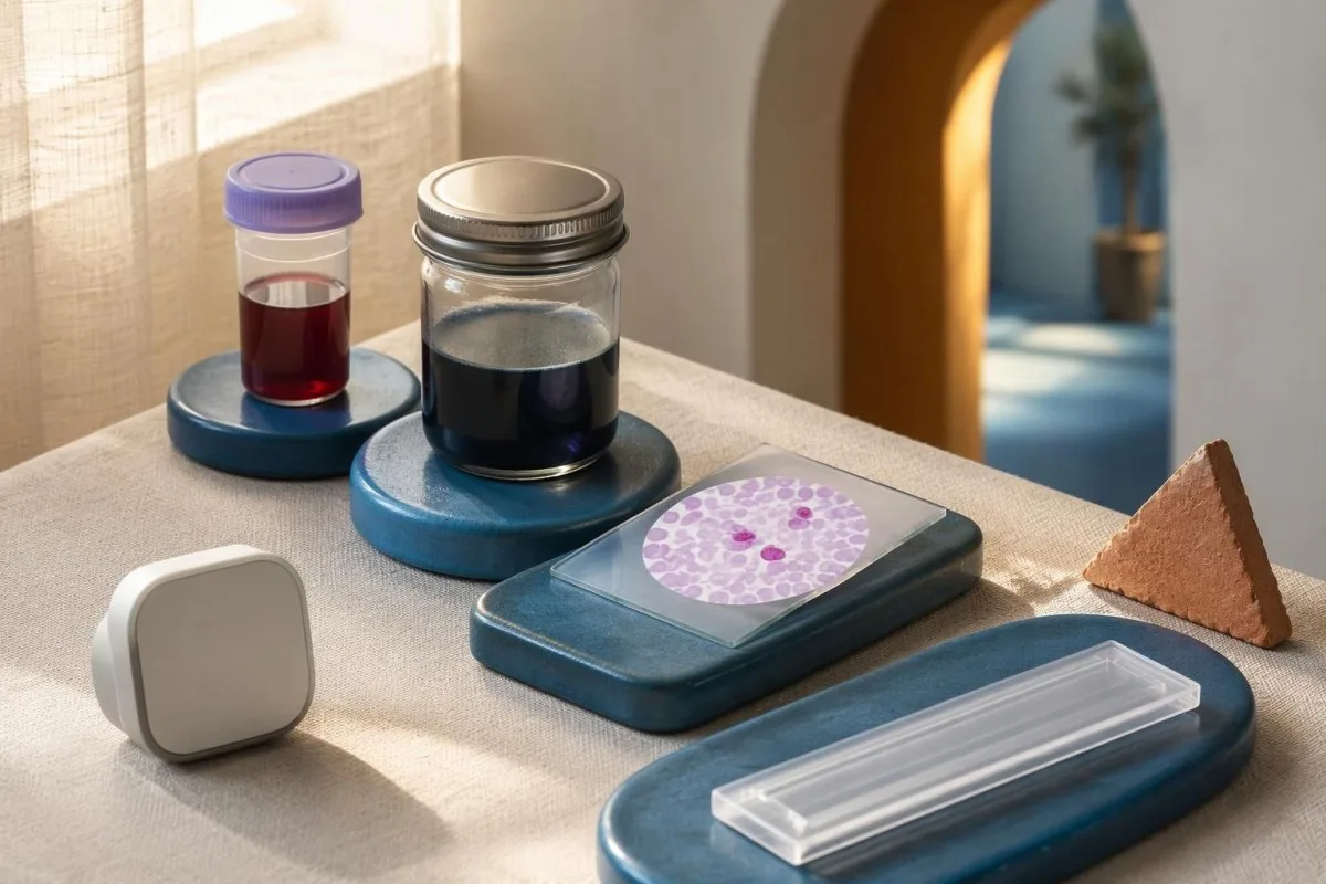
Postpartum sampling commonly occurs approximately 30 minutes to 2 hours after delivery, although the local laboratory specifies its accepted interval. A sample taken immediately at birth may not yet represent well-mixed circulation, and a later specimen can be affected by cell clearance or intervening treatment.
Rh immune globulin is generally administered as soon as practical and within 72 hours of an eligible sensitizing event or delivery. Collecting a specimen before prophylaxis is often preferred, but arranging the assay must not unnecessarily delay indicated treatment; Qureshi et al. (2014) emphasize prompt prophylaxis within a structured laboratory pathway.
The laboratory needs the event time, gestational age, maternal RhD type, and details of any recent anti-D or transfusion. There is usually no fasting requirement for this stain, unlike some metabolic tests; our ຄູ່ມືການກະກຽມຄັ້ງທຳອິດ helps separate routine preparation from the time-sensitive details of pregnancy testing.
Antenatal testing after trauma has 2 purposes that should not be confused: estimating exposure for prophylaxis and supporting assessment of fetal loss. A negative maternal stain does not cancel the need for obstetric observation when the injury, contractions, pain, or fetal monitoring justify it.
ທ່ານແປຄວາມໝາຍຂອງເປີເຊັນ ແລະປະລິມານທີ່ຄາດຄະເນແນວໃດ?
Kleihauer-Betke test interpretation requires identifying whether the report gives a fetal-cell percentage, fetal whole-blood volume, or fetal red-cell volume. Under a common 5,000 mL maternal-volume assumption, fetal-cell percentage × 50 estimates fetal whole blood in mL; 0.1% therefore corresponds to approximately 5 mL.

The basic calculation is fetal-cell fraction × estimated maternal blood volume. For example, 0.4% becomes 0.004 × 5,000 mL = 20 mL of fetal whole blood; entering 0.4 rather than 0.004 into a fraction-based calculator produces a 100-fold error.
Fetal whole blood and fetal red cells are not interchangeable volumes. Using an illustrative fetal hematocrit of 50%, 20 mL of fetal whole blood contains about 10 mL of red cells, but laboratories may use a different validated conversion; check the report's exact wording before comparing it with a dosing threshold.
A report saying only positive provides less information than one reporting 0.4% and an estimated 20 mL. Our guide to ຜົນການວິນິດໄສທຽບກັບຜົນການວັດແທກ explains this distinction: a detection flag answers whether something was found, while a quantitative result supports a calculation.
In Kantesti educational content, we keep 0.4%, 20 mL of fetal whole blood, and 10 mL of fetal red cells separate because each can appear on a report. Ask whether the laboratory used a fixed maternal volume, a patient-specific estimate, or an assay-specific formula; the printed volume is an estimate, not blood collected directly from the fetus.
ໃຜຄວນໄດ້ຮັບອິມມູໂນໂກລບູລິນ Rh ຫຼັງຈາກໄດ້ຜົນບວກ?
ອິມມູໂນໂກລບູລິນ Rh, also called anti-D immunoglobulin, is generally indicated for an eligible RhD-negative mother without established immune anti-D when RhD-positive fetal-cell exposure is possible. A positive stain does not itself determine eligibility, and a standard 300-microgram dose prevents sensitization rather than treating fetal anemia.

An RhD-positive mother generally does not need anti-D prophylaxis, although she can still experience clinically significant fetomaternal hemorrhage. Conversely, an unsensitized RhD-negative mother may need prophylaxis even when a stain detects 0% fetal cells, because small sensitizing exposures can fall below the assay's reliable detection level.
Established immune anti-D changes management because prophylactic anti-D does not reverse existing sensitization. ACOG Practice Bulletin No. 181 (2017) distinguishes prevention from management of alloimmunization; a positive antibody screen after a recent 300-microgram injection may instead reflect passive anti-D, which requires laboratory interpretation.
Maternal antibodies and antibodies coating a baby's cells are also different measurements. A newborn's 直接抗球蛋白試驗呈陽性 can help investigate antibody-related red-cell effects, but it does not measure the mL of fetal blood transferred into the mother's circulation.
Eligibility can be less straightforward with weak or partial D typing results. Molecular testing may classify some weak D types, particularly types 1, 2, and 3, as not needing prophylaxis under established guidance, while other variants require specialist decisions; do not decide from a shortened blood-group label alone.
ຢາຕ້ານ D ຂະໜາດໜຶ່ງຄຸ້ມຄອງເລືອດທາລົກໄດ້ປະລິມານເທົ່າໃດ?
A 300-microgram anti-D dose, equivalent to 1,500 IU, typically covers 30 mL of fetal whole blood or 15 mL of fetal red cells. Other countries and products use different routine doses, so the local transfusion protocol—not the number of positive cells alone—determines additional prophylaxis.

US practice commonly uses 300 micrograms after delivery of an RhD-positive infant to an eligible mother, with extra doses when the measured exposure exceeds coverage. ACOG (2017) describes the 30 mL fetal-whole-blood coverage principle; this threshold reflects prophylactic capacity, not a universal boundary between harmless and dangerous fetal loss.
The BCSH guideline by Qureshi et al. (2014) describes at least 500 IU after eligible events from 20 weeks and after eligible delivery, with testing to identify exposures requiring more anti-D. A 500 IU dose covers approximately 4 mL of fetal red cells in that framework; product characteristics and current local guidance must still be checked.
Additional-dose calculations may use an IU-per-mL rule, whole-vial dosing, and a safety allowance for measurement uncertainty. Some UK intramuscular protocols use at least 125 IU per mL of fetal red cells, while intravenous preparations may have different instructions; neither route nor dose should be inferred from an internet formula.
Blood group labels do not change the fundamental whole-blood versus red-cell distinction: 15 mL of fetal red cells and 30 mL of fetal whole blood represent equivalent coverage only under the assumed 50% hematocrit. Our blood group and hematology guide provides background without replacing the blood bank's dose authorization.
ຕົວຢ່າງປະກອບ: ຜົນ 0.8% ອາດໝາຍເຖິງຫຍັງ?
ບຸກຄົນສົມມຸດ 0.8% fetal-cell result estimates 40 mL of fetal whole blood when maternal blood volume is assumed to be 5,000 mL. Dividing 40 mL by 30 mL gives 1.33 standard 300-microgram dose equivalents, but the final prescribed amount includes the laboratory's rounding and safety rules.
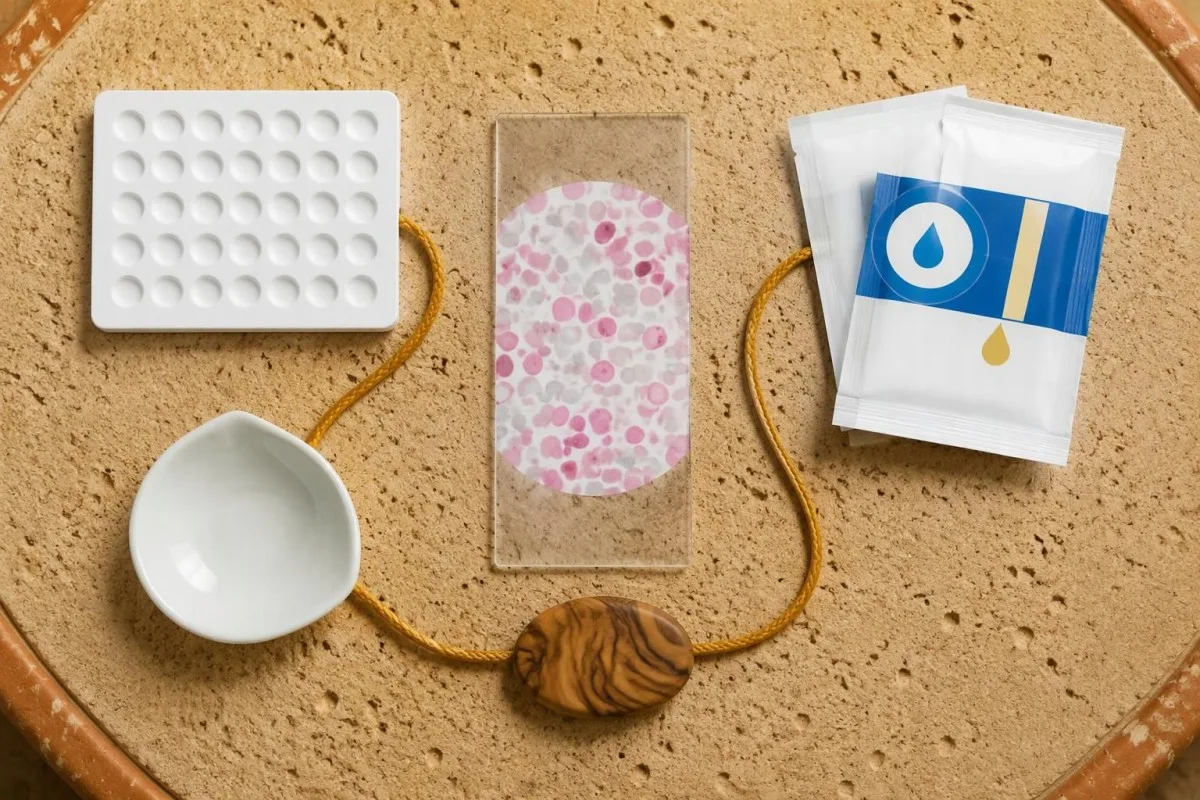
First, convert the percentage correctly: 0.8% = 0.008, and 0.008 × 5,000 mL = 40 mL. This example assumes the counted cells are genuinely fetal and the maternal-volume estimate is appropriate; either assumption can fail, so the calculation does not independently validate the laboratory result.
Next, calculate nominal coverage: 40 ÷ 30 = 1.33 standard doses. Under a common US safety convention, a decimal below 0.5 is rounded down before adding 1 vial, giving 2 vials in this example; other laboratories use different validated rounding or individualized dosing.
A rounding allowance intentionally exceeds the arithmetic minimum because staining and counting are imprecise. Under that same convention, an estimate of 60 mL gives 2 dose equivalents and then 1 extra vial, illustrating why the prescription can look larger than simple division suggests.
Anti-D is an immunoglobulin medicine, not a replacement transfusion for the baby. If fetal anemia requires treatment, compatibility testing and specialist fetal or neonatal care form a separate pathway; our transfusion compatibility explanation describes why giving 2 prophylactic vials to the mother does not restore fetal circulating volume.
ເປັນຫຍັງປະລິມານການຕົກເລືອດທີ່ປະເມີນໄດ້ຈຶ່ງບໍ່ແມ່ນຄ່າທີ່ແນ່ນອນ?
ໄດ້ Kleihauer-Betke volume estimate is uncertain because manual counting, staining quality, cell classification, and estimated maternal blood volume all affect the answer. When 2 fetal-type cells are counted among 2,000 cells, the reported fraction is 0.1%; a difference of just 1 counted cell substantially changes that estimate.
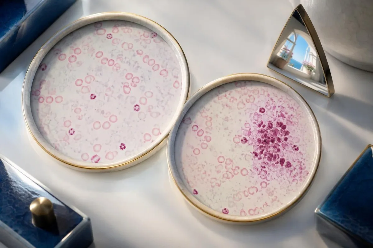
Rare-cell sampling produces unavoidable statistical variation, even with good technique. At a true fraction of 0.1%, a count of 2,000 cells would contain only about 2 fetal cells on average, whereas 10,000 cells would contain about 10; larger counts help, but they do not eliminate misclassification.
Maternal blood volume is another source of error. The same hypothetical 0.4% fraction estimates 16 mL with a 4,000 mL maternal volume and 24 mL with a 6,000 mL volume, rather than the 20 mL obtained using a fixed 5,000 mL assumption.
Red-cell percentages do not translate perfectly into whole-blood volumes when maternal and fetal hematocrits or cell sizes differ. A hypothetical maternal hematocrit of 30% and fetal hematocrit of 50% illustrates why some laboratory methods apply corrections; our การเปรียบเทียบฮีมาโตคริตและฮีโมโกลบิน explains the underlying measures.
ແຄນເທສຕີ ເປັນ ແພລດຟອມການຕີຄວາມໝາຍ biomarker ຂອງ AI that can help explain why a calculated 20 mL is an estimate rather than an exact measurement. The practical question is whether the uncertainty could change prophylaxis or fetal assessment; unexpectedly large, borderline-for-dosing, or technically doubtful results warrant discussion with the transfusion laboratory.
ຮີໂມໂກລບິນຂອງທາລົກໃນຄັນຈາກແມ່ ສາມາດເຮັດໃຫ້ຜົນບວກທີ່ຊວນໃຫ້ເຂົ້າໃຈຜິດໄດ້ຫຼືບໍ່?
Maternal HbF-containing red cells, known as F cells, can retain the Kleihauer-Betke stain and be mistaken for fetal cells. Pregnancy, hereditary persistence of fetal hemoglobin, some hemoglobin disorders, and HbF-increasing treatment can complicate interpretation; an HbF result of 2% does not mean 2% of circulating cells are fetal.
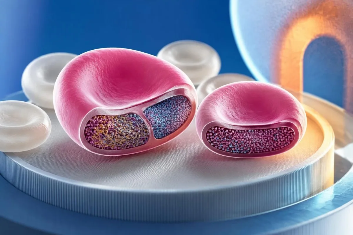
Hemoglobin analysis measures the proportion of hemoglobin types, while Kleihauer-Betke testing counts stained cells. For example, a hypothetical HbF concentration of 3% could be distributed across many maternal F cells, each containing some HbF, rather than confined to 3% distinctly fetal cells.
A history of thalassemia, sickle cell disease, or hereditary persistence of HbF should be given to the laboratory before interpreting an apparent large transfer. Our hemoglobin electrophoresis guide explains HbA, HbS, HbC, and HbF patterns; none of those fractions alone provides a hemorrhage volume in mL.
A low maternal MCV can support investigation for iron deficiency or thalassemia, but it does not prove HbF interference. A hypothetical MCV of 68 fL requires its own workup, described in our low MCV interpretation guide; only some underlying disorders produce enough maternal HbF to affect this stain.
Manual stain appearance may suggest maternal F cells, but visual distinction is not always reliable. Sandler and Sathiyamoorthy (2010) review this limitation of acid-elution testing; if an apparent 1.5% fetal-cell result conflicts with the pregnancy assessment or a known HbF disorder, a validated confirmatory method may prevent substantial overestimation.
ເມື່ອໃດທີ່ flow cytometry ຊ່ວຍຢືນຢັນການຖ່າຍໂອນເຊວຂອງທາລົກ?
ການກວດການໄຫຼຂອງຈຸລັງ (flow cytometry) helps when a Kleihauer-Betke result is unexpectedly large, technically uncertain, or potentially confounded by maternal HbF. A validated 2-marker assay can distinguish fetal cells from maternal F cells more effectively than HbF staining alone, while anti-D-based methods require a suitable maternal–fetal RhD difference.

Anti-HbF flow cytometry measures fluorescence in individual cells and can analyze far more cells than a manual count. However, an assay using only 1 HbF marker may still count maternal F cells; greater numerical precision does not automatically mean greater certainty about fetal origin.
Some dual-marker assays combine HbF with carbonic anhydrase, an adult-cell marker that is lower in fetal erythrocytes. The 2-dimensional pattern can separate HbF-containing maternal cells from fetal cells, but the laboratory must validate its gating because cell populations can overlap and developmental stage affects marker expression.
Anti-D flow cytometry identifies RhD-positive cells in an RhD-negative maternal background when that contrast is present. It cannot provide the same discrimination if both mother and fetus are RhD-negative or both are RhD-positive, and recent anti-D treatment or complex D typing should be discussed before choosing this method.
Flow cytometry is a method family, not 1 interchangeable test: a PNH flow cytometry assay uses different markers and answers a different question. Kantesti's ຂໍ້ມູນການກວດສອບທາງວິຊາການ concerns our interpretation system, not accreditation of a hospital's fetal-cell assay; request the actual laboratory method when confirmation matters.
ຜົນບວກໝາຍຄວາມວ່າທາລົກເລືອດຈາງບໍ?
A positive Kleihauer-Betke result does not diagnose fetal anemia; it estimates transferred cells in maternal circulation. Fetal assessment may include heart-rate monitoring and middle cerebral artery Doppler, where a peak systolic velocity around or above 1.5 multiples of the median raises concern for moderate-to-severe anemia in appropriate settings.
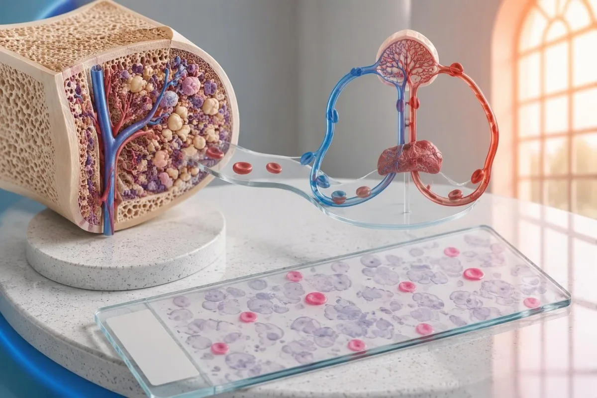
The same estimated loss can have different consequences at different gestational ages. In a purely hypothetical fetus with a 100 mL circulating volume, a 20 mL loss represents 20% of that volume; this arithmetic is illustrative, not a method for calculating an individual fetus's circulation from a maternal stain.
Middle cerebral artery Doppler measures flow velocity rather than hemoglobin directly. The 1.5 MoM threshold is a screening signal, not a stand-alone diagnosis, and performance can be less reliable after intrauterine transfusion or later in pregnancy; a fetal medicine specialist integrates gestational age and other findings.
Reduced fetal movement or significant abdominal trauma requires prompt obstetric assessment even with a negative or pending stain. Severe headache, upper abdominal pain, or other pregnancy warning symptoms may instead suggest conditions such as ໂຣກ HELLP, which follows a different laboratory pathway and should not be explained away by 1 positive result.
Fetomaternal transfer is not synonymous with maternal hemorrhage or a clotting disorder. When obstetric complications raise concern for ภาวะลิ่มเลือดกระจายทั่วไปในหลอดเลือด, platelet count, fibrinogen, and clotting times answer additional questions; a 0.5% fetal-cell fraction neither confirms nor excludes DIC.
ຜົນການກວດ Kleihauer-Betke ທີ່ເປັນລົບ ແລະຜົນກວດຊ້ຳໝາຍຄວາມວ່າແນວໃດ?
A negative Kleihauer-Betke result means fetal-type cells were not detected above that laboratory's reporting limit in that specimen. It does not prove that transfer was exactly 0 mL, exclude every clinically relevant exposure, or replace fetal assessment; repeat testing is chosen according to the event, treatment, and assay.
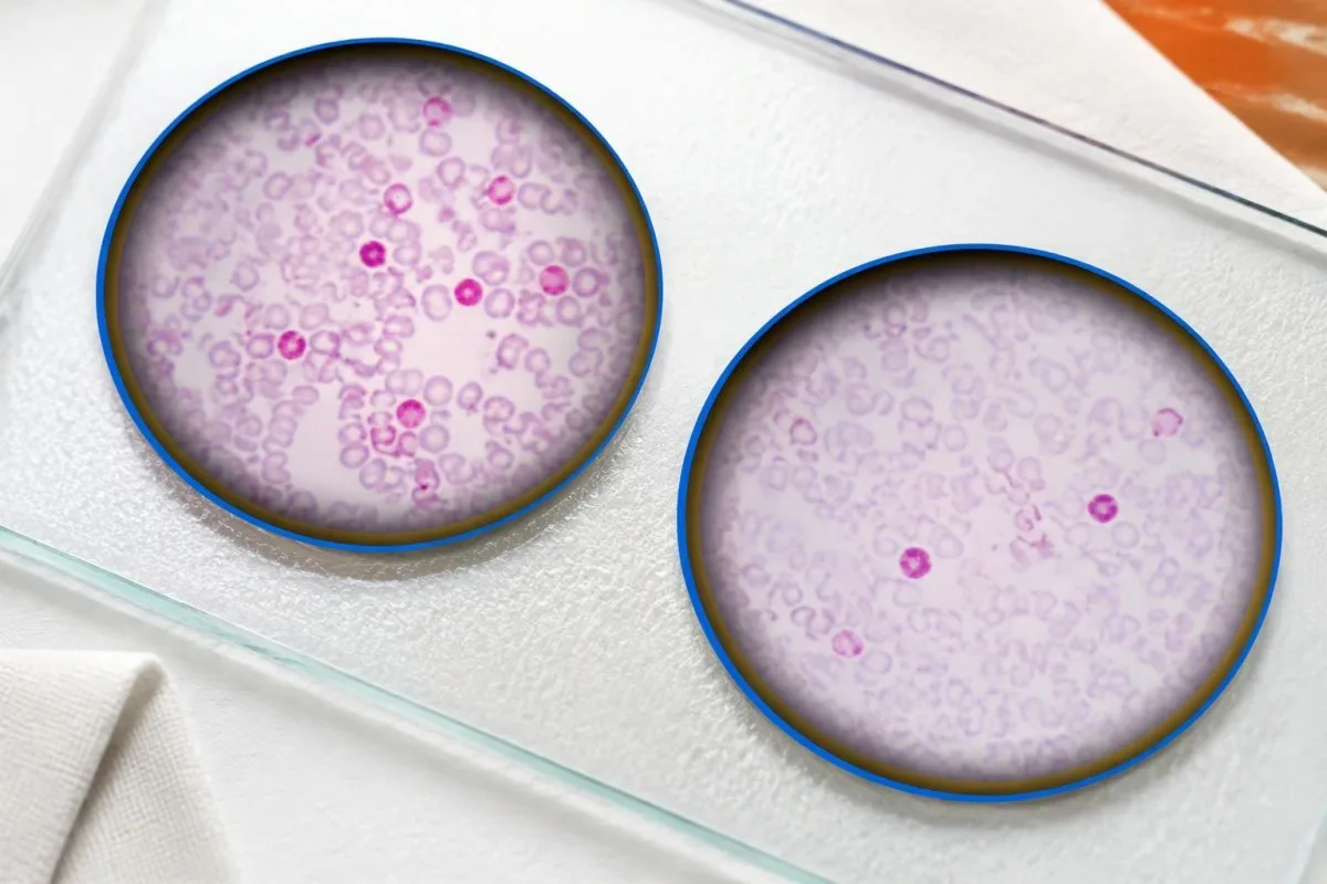
The test is less reliable for very small transfers than for larger ones, particularly when few cells are counted manually. A mother eligible for routine postpartum anti-D may still need the standard dose despite a reported 0.0%, because the assay primarily identifies exposure beyond routine coverage rather than eliminating all sensitization risk.
Large confirmed transfers may require follow-up testing to show that RhD-positive fetal cells have cleared after sufficient prophylaxis. Some UK protocols reassess approximately 48 hours after intravenous anti-D or 72 hours after intramuscular anti-D; the transfusion service chooses the interval and assay rather than applying a universal repeat schedule.
A lower percentage on repeat testing does not always mean the original estimate was wrong. Cell clearance, dilution, and sampling variation can change a hypothetical 0.4% result to 0.2%; our guide to ການປ່ຽນແປງຂອງການກວດເລືອດຕາມເວລາ explains why chronology matters when comparing laboratory values.
A new sensitizing event needs a new clinical review even if testing was completed 1 day earlier. Previous prophylaxis, its dose, the interval since administration, and the new exposure all matter; patients should not assume that an earlier negative stain or routine antenatal injection covers every subsequent event.
ທ່ານຄວນຖາມທີມດູແລຂອງທ່ານເລື່ອງຜົນການກວດແນວໃດ?
Ask your care team for the fetal-cell percentage, estimated volume and its units, maternal RhD and antibody status, and the planned action. For a result such as 0.6%, clarify whether the estimate represents 30 mL of fetal whole blood or a separately calculated red-cell volume before discussing prophylaxis.

ແຄນເທສຕີ ເປັນ ແພລດຟອມການອ່ານຜົນກວດເລືອດຂອງ AI operated by Kantesti Ltd, whose ຂໍ້ມູນອົງກອນ identifies UK Company No. 17090423. Our role is educational interpretation: explaining 0.6%, 30 mL, and their assumptions without replacing the laboratory's fetal-cell confirmation or your clinician's treatment decision.
A useful discussion separates 3 tasks: confirming cell origin, calculating prophylaxis when indicated, and assessing the fetus or newborn when clinically necessary. Our AI report explanation guide describes source checking and limitations; an AI-generated dose calculation is not an authorized prescription.
If maternal HbF is known to be elevated, ask whether the laboratory considered F-cell interference and whether a 2-marker flow assay would change management. Also provide the time of the event, any recent anti-D, prior transfusions, and hemoglobin-disorder results; missing context can matter more than an extra decimal place.
ຂອງພວກເຮົາ ຄູ່ມືເຕັກໂນໂລຊີການຕີລາຄາ explains how AI supports reading laboratory information, but no software can assess fetal movement or perform Doppler imaging. Contact maternity services promptly for concerning symptoms, and contact the prescribing team if the anti-D plan is unclear rather than waiting until the 72-hour window is nearly over.
สิ่งพิมพ์วิจัยและหลักฐานเบื้องหลังการตีความ
The clinical basis for Kleihauer-Betke interpretation comes from transfusion laboratory methods and obstetric anti-D guidance, not unrelated biomarker publications. The 2017 ACOG bulletin, 2014 BCSH guideline, and 2010 laboratory-methods review below address prevention, dose coverage, and assay limitations; local protocols remain necessary.

ACOG Practice Bulletin No. 181 (2017) supports RhD alloimmunization prevention and the relationship between 300 micrograms of anti-D and 30 mL of fetal whole blood. Qureshi et al. (2014) describe a UK approach using different routine doses and red-cell-volume thresholds; those differences explain why prescriptions from 2 countries may not look identical.
Sandler and Sathiyamoorthy (2010) review acid-elution and flow-based laboratory methods, including limitations relevant to maternal HbF. The enduring distinction is between detection, quantification, and identification: counting 10 stained cells more accurately does not prove they are fetal unless the method can discriminate their origin.
Two Kantesti publications are listed separately in the DOI references: the complement and ANA guide and the Nipah virus diagnostic guide. These 2 publications concern other laboratory topics and are not evidence for Kleihauer-Betke accuracy, fetal-anemia thresholds, or anti-D dosing; a DOI identifies a deposited work but does not itself establish peer review or clinical validation.
ຂອງພວກເຮົາ ມາດຕະຖານເນື້ອຫາທາງການແພດ distinguish educational explanations from individualized medical decisions. For related laboratory context, the ຄູ່ມືການກວດການກໍ່ຕົວຂອງເລືອດ covers aPTT and other clotting markers, but those tests cannot substitute for fetal-cell quantification; ask which of these 2 diagnostic questions your ordered panel is intended to answer.
ຄໍາຖາມທີ່ຖືກຖາມເລື້ອຍໆ
ຜົນກວດ Kleihauer-Betke ເປັນບວກໝາຍຄວາມວ່າຢ່າງໃດ?
ຜົນກວດ Kleihauer-Betke ເປັນບວກ ໂດຍທົ່ວໄປໝາຍຄວາມວ່າເມັດເລືອດແດງຂອງທາລົກໄດ້ເຂົ້າສູ່ກະແສເລືອດຂອງແມ່ ເຊິ່ງບົ່ງຊີ້ເຖິງການຕົກເລືອດຈາກທາລົກສູ່ແມ່. ສານຍ້ອມສີນີ້ກວດພົບເຊລທີ່ຍັງຄົງມີເຮໂມໂກລບິນຂອງທາລົກ ດັ່ງນັ້ນ ບາງຄັ້ງເຊລຂອງແມ່ທີ່ມີ HbF ອາດເບິ່ງຄ້າຍກັບເຊລຂອງທາລົກ. ເມື່ອໃຊ້ສົມມຸດຖານທົ່ວໄປວ່າປະລິມານເລືອດຂອງແມ່ເທົ່າກັບ 5,000 mL, ຜົນທີ່ພົບເຊລຂອງທາລົກ 0.2% ຈະປະເມີນວ່າມີເລືອດທັງໝົດຂອງທາລົກປະມານ 10 mL. ແພດໃຊ້ຜົນກວດນີ້ປະກອບກັບສະຖານະ RhD, ການກວດຫາແອນຕິບໍດີ ແລະການປະເມີນການຖືພາ.
ຜົນກວດ Kleihauer-Betke ເປັນບວກ ໝາຍຄວາມວ່າລູກຂອງຂ້ອຍກຳລັງຕົກຢູ່ໃນອັນຕະລາຍບໍ?
A positive Kleihauer-Betke test does not by itself show that the baby is anemic or in immediate danger. Risk depends on how much blood was lost relative to the fetus's circulation, how quickly it was lost, and the fetal assessment. Middle cerebral artery Doppler velocity around or above 1.5 MoM can raise concern for anemia in appropriate settings, but it is not a stand-alone diagnosis. Reduced fetal movement, significant abdominal trauma, or vaginal bleeding warrants prompt maternity assessment regardless of the test result.
ເປີເຊັນ Kleihauer-Betke ຖືກແປງເປັນປະລິມານການຕົກເລືອດແນວໃດ?
A common calculation estimates fetal whole-blood volume in mL by multiplying the reported fetal-cell percentage by 50, assuming 5,000 mL of maternal blood. For example, 0.8% × 50 estimates 40 mL of fetal whole blood. This shortcut does not directly calculate fetal red-cell volume and may differ from a laboratory's validated, patient-specific method. The result remains an estimate because counting, hematocrit differences, and maternal blood-volume assumptions introduce uncertainty.
ຕ້ອງໃຊ້ອິມມູນໂກລບູລິນ Rh ຫຼາຍປານໃດຫຼັງຈາກຜົນກວດເປັນບວກ?
ຂະໜາດ Rh immune globulin ທີ່ຕ້ອງໃຊ້ຂຶ້ນກັບຄຸນສົມບັດການມີສິດໄດ້ຮັບ, ປະລິມານເລືອດຂອງທາລົກທີ່ຄາດວ່າປົນເຂົ້າມາ, ຂອບເຂດການຄຸ້ມຄອງຂອງຜະລິດຕະພັນ ແລະ ຂັ້ນຕອນການໃຫ້ເລືອດຂອງທ້ອງຖິ່ນ. ຂະໜາດມາດຕະຖານ 300 ໄມໂຄຣກຣາມ, ເທົ່າກັບ 1,500 IU, ໂດຍທົ່ວໄປຄຸ້ມຄອງເລືອດເຕັມສ່ວນຂອງທາລົກ 30 mL ຫຼື ເມັດເລືອດແດງຂອງທາລົກ 15 mL. ການປົນເຂົ້າມາໃນປະລິມານທີ່ຫຼາຍກວ່ານີ້ອາດຕ້ອງໃຫ້ຂະໜາດເພີ່ມ ໂດຍມີການເຜື່ອສ່ວນເພີ່ມເພື່ອຄຳນຶງເຖິງຄວາມບໍ່ແນ່ນອນຂອງການກວດວິເຄາະ. ທະນາຄານເລືອດ ຫຼື ແພດຜູ້ສັ່ງຢາຕ້ອງເປັນຜູ້ອະນຸມັດຂະໜາດຢາ; anti-D ປ້ອງກັນການກະຕຸ້ນໃຫ້ເກີດພູມຕ້ານທານ ແຕ່ບໍ່ໄດ້ຮັກສາພາວະເລືອດຈາງຂອງທາລົກໃນຄັນ.
ການກວດ Kleihauer-Betke ສາມາດໃຫ້ຜົນບວກປອມເນື່ອງຈາກ HbF ຂອງແມ່ໄດ້ບໍ?
ເມັດເລືອດແດງຂອງແມ່ທີ່ມີ HbF ອາດຕິດສີຄືກັບເຊວຂອງທາລົກໃນຄັນ ແລະເຮັດໃຫ້ຜົນ Kleihauer-Betke ສູງເກີນຈິງ. ການຖືພາ, ພາວະ HbF ຄົງຢູ່ສືບທອດ, ຄວາມຜິດປົກກະຕິຂອງເຮໂມໂກລບິນບາງຊະນິດ ແລະການປິ່ນປົວທີ່ເພີ່ມ HbF ອາດເປັນສາເຫດຂອງການລົບກວນນີ້. ສັດສ່ວນ HbF 3% ຂອງແມ່ແມ່ນຄ່າວັດແທກເຮໂມໂກລບິນ ບໍ່ແມ່ນຫຼັກຖານວ່າ 3% ຂອງເຊວທີ່ໝູນວຽນຢູ່ໃນເລືອດເປັນເຊວຂອງທາລົກ. ການກວດ flow cytometry ທີ່ຜ່ານການຢືນຢັນວ່າໃຊ້ແຍກເຊວ F ຂອງແມ່ອອກຈາກເຊວຂອງທາລົກໄດ້ ອາດຊ່ວຍຊີ້ແຈງຜົນກວດທີ່ຍັງບໍ່ແນ່ນອນ.
ຜົນກວດ Kleihauer-Betke ເປັນລົບ ໝາຍຄວາມວ່າຂ້ອຍບໍ່ຈຳເປັນຕ້ອງໄດ້ຮັບ anti-D ບໍ?
A negative Kleihauer-Betke test does not automatically remove the need for anti-D prophylaxis in an eligible RhD-negative mother. A reported 0.0% means no fetal-type cells were detected above the method's reporting limit, not that exposure was exactly zero. Routine prophylaxis can still be indicated after an eligible event or delivery, while the assay helps identify exposures requiring additional coverage. Anti-D is generally given promptly and within 72 hours, so arranging testing should not unnecessarily delay indicated treatment.
ຮັບຄຳອະທິບາຍຜົນກວດເລືອດຂອງທ່ານ
ອັບໂຫຼດລາຍງານຜົນກວດຂອງທ່ານ ແລະ AI ຂອງ Kantesti ຈະອະທິບາຍ biomarker ແຕ່ລະຕົວດ້ວຍພາສາເຂົ້າໃຈງ່າຍພາຍໃນບໍ່ກີ່ວິນາທີ. ມີຜູ້ໃຊ້ຫຼາຍກວ່າ 2 ລ້ານຄົນທົ່ວໂລກ.
ເອກະສານອ້າງອີງ
- Klein, T., Mitchell, S., & Weber, H. (2026). ຄູ່ມືການກວດເລືອດ C3 C4 Complement ແລະຄ່າໄຕເຕີ ANA. ການຄົ້ນຄວ້າທາງການແພດດ້ວຍ AI ຂອງ Kantesti. ດອຍ
- Klein, T., Mitchell, S., & Weber, H. (2026). ການກວດເລືອດຫາເຊື້ອໄວຣັສ Nipah: ຄູ່ມືການກວດພົບໄວ ແລະການວິນິດໄສ 2026. ການຄົ້ນຄວ້າທາງການແພດດ້ວຍ AI ຂອງ Kantesti. ດອຍ
- ວິທະຍາໄລສູຕິແພດ ແລະ ນະລີເວດຂອງອາເມລິກາ (2017). Practice Bulletin No. 181: Prevention of Rh D Alloimmunization. ສູຕິສາດ ແລະ ນະລີເວດວິທະຍາ. ດອຍ
- Qureshi H et al. (2014). BCSH guideline for the use of anti-D immunoglobulin for the prevention of haemolytic disease of the fetus and newborn. Transfusion Medicine. ດອຍ
- Sandler SG and Sathiyamoorthy S (2010). Laboratory methods for Rh immunoprophylaxis: a review. Immunohematology.
- 2 ລ້ານ+ການກວດທີ່ວິເຄາະ
- 127+ປະເທດ
- 100+ພາສາ
ກ່ຽວກັບທີມແພດ


ສັນຍານຄວາມໄວ້ໃຈ E-E-A-T
ປະສົບການ
ການທົບທວນຄລີນິກຂອງແພດຜູ້ນຳພາ ກ່ຽວກັບຂັ້ນຕອນການຕີຄວາມໝາຍຜົນການກວດໃນຫ້ອງທົດລອງ.
ຄວາມຊ່ຽວຊານ
ວິຊາການແພດທົດລອງ (ການແພດທາງຫ້ອງທົດລອງ) ເນັ້ນໃສ່ວ່າຕົວຊີ້ວັດ (biomarkers) ມີພຶດຕິກຳແນວໃດໃນບັນບົດທາງຄລີນິກ.
ຄວາມເປັນອຳນາດ
ກວດທົບທວນທາງການແພດໂດຍ ດຣ. Thomas Klein, ໂດຍມີສ່ວນຮ່ວມຈາກ ດຣ. Sarah Mitchell ແລະ ສາດສະດາຈານ ດຣ. Hans Weber.
ຄວາມໜ້າເຊື່ອຖື
ການຕີຄວາມໝາຍອີງຕາມຫຼັກຖານດ້ວຍເສັ້ນທາງຕິດຕາມທີ່ຊັດເຈນ ເພື່ອຫຼຸດການຕົກໃຈ.




ຂໍ້ປະຕິເສດຄວາມຮັບຜິດຊອບທາງການແພດ
ບົດຄວາມນີ້ມີຈຸດປະສົງເພື່ອການສຶກສາເທົ່ານັ້ນ ແລະບໍ່ແມ່ນຄຳແນະນຳທາງການແພດ. ຄວນປຶກສາຜູ້ໃຫ້ບໍລິການດ້ານສຸຂະພາບທີ່ມີຄຸນວຸດທິສະເໝີ ສຳລັບການວິນິດໄຊ ແລະ ການຕັດສິນໃຈດ້ານການຮັກສາ.
ໄດ້ຮັບການເຜີຍແຜ່ · ຕິດຕໍ່ທີມບັນນາທິການ
ຈັດພິມໂດຍ Kantesti Ltd, ຈົດທະບຽນໃນອັງກິດ ແລະເວວສ໌, ເລກທີບໍລິສັດ. 17090423, ລອນດອນ, ສະຫະລາຊະອານາຈັກ.
