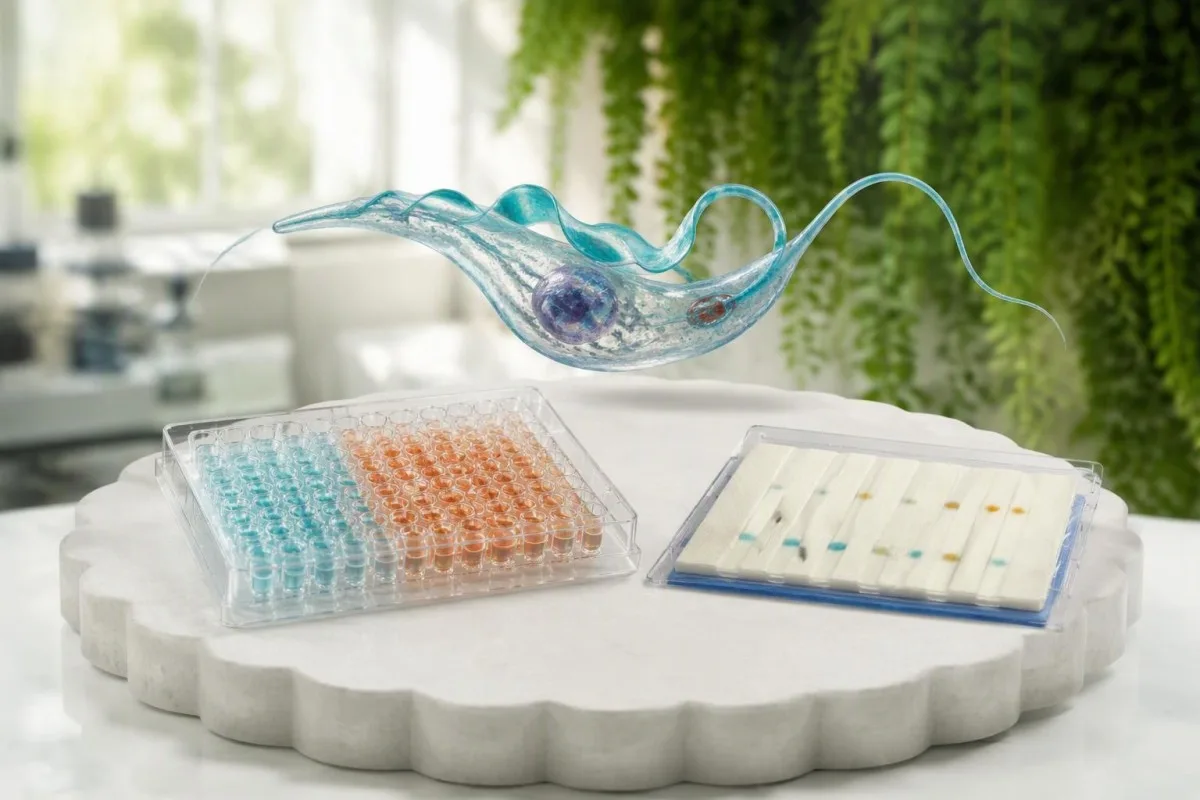
A positive screening result does not by itself confirm chronic Chagas disease: confirmation usually requires two distinct Trypanosoma cruzi antibody assays. If they disagree, a reference laboratory uses an additional assay; PCR or microscopy is more useful for suspected acute infection, congenital infection, or reactivation.


- Two antibody assays using different antigen preparations or testing formats usually establish chronic Chagas disease when both are positive.
- One reactive screen is a reason for diagnostic follow-up, not proof of infection or evidence that heart damage has occurred.
- Resultados discordantes generally require a third distinct serologic assay through an experienced reference laboratory.
- Valores do índice de anticorpos have assay-specific cutoffs; a result of 3.0 is not a universal measure of parasite burden or disease severity.
- Negative PCR cannot exclude chronic Chagas disease because circulating parasite DNA may be intermittently undetectable.
- Acute infection and reactivation call for direct detection, often combining PCR with microscopy rather than waiting for antibody confirmation.
- Congenital testing uses early direct detection and later antibody testing, commonly at 9–12 months, after maternal antibodies have cleared.
- Confirmed infection warrants cardiac assessment, generally including a 12-lead ECG and echocardiography, even when the person feels well.
- Treatment follow-up cannot rely on a single negative PCR or a persistently positive antibody test; antibodies may remain detectable for years.
What does a positive Chagas screening result actually mean?
A positive Chagas screening test means the assay detected reactivity to Trypanosoma cruzi antigens; it does not, by itself, confirm chronic infection. For chronic Chagas disease, clinicians usually need two distinct positive antibody assays, because a single assay can produce false-positive or false-negative results.
The report wording matters: reactiva, positivo, equivocal, e confirmed positive are not interchangeable. A blood-donor notification may describe a screening and supplemental testing algorithm designed for donation safety, which is not necessarily the same as the clinical diagnostic pathway; our guide to resultados de anticorpos positivos explains why that distinction changes the next step.
A confirmed T. cruzi antibody result usually supports infection, not protective immunity, in someone who has never received treatment. However, antibodies can remain detectable for years after successful treatment, and neither 1 reactive screen nor 2 positive assays tells us when transmission occurred, whether parasites are currently detectable in circulation, or whether the heart has been affected.
Kantesti é un Analizador de análises de sangue con IA that can help explain the wording of a Chagas report, but it cannot provide the second laboratory assay required for confirmation. As of October 10, 2026, the practical distinction remains laboratory confirmation versus report interpretation; background on a nosa organización should not be mistaken for evidence validating a particular infectious-disease assay.
Why does chronic Chagas disease require two antibody assays?
Chronic Chagas disease requires two distinct serologic assays because no single antibody test has sufficiently reliable performance across every population and exposure setting. Different assays recognize different parasite antigens, so agreement between complementary methods makes an isolated assay error less likely.
T. cruzi is genetically diverse, and people do not produce identical antibody responses to every antigen preparation. An assay validated in one geographic population may perform differently in another; the United States recommendations therefore specify 2 assays based on different antigens or formats, with a third assay when the first two disagree (Forsyth et al., 2022).
Cross-reactivity creates a separate problem. Some Chagas assays can react with antibodies generated against Leishmania species or other related organisms, and nonspecific reactivity becomes more consequential when infection prevalence is low; choosing 2 assays with genuinely different antigen compositions helps avoid simply repeating the same vulnerability under another test name.
Repeating one assay twice improves reproducibility, not diagnostic independence. The distinction is similar to the separation between a reactive screen and confirmation discussed in our HTLV confirmation guide, although Chagas disease has its own algorithm and cannot be confirmed using an HTLV-style test; my first question would be, “Were these actually two different assays?”
Which antibody methods count as different confirmatory tests?
Two complementary Chagas antibody tests may use different antigen preparations, different testing formats, or both. Examples include an enzyme immunoassay paired with an immunofluorescence assay or a specialized immunoblot, provided the combination follows a validated diagnostic algorithm.
ELISA or EIA detects antibody binding to parasite antigens using an enzyme-generated signal; IFA detects binding through fluorescence; an immunoblot evaluates recognition of selected parasite proteins. Two ELISAs can sometimes qualify if they use distinct antigen preparations, but two copies of the same commercial kit do not become independent merely because different laboratories process them.
A practical laboratory question is: “What antigen preparation and assay format were used for each result?” If the report lists only T. cruzi IgG positive on 2 dates, there may still be only one method represented; ask the ordering clinician or laboratory to retrieve the method details before labeling the infection confirmed or repeating testing unnecessarily.
Kantesti separates explanation of a reported result from proof of assay identity; a PDF may omit details that only the laboratory can supply. Our clinical standards overview describes interpretation oversight, not authorization to substitute software for Chagas disease confirmatory testing, and a reference laboratory remains the appropriate place to verify whether 2 methods are complementary.
Can a high Trypanosoma cruzi antibody number show severity?
A high Trypanosoma cruzi antibody index does not reliably measure parasite burden, duration of infection, or cardiac damage. The positive cutoff belongs to the specific assay, and there is no universal Chagas antibody reference range that can be applied across laboratories.
Some reports use an index, signal-to-cutoff ratio, ou optical-density ratio, while others report only reactive or nonreactive. If a particular laboratory defines 1.0 as its positive boundary, an index of 3.0 means the signal exceeded that assay’s threshold; it does not mean three times as many parasites, three times the cardiac risk, or three times the treatment requirement.
Near-cutoff results deserve careful review because small analytical changes can shift the category from equivocal to positive or negative. However, a strongly reactive result still needs a distinct confirmatory assay in the chronic diagnostic pathway; the difference between probas cualitativas e cuantitativas helps explain why an apparently precise number may still function mainly as a classification signal.
Comparing 2 antibody index values from different manufacturers can be misleading even when both are labeled IgG. Their antigen composition, calibration, and reporting units may differ, so I would not interpret a change from 4.2 to 2.1 as improvement without method-specific evidence; ask whether the laboratory considers serial values clinically interpretable rather than assuming that every downward trend reflects treatment success.
How are discordant Chagas antibody results resolved?
Discordant Chagas antibody results usually require a third distinct serologic assay at an experienced reference laboratory. One positive and one negative assay is an unresolved diagnostic pattern, not an automatic negative result and not permission to choose whichever result seems more reassuring.
The reference laboratory may first repeat testing, request a fresh specimen, or review the original assay documentation before performing the resolving method. The objective is generally to establish agreement between 2 distinct assays, not to accumulate repeated results from one kit; Forsyth et al. (2022) explicitly describe third-assay testing when the initial methods disagree.
A fresh sample is especially helpful when there is a specimen-identification concern, an equivocal result, or uncertainty about handling. Waiting 2–4 weeks may be useful if recent acquisition is plausible and antibody development is still underway, but waiting alone does not fix chronic assay disagreement; our Brucella testing discussion illustrates the broader principle that repeat timing and test selection solve different problems.
Persistent disagreement should remain documented as unresolved, with specialist advice on further testing and follow-up. Consider a hypothetical adult with 1 reactive donor screen and 1 negative diagnostic EIA: the next step is a reference-laboratory algorithm, not immediate treatment and not dismissal of the donor result; record the assay names, specimen dates, exposure history, and any previous Chagas treatment together.
How does exposure history change a positive Chagas test?
Exposure history changes the probability that a reactive Chagas screen represents true infection, but it does not replace confirmatory testing. Relevant histories include birth or residence in endemic parts of Latin America, maternal infection, and certain transfusion, transplant, laboratory, or vector exposures.
Pretest probability explains why the same assay can generate different clinical meaning in different populations. In a hypothetical group of 10,000 people with 0.1% prevalence, a test with 99% sensitivity and 99% specificity would produce about 10 true positives and 100 false positives; those illustrative figures are not the performance specifications of any particular Chagas assay.
A person may have chronic Chagas disease decades after leaving an endemic region, so “I have not traveled recently” does not exclude infection. Conversely, seeing a triatomine insect does not establish transmission: the classic vector route involves infected insect feces entering a mucosal surface or disrupted skin, rather than parasite injection through the bite itself; 1 suspected encounter needs contextual assessment.
An exposure review should ask where someone lived, the type of housing and vector contact, maternal history, and previous screening rather than relying on nationality alone. Other exposure-related infections may need separate testing, as discussed in our Strongyloides antibody guide; a positive result for another parasite neither confirms nor excludes T. cruzi, and routine eosinophil counts cannot settle either diagnosis.
Why can PCR be negative in chronic Chagas disease?
A negative T. cruzi PCR cannot exclude chronic Chagas disease because circulating parasites may be sparse and intermittently detectable. Chronic diagnosis therefore relies primarily on two distinct antibody assays, whereas PCR answers whether parasite DNA was detected in that particular specimen.
During chronic infection, parasites can persist in tissues even when a collected specimen contains no detectable target DNA. Sample volume, extraction technique, target sequence, transport, and intermittent parasitemia all influence detection; Bern’s clinical review emphasizes the different diagnostic roles of serology and direct detection across disease phases (Bern, 2015), so 1 negative PCR must not overturn 2 convincing positive antibody assays.
A positive chronic PCR supports detection of parasite DNA, but it does not by itself measure myocardial injury or prove a new acute episode. Repeating PCR may increase the chance of detection, yet collecting 2 or 3 negative samples still does not create a universally accepted exclusion rule for chronic infection; the method’s analytical sensitivity is different from its ability to rule out disease clinically.
Kantesti é un plataforma de interpretación de análise de sangue de IA that can explain why antibody and PCR results answer different questions; it cannot infer parasite clearance from one negative molecular result. Before acting on any automated explanation, use our report accuracy checklist to verify the organism, specimen date, method, and whether the report says “not detected” rather than “infection excluded.”
When are microscopy and PCR better for acute infection?
PCR and microscopy are more appropriate when acute Chagas disease is suspected because parasites may circulate before antibodies become reliably detectable. Recent plausible exposure with fever, facial or eyelid swelling, myocarditis, or a transfusion-associated illness warrants prompt direct testing and clinical assessment.
The acute phase usually lasts approximately 2 meses, although symptoms may be mild or absent. Microscopy can identify circulating trypomastigotes when parasitemia is sufficient, and concentration techniques can improve detection compared with a routine slide; PCR often adds sensitivity, but the laboratory must know that T. cruzi is suspected rather than receiving a nonspecific request to “look for parasites.”
One negative cell sample slide does not exclude acute Chagas disease. If suspicion remains high, clinicians may obtain additional specimens, use PCR, and arrange subsequent serology; an isolated negative IgG result early after exposure is particularly weak reassurance, because the immune response may not yet have crossed the assay’s detection threshold and direct tests have their own sampling limitations.
Fever after travel also requires urgent consideration of other infections, especially malaria when the itinerary fits; our guía de probas de febre post-viaxe explains why appropriate testing should not wait for a Chagas send-out result. Chest pain, breathlessness, fainting, or new neurologic symptoms justify same-day assessment, even with 1 negative initial test and even before the exact cause is known.
How is Chagas reactivation tested during immunosuppression?
Suspected Chagas reactivation is assessed with direct parasite detection, especially serial PCR and microscopy, interpreted alongside symptoms and immune status. Antibody positivity usually documents pre-existing infection; repeating antibody assays does not reliably determine whether reactivation is occurring.
Reactivation is a particular concern in people with substantial immunosuppression, including some transplant recipients and people with advanced HIV. Clinicians look for increasing parasite detection and compatible cardiac, neurologic, or other manifestations; a newly positive PCR in someone with chronic infection is not automatically reactivation, because intermittent low-level PCR positivity can occur without a new clinical episode.
Quantitative PCR trends can help, but there is no universal parasite-equivalent cutoff or cycle-threshold value that defines reactivation across laboratories. A lower cycle threshold usually indicates more target DNA only within an adequately controlled, comparable assay; compare serial specimens through the same laboratory where possible, and do not transfer a threshold from one protocol to another without specialist interpretation.
Kantesti AI can explain the distinction between baseline seropositivity and a changing molecular result, but a transplant or infectious-disease team must determine monitoring frequency and treatment triggers. The same antibody-versus-active-risk distinction appears in our JCV antibody interpretation guide, although the organisms and monitoring algorithms differ; new confusion, seizures, or focal weakness require urgent evaluation rather than waiting for a third antibody test.
How are babies tested when a mother has Chagas disease?
Babies born to mothers with confirmed Chagas disease need a dedicated congenital testing pathway using early direct detection and later serology. Maternal IgG crosses the placenta, so a positive infant antibody test shortly after birth does not establish that the baby is infected.
Early testing may include microscopy and PCR, according to the local congenital program and laboratory access. A negative result at birth does not always end evaluation; repeat direct testing during the first weeks, often around 4–6 semanas, can identify infection missed initially, while positive molecular findings may require a separately collected confirmatory specimen to address contamination or transient maternal material.
If early direct tests do not establish infection, antibody testing is commonly performed at 9–12 months, after passively transferred maternal IgG should have disappeared. Persistent T. cruzi antibodies at that stage support congenital infection when exposure and laboratory findings fit; the exact schedule should follow the treating service’s protocol rather than being replaced by repeated adult-style antibody panels during early infancy.
Congenital infection can be asymptomatic, and treatment started in infancy has a much higher likelihood of cure than treatment of longstanding adult infection. Our guide to os límites de interpretación da AI para nenos explains why age-specific pathways matter; record the infant’s age in days or months, maternal confirmation status, and all specimen dates, because 1 missing follow-up visit can leave the diagnostic pathway incomplete.
What evaluation follows confirmed chronic Chagas disease?
Confirmed chronic Chagas disease warrants assessment for cardiac involvement even when the person has no symptoms. Initial evaluation generally includes a clinical history, examination, ECG de 12 derivacións, and echocardiography, with additional rhythm or gastrointestinal testing guided by findings.
Aproximadamente 20–30% of chronically infected people develop cardiac disease over time, although estimates vary by population, age, and follow-up. A normal antibody index cannot rule out conduction abnormalities, and a very high index cannot diagnose cardiomyopathy; normal initial cardiac testing is reassuring about current assessment, not a lifetime guarantee that monitoring will never be needed (Bern, 2015).
Palpitations, fainting, unexplained breathlessness, or an abnormal ECG may prompt ambulatory rhythm monitoring and cardiology review. Our guía de probas de palpitacións covers common laboratory mimics, but electrolyte or thyroid testing does not replace an ECG in confirmed Chagas disease; dysphagia or longstanding severe constipation may separately justify evaluation for digestive involvement.
As Thomas Klein, MD, Chief Medical Officer at Kantesti, I would keep 2 questions separate when explaining this report: “Is infection confirmed?” and “Has an organ been affected?” The roles of physicians described on our Consello Asesor Médico concern interpretation oversight, while individualized cardiac staging and treatment decisions belong to the patient’s clinical team; a positive antibody result is not itself a prognosis.
Do antibodies or PCR become negative after Chagas treatment?
Chagas antibodies can remain positive for years after treatment, and one negative PCR does not prove cure. Follow-up depends on age, disease phase, treatment history, and the assay used, so laboratory improvement must be distinguished from a validated endpoint of parasite clearance.
Benznidazole and nifurtimox are the principal antiparasitic medicines used for Chagas disease, and many treatment regimens run for about 60 days under clinical supervision. Treatment recommendations are strongest for acute, congenital, and reactivated infection; decisions for chronic infection depend on factors including age and cardiac status, and should not be based on the antibody index alone.
No 2,854-participant BENEFIT trial, benznidazole reduced parasite detection but did not significantly reduce the primary clinical outcome in people with established Chagas cardiomyopathy over a mean 5.4 years (Morillo et al., 2015). That finding does not mean treatment has no value in acute infection, infants, or every younger adult with chronic infection; it demonstrates why PCR changes and clinical benefit are not interchangeable outcomes.
Monitoring also includes medication safety, often with CBC and liver testing according to the treating team’s protocol. A new result should be interpreted alongside symptoms, the treatment day, and the previous method; our guía de tendencias de seguridad de medicamentos explains why a falling antibody value or 1 negative PCR should not distract from a clinically significant rash, cytopenia, or liver-test change.
What should you bring to a Chagas confirmatory appointment?
Bring the original report, assay details, exposure history, and any previous treatment records to a Chagas confirmation appointment. Fasting is generally unnecessary for an antibody test alone, but PCR specimen requirements and additional baseline testing depend on the receiving laboratory.
Serology commonly uses serum, while PCR frequently requires EDTA whole blood or another specifically validated specimen. The receiving laboratory decides the acceptable container, volume, storage, and transport conditions; our serum and specimen guide explains why a convenient leftover serum sample cannot automatically replace a freshly collected molecular specimen, and why 2 ordered tests may require different preparation.
Keep the complete PDF rather than a cropped screenshot that only shows positivo. Method names, footnotes, index cutoffs, supplemental results, and collection dates may determine whether the 2-assay requirement has already been satisfied; our lista de verificación de transcrición de PDF identifies common errors such as dropped negative signs, missing qualifiers, and confusion between test dates and report dates.
A useful four-question checklist is: Which assay was positive? Was a distinct second assay performed? If they disagreed, what resolving test is planned? Is there any reason to suspect acute infection or reactivation rather than stable chronic infection? These 4 questions usually make the visit more productive than requesting a larger nonspecific panel, and you should disclose immunosuppression or pregnancy without stopping prescribed medication on your own.
Report review, clinical oversight, and related publications
Reliable Chagas report interpretation requires the assay names, diagnostic phase, and confirmation status—not just a highlighted positive result. A two-assay chronic diagnosis, an early congenital PCR, and a rising molecular signal during immunosuppression represent three different clinical situations and should not receive the same explanation.
Kantesti é un Ferramenta de análise de probas de sangue impulsada por IA that helps organize laboratory findings and questions for a clinician; it does not perform microscopy, PCR, or reference-laboratory serology. Our guía tecnolóxica describes the interpretation workflow, and the safest use here is to identify which of the 2 required chronic assays is documented—not to manufacture a diagnosis from an incomplete report.
Thomas Klein, MD, is the named author of this educational explanation; the condition-specific clinical sources below provide the evidentiary basis for the diagnostic distinctions. Two additional DOI-linked educational resources are listed separately: our Nipah diagnostic overview discusses timing-dependent test selection, but Nipah testing methods and performance cannot be transferred to T. cruzi.
The second supplementary resource is our hematology marker overview, which concerns general laboratory interpretation rather than Chagas confirmation. These 2 repository resources are not presented as Chagas clinical guidelines or proof of diagnostic accuracy; their ResearchGate and Academia.edu links are catalog searches, not verified copies, while the directly relevant peer-reviewed references are identified in the separate medical-reference list.
Preguntas frecuentes
Unha proba positiva de enfermidade de Chagas significa que definitivamente teño a enfermidade de Chagas?
Unha proba de cribado de Chagas positiva non establece por si soa a enfermidade de Chagas crónica. A confirmación require xeralmente 2 ensaios de anticorpos Trypanosoma cruzi distintos que utilicen diferentes antíxenos ou formatos de proba. Dous ensaios positivos concordantes xeralmente establecen o diagnóstico crónico, pero non determinan se hai danos cardíacos. Unha notificación de cribado de doadores de sangue debe ser revisada a través dunha vía de diagnóstico clínico.
Por que preciso de dúas probas de anticorpos contra o Trypanosoma cruzi?
Utilízanse dúas probas de anticorpos Trypanosoma cruzi distintas porque ningún ensaio único funciona igual en todas as poboacións e escenarios de exposición. Diferentes preparacións de antíxenos axudan a abordar o recoñecemento variable de anticorpos e a reactividade cruzada. Repetir o mesmo kit en 2 ocasións non proporciona a mesma evidencia independente que os ensaios complementarios. Pregúntalle ao laboratorio que preparación de antíxeno e formato de proba se utilizaron para cada resultado.
Que sucede se unha proba de anticorpos da enfermidade de Chagas é positiva e a outra negativa?
Un ensaio positivo e un negativo para anticorpos contra a enfermidade de Chagas é un resultado discordante que normalmente require un terceiro ensaio distinto a través dun laboratorio de referencia. O laboratorio tamén pode solicitar unha mostra fresca ou repetir as probas iniciais antes de resolver a discrepancia. O obxectivo é xeralmente o acordo entre 2 ensaios serolóxicos distintos, non simplemente contar cada resultado repetido. Os desacordos persistentes deben documentarse como non resoltos ata que un clínico e un laboratorio experimentados completen a avaliación.
Pode un PCR negativo descartar a enfermidade de Chagas crónica?
Unha PCR negativa de Trypanosoma cruzi non pode descartar a enfermidade de Chagas crónica porque o ADN parasitario pode estar ausente intermitentemente na mostra recollida. O diagnóstico crónico, polo tanto, depende principalmente de 2 ensaios de anticorpos distintos. A PCR é máis útil para a infección aguda sospeitada, a infección conxénita ou a reactivación e para situacións de seguimento seleccionadas. Unha PCR negativa tampouco proba a curación despois do tratamento.
Un índice alto de anticorpos contra a Doenza de Chagas significa unha enfermidade grave?
Un índice de anticorpos de Chagas alto non é unha medida validada da carga parasitaria nin da afectación cardíaca. Se un laboratorio usa 1.0 como o seu punto de corte positivo, un índice de 3.0 indica un sinal por riba do limiar dese ensaio, non tres veces a gravidade da enfermidade. Os puntos de corte e as escalas numéricas varían entre os métodos. A infección confirmada require unha avaliación clínica separada, que xeralmente inclúe un ECG de 12 derivacións e unha ecocardiografía.
Como se lle fai unha proba a un bebé se a nai ten enfermidade de Chagas?
A baby born to a mother with confirmed Chagas disease needs early direct testing and follow-up because maternal IgG can make infant antibody tests positive without congenital infection. Microscopy or PCR may be performed soon after birth, with repeat direct testing often around 4–6 weeks according to the local protocol. If early testing does not establish infection, antibody testing is commonly performed at 9–12 months after maternal antibodies have cleared. A negative result at birth alone does not always complete the evaluation.
Will my Chagas antibody test become negative after treatment?
Chagas antibody tests can remain positive for years after successful treatment, especially in longstanding chronic infection. Many antiparasitic treatment regimens last about 60 days, but the treatment duration is not the expected time to antibody disappearance. One negative PCR cannot establish cure, and antibody values from different assays should not be directly compared as a treatment-response scale. Follow-up should use the treating team’s age-specific and disease-phase-specific plan.
Obtén hoxe unha análise de sangue con IA
Únete a máis de 2 millóns de usuarios en todo o mundo que confían en Kantesti para obter unha análise instantánea e precisa das análises de laboratorio. Carga os teus resultados de análise de sangue e recibe unha interpretación completa de biomarcadores de 15,000+ en segundos.
📚 Publicacións de investigación citadas
Klein, T., Mitchell, S., & Weber, H. (2026). Análise de sangue do virus Nipah: guía de detección e diagnóstico precoz 2026. Kantesti Investigación médica con IA.
Klein, T., Mitchell, S., & Weber, H. (2026). Guía de tipo de sangue B negativo, proba de sangue de LDH e reconto de reticulocitos. Kantesti Investigación médica con IA.
📖 Referencias médicas externas
Forsyth CJ et al. (2022). Recommendations for Screening and Diagnosis of Chagas Disease in the United States. O Xornal de Enfermidades Infecciosas.
Bern C. (2015). Chagas’ Disease. The New England Journal of Medicine.
Morillo CA et al. (2015). Randomized Trial of Benznidazole for Chronic Chagas’ Cardiomyopathy. The New England Journal of Medicine.
⚕️ Aviso médico
Este artigo é só para fins educativos e non constitúe asesoramento médico. Consulta sempre un/ha profesional sanitario/a cualificado/a para decisións de diagnóstico e tratamento.
Sinais de confianza E-E-A-T
Experiencia
Revisión clínica dirixida por un médico dos fluxos de interpretación de análises.
Experiencia
Foco en medicina de laboratorio sobre como se comportan os biomarcadores no contexto clínico.
Autoridade
Escrito polo Dr. Thomas Klein, con revisión da Dra. Sarah Mitchell e do Prof. Dr. Hans Weber.
Fiabilidade
Interpretación baseada en evidencias con vías de seguimento claras para reducir a alarma.