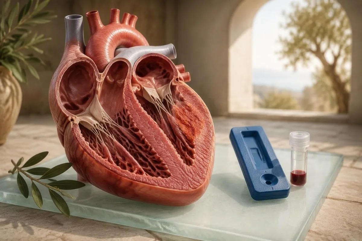
Faodaidh troponin milleadh fèithe cridhe aithneachadh, ach cha chruthaich e gur e myocarditis an t-adhbhar. Tha an dearbhadh a’ tighinn bho bhith a’ maidseadh toraidhean fala le comharraidhean, toraidhean ECG, echocardiography, agus mar as trice MRI cairdiach.


- Troponin os cionn na 99mh-ceud percentile crìoch iomraidh àrd an leabharlainn a’ nochdadh milleadh air a’ mhucardium, chan e dearbhadh sònraichte de myocarditis.
- Troponin àrd-mhothachail dh’ fhaodadh iad a bhith àbhaisteach ann am myocarditis tlàth no le dàil, mar sin chan eil toradh àbhaisteach ga dùnadh a-mach gu tur.
- CRP os cionn 10 mg/L a’ toirt taic do sè siostamach gnìomhach ach chan urrainn dha sè anns a’ chridhe a chomharrachadh gu sònraichte.
- Deuchainn sreathach (serial testing) tha cudromachd ann: tha pàtran troponin ag èirigh no a’ tuiteam thairis air 1-3 uairean nas fhiosaiche na aon toradh iomallach.
- MRI Cairdiach is e am prìomh dheuchainn neo-ionnsaigh a thathas a’ cleachdadh gus taic a thoirt do myocarditis nuair a tha comharraidhean, ECG, agus troponin a’ togail dragh.
- Galar coronach feumar beachdachadh air ann an inbhich le pian broilleach agus troponin àrdachadh, gu sònraichte às deidh aois 40 no le factaran cunnairt shoithichean.
- Measadh èiginneach tha feum air airson cuideam broilleach, anail gann aig fois, fàilneachadh, palpitations leantainneach, no troponin àrdachadh le comharraidhean draghail.
An urrainn do dheuchainn fala airson myocarditis sè cridhe a dhearbhadh?
Chan urrainn do aon deuchainn fala air sàr-chridheachd a dhearbhadh no a dhiùltadh. Lorgar leòintean air fèith a' chridhe le troponin, ach lorgar sè san àite sam bith air a' bhrìgh le CRP agus ESR; cha bhith gin dhiubh sin a' stèidheachadh na h-adhbhar. Mar an Dotair Thomas Klein, tha mi ag innse do dh'euslaintich gu bheil obair fala a' tòiseachadh an rannsachaidh, ach mar as trice, is e an MRI cairdiach agus an ìomhaigh chlinigeach a chuireas crìoch air.
A deuchainn fala air sàr-chridheachd na fhianais thaice, chan e binn. Faodaidh troponin èirigh air sgàth sàr-chridheachd, ionnsaigh air a' chridhe, buillean luath, embolism sgamhain, bruthadh-fala mòr, tinneas dubhaig, sepsis, no eacarsaich rò-dhian. Tha aithisg suidheachadh 2013 bho Chomann Cridhe na h-Eòrpa a' toirt cunntas air sàr-chridheachd mar staid chlinic-path-eòlasach seach mar dhiagnosis a chaidh a dhèanamh bho aon luach obair-lann (Caforio et al., 2013). Airson ath-bhreithneachadh ann an cànan sìmplidh air diofar modhan tomhais, faic toraidhean troponin I agus T.
Nam eòlas clionaigeach, is e an suidheachadh as miosa mealladh a bhith a' faicinn neach òg le pian mòr anns a' bhroilleach às dèidh galar viral agus troponin a tha air èirigh beagan. Faodaidh am pàtran sin freagairt air sàr-chridheachd, ach faodaidh e cuideachd a bhith na pericarditis, cnap-starra corranach, no àrdachadh neo-cheangailte ann an troponin às dèidh obair chruaidh. Feumaidh ràdh eadar-dhealaichte a bhith aig fear 52-bliadhna a tha a' ruith le hs-cTnT de 38 ng/L às dèidh rèis marathon bho fhear 22-bliadhna le 38 ng/L, fiabhras, giorrad anail ùr, agus atharrachaidhean ECG.
Tha Kantesti na Anailisiche deuchainn fala AI a leughas troponin còmhla ri gnìomh dubhaig, comharran sè, buill-bòrd grùthan, agus toraidhean roimhe seach a bhith a' làimhseachadh aon luach a tha air a chomharrachadh mar dhiagnosis. Faodaidh an siostam againn cuideachadh le bhith ag eagrachadh a' phàtrain airson clionaiche, ach chan urrainn dha a bhith a' cur an àite measadh èiginn, ECG, no ìomhaigh. Riaghailt phractaigeach: ma tha comharran broilleach an làthair, na feith airson mìneachadh air-loidhne.
Dè tha ìrean troponin airson myocarditis a’ ciallachadh gu dearbh?
Tha troponin a tha nas àirde na an ìre iomraidh as àirde a tha sònraichte don dòigh tomhais a' ciallachadh gu bheil leòn air fèith a' chridhe an làthair, ach cha bhith e a' comharrachadh sàr-chridheachd mar an adhbhar. Bidh a' mhòr-chuid de dhòighean tomhais àrd-chugallach a' cleachdadh an 99mh ceud airson sluagh iomraidh fallain, mar as trice timcheall air 14 ng/L airson hs-cTnT agus timcheall air 4-20 ng/L airson hs-cTnI a rèir an neach-dèanamh.
Chan eil luach de hs-cTnT 16 ng/L faodaidh gun tèid e thairis air gearradh aon obair-lann beagan, ach hs-cTnI 1,500 ng/L tha e gu math àrd; cha bhith gin dhiubh sin leotha fhèin a' measadh cho dona 'sa tha sàr-chridheachd. Chan urrainn do obair-lannan luachan troponin I agus troponin T a mhalairt gu sàbhailte oir tha na dòighean tomhais aca a' cleachdadh diofar antibodies, calibrations, agus crìochan iomraidh. Is e an ìre iomraidh as àirde a tha air aithisg obair-lann an àireamh a tha cudromach.
Bidh an cùrsa ùine gu tric a' giùlan barrachd fiosrachaidh clionaigeach na an t-àm as àirde. Mar as trice, tòisichidh troponin a' èirigh 2-4 uairean às dèidh leòn gu h-obann, agus bidh clionaichean ga ath-aithris gu tric an dèidh 1-3 uairean ann an slighean èiginn. Tha àrdachadh no tuiteam soilleir a' toirt taic do phròiseas obann; faodaidh àrdachadh beag seasmhach tachairt le tinneas dubhaig cronach, tinneas cridhe structarail, no cuideam miocàrdiach fad-ùine. Tha an stiùireadh deuchainn fala airson pian bhoilg a' mìneachadh carson a thèid sampallan sreathach a chleachdadh.
Chan eil troponin àbhaisteach a' cur às do sàr-chridheachd gu earbsach. Faodaidh raointean beaga fòcasach de fhreagairt clò, deuchainn fadalach às dèidh na h-ùine as àirde, agus dòighean tomhais nas lugha de chugallachd uile toradh àbhaisteach a thoirt seachad. Air an làimh eile, chan eil luachan glè àrd a' ciallachadh milleadh neo-sheasmhach gu fèin-obrachail - tha àrdachaidhean follaiseach aig cuid de dh'euslaintich le sàr-chridheachd obann fhathast a' faighinn air ais gnìomh pumpaidh àbhaisteach.
Carson a dh’ èiricheas troponin ann am myocarditis agus suidheachaidhean eile
Troponin rises when cardiac muscle cells release intracellular proteins after injury or intense cellular stress. Myocarditis is one cause, but coronary blockage remains the diagnosis clinicians must urgently exclude when chest pain and raised troponin occur together.
In myocarditis, immune activity within the myocardium can disrupt cell membranes and release cardiac troponin I or T into circulation. In a heart attack, blocked coronary flow causes the same laboratory signal through a different mechanism. That overlap is why an elevated troponin should never be casually labelled “viral myocarditis” without considering coronary disease, particularly in people over 40 or those with diabetes, smoking exposure, or high ApoB.
Symptoms help but are imperfect. Myocarditis may cause central chest ache, breathlessness, reduced exercise capacity, skipped beats, or no symptoms at all. A person with reproducible pain when pressing the chest wall and a normal ECG is less likely to have myocardial injury, yet this cannot be confirmed remotely. New chest discomfort plus sweating, nausea, breathlessness, or fainting warrants emergency care.
Kantesti’s stiùireadh airson comharran bith-eòlasach deuchainn fala places a troponin result within its assay units and reference interval, which helps avoid a common error: comparing a value in ng/L with an internet cutoff reported in ng/mL. One ng/mL equals 1,000 ng/L; unit confusion can make a result look a thousand-fold more alarming than it is.
An urrainn do CRP, ESR, no CBC myocarditis a nochdadh?
CRP, ESR, and white-cell counts can support an inflammatory illness, but none can locate inflammation in the heart. A CRP above 10 mg/L is often considered elevated in routine practice, although the result can rise from a cold, dental infection, autoimmune flare, obesity, or recent intense exercise.
Myocarditis CRP may be normal, mildly raised, or substantially elevated. CRP rises within about 6-12 hours after inflammatory signalling and usually falls faster than ESR when the trigger settles. A CRP of 48 mg/L with fever and chest symptoms adds weight to an inflammatory diagnosis, but it does not distinguish myocarditis from pneumonia, influenza, or bacterial infection. Read more about symptom context in high hs-CRP results.
ESR is slower and less specific. An ESR above 20-30 mm/hour may persist for days to weeks because it is influenced by fibrinogen, anaemia, age, pregnancy, and immunoglobulin levels. I rarely use ESR alone to make a decision about suspected myocarditis; it is more useful when checking whether a broad inflammatory pattern has persisted across time.
A CBC can show mild leukocytosis, lymphocyte changes, or normal findings. White cells above 11.0 × 10⁹/L can accompany infection or stress, yet many confirmed myocarditis cases have normal counts. The useful clinical question is whether CRP and WBC disagree—a pattern explored in our WBC versus CRP guide—rather than assuming either confirms cardiac involvement.
A bheil CK-MB, CK, LDH, agus myoglobin fhathast a’ cuideachadh?
CK-MB, total CK, LDH, and myoglobin are less specific than troponin for heart-muscle injury and cannot diagnose myocarditis. They can occasionally clarify whether strenuous exercise or generalized skeletal-muscle injury is contributing to an abnormal result.
CK-MB is found in cardiac tissue but also in skeletal muscle, so a raised result can occur after vigorous exercise, trauma, or muscle inflammation. Some laboratories report a CK-MB index, calculated relative to total CK, but high-sensitivity troponin has largely replaced it for acute myocardial injury assessment. Our explanation of what CK-MB means covers its remaining niche.
Iomlan creatine kinase can rise dramatically after resistance training, statin-associated muscle injury, seizures, or rhabdomyolysis. A CK of 2,000 IU/L after an unfamiliar endurance event can coexist with a small troponin rise and may point toward skeletal-muscle stress, but clinicians still need to rule out cardiac injury if symptoms are present. CK values above 5,000 IU/L raise concern for significant muscle breakdown and kidney stress.
LDH and myoglobin are even broader markers of tissue turnover. In my practice, they are most helpful when the panel also includes AST, potassium, creatinine, and urine findings—especially after exertion. A high creatine kinase pattern should be interpreted as a muscle-safety issue, not casually attributed to myocarditis.
Dè tha BNP agus NT-proBNP a’ cur ris an teagamh mu myocarditis
BNP and NT-proBNP indicate cardiac wall stress and possible heart failure; they do not prove inflammation or myocarditis. NT-proBNP below 125 ng/L is often used in stable outpatient adults to make chronic heart failure less likely, while acute-care thresholds are higher and age-dependent.
Myocarditis can impair the heart’s pumping or relaxation function, causing BNP no NT-proBNP to rise as cardiac chambers stretch. A raised NT-proBNP alongside breathlessness, ankle swelling, a fast heart rate, and an abnormal echocardiogram is more concerning than the same value in isolation. Kidney impairment, atrial fibrillation, older age, and pulmonary embolism can also raise NT-proBNP.
A low natriuretic peptide is reassuring but not absolute. Very early myocarditis, focal inflammation, obesity, and preserved heart function can all yield a normal value. The numerical cutoff must match the setting: an outpatient screening threshold should not be used to dismiss someone with acute breathlessness in an emergency department.
Kantesti AI ’s e àrd-ùrlar mìneachaidh biomarcadairean AI that can show how NT-proBNP, creatinine, sodium, and troponin move together across separate draws. That longitudinal view is useful because a falling NT-proBNP over days may reflect improving haemodynamics, whereas a rising trend merits prompt clinical review. See NT-proBNP cutoffs and symptoms airson co-theacsa a rèir an toraidh.
Carson a dh’ atharraicheas toraidhean dubhaig, grùthan agus electrolytes an leughadh
Creatinine, eGFR, potassium, AST, ALT, and sodium can change how clinicians interpret a raised troponin. Kidney disease can produce chronic low-level troponin elevation, while potassium abnormalities can trigger rhythm problems that mimic or complicate myocarditis.
An eGFR below 60 mL/min/1.73 m² is associated with more frequent baseline hs-troponin elevation, especially for troponin T. This does not mean the result should be ignored; clinicians look harder at the delta, symptoms, ECG, and prior baseline. A dynamic increase remains clinically meaningful even when kidney function is reduced.
Potassium below 3.0 mmol/L or above 6.0 mmol/L can increase arrhythmia risk and needs prompt medical attention, especially with palpitations or weakness. Sodium below 130 mmol/L in a person with breathlessness may signal significant fluid imbalance or heart failure, though many non-cardiac illnesses can cause it. The iùl air a’ phannal bunaiteach metabolach explains these tests in plain language.
AST can rise with cardiac or skeletal-muscle injury, whereas ALT is more liver-specific. A disproportionate AST rise after a race may be muscular; AST and ALT elevations with jaundice, bilirubin changes, or medication exposure need a broader liver work-up. Context is everything here—one isolated enzyme almost never tells the whole story.
Am bu chòir antibodies bhìorasach no deuchainnean fala fèin-ghalarach òrdachadh?
Routine viral antibody panels rarely confirm the cause of myocarditis, and positive antibodies often show past exposure rather than active cardiac infection. Autoimmune testing is targeted when the history, examination, or other organs suggest systemic inflammatory disease.
Most adults have antibodies to common viruses such as Epstein-Barr virus, cytomegalovirus, and coxsackievirus. A positive IgG usually reflects previous exposure; it does not prove that a virus is affecting the heart now. Even IgM can be falsely positive or persist longer than expected. The EBV antibody pattern guide shows why antibody timing is tricky.
Clinicians may order ANA, ENA antibodies, ANCA, rheumatoid factor, complement levels, thyroid testing, or eosinophil counts when there are clues such as rash, joint swelling, asthma, sinus disease, kidney changes, recurrent inflammation, or drug exposure. Eosinophilic myocarditis is uncommon but clinically important because it may need urgent specialist-directed treatment.
Cardiac MRI and, in selected unstable or treatment-changing cases, endomyocardial tissue examination are better tools for characterising myocardial involvement than broad viral serology. The evidence is honestly mixed on exactly which ancillary tests every patient needs; the work-up should follow the phenotype, not a fixed shopping list.
Dè na deuchainnean as urrainn dearbhadh no taic làidir a thoirt do myocarditis?
Cardiac MRI is the leading non-invasive test for supporting myocarditis because it can show oedema and non-ischaemic tissue injury patterns. ECG, echocardiography, coronary assessment when appropriate, and sometimes endomyocardial biopsy complete the diagnostic pathway.
The updated Lake Louise cardiac MRI criteria use at least one T2-based marker of oedema and one T1-based marker of non-ischaemic injury, such as late gadolinium enhancement or abnormal T1 mapping. Ferreira and colleagues reported that combining these tissue markers improves diagnostic assessment compared with older MRI approaches (Ferreira et al., 2018). MRI is strongest when performed close to the symptomatic period, although timing depends on stability and local access.
An ECG may show ST-T changes, PR depression, conduction delay, or rhythm disturbance, but a normal ECG does not exclude myocarditis. Echocardiography evaluates ejection fraction, chamber size, valve function, and pericardial fluid; it can be entirely normal in mild focal disease. A reduced left-ventricular ejection fraction below 50% changes urgency and follow-up planning.
Endomyocardial biopsy is not routine for everyone. It becomes more relevant with cardiogenic shock, rapidly worsening heart failure, high-grade heart block, sustained ventricular arrhythmia, or suspicion of a subtype that would alter treatment. This distinction matters: blood tests can raise suspicion, but only carefully selected tissue analysis can directly identify myocardial inflammatory cell patterns.
Cuin a bu chòir deuchainnean fala airson myocarditis ath-aithris?
Repeat troponin timing depends on symptoms and setting: emergency pathways commonly repeat high-sensitivity troponin after 1-3 hours, while outpatient follow-up may occur over days or weeks. Repeating a test without a clinical plan can create noise rather than clarity.
For acute chest symptoms, clinicians commonly use an immediate sample and a second sample at 1 or 2 hours, depending on the local validated algorithm. The absolute change in ng/L matters more than a percentage change when starting values are low. Do not use a home-timed repeat test to manage active chest pain—emergency teams need the ECG and vital signs at the same time.
After confirmed or probable myocarditis, troponin may normalise within days to weeks, but recovery is uneven. CRP often improves earlier than MRI abnormalities, and symptoms can improve before electrical irritability has fully settled. I advise patients to record the date, symptoms, exercise level, medications, and illness around each blood draw; our iùl loidhne-tìm na h-obrach-lann explains why those details matter.
Dr. Thomas Klein’s practical rule is that a repeat result should answer a specific question: “Is injury continuing?”, “Is this baseline elevation?”, or “Is recovery on track?” A troponin test repeated after heavy exercise, dehydration, or a different assay method may be difficult to compare. Ideally, follow-up uses the same laboratory and assay.
An urrainn do eacarsaich troponin àrdachadh gun myocarditis?
Yes, prolonged strenuous exercise can cause a temporary troponin rise without clinical myocarditis, particularly after marathons, ultradistance events, or intense cycling. Symptoms, ECG changes, recovery kinetics, and imaging determine whether the elevation is benign exertional release or true myocardial injury.
Post-exercise troponin elevations often peak within 2-6 hours and fall toward baseline within 24-48 hours, whereas sustained or rising values are more concerning. The overlap is not perfect, so symptoms matter enormously. Chest pain, collapse, unusual shortness of breath, or persistent palpitations after exercise should never be dismissed as “just training.”
During suspected myocarditis, vigorous exercise is usually restricted because inflamed myocardium may be more vulnerable to dangerous arrhythmias. Many sports-cardiology protocols reassess athletes after 3-6 mìosan with symptoms, biomarkers, ECG monitoring, ventricular function, and sometimes MRI guiding return. The exact timeline varies with severity and residual findings.
A high CK after exercise can muddy the picture, especially when AST rises too. Our stiùireadh deuchainn fala airson lùth-chleasaichean seasmhachd helps distinguish training-related lab shifts from patterns that need medical review. Rest is not merely cautious advice here—it is part of risk reduction while the diagnosis is unresolved.
Nuair a dh’ fheumas troponin àrdachadh cùram èiginn
A raised troponin with active chest pressure, breathlessness at rest, fainting, confusion, sustained palpitations, or new weakness needs emergency assessment now. The result may reflect myocarditis, heart attack, pulmonary embolism, severe arrhythmia, or another condition that cannot be safely sorted out online.
Call emergency services rather than driving yourself if chest symptoms are severe, persistent beyond 10-15 minutes, accompanied by collapse, or associated with blue lips, severe breathlessness, or a racing irregular heartbeat. Troponin is not a measure of pain intensity; a person can have a dangerous condition with a modest elevation or little pain.
There are quieter warning patterns too: resting heart rate persistently above 120 beats per minute, new inability to climb one flight of stairs, swelling, reduced urine output, or palpitations with dizziness. A recent viral illness does not make a cardiac cause less urgent. In fact, it can increase suspicion for myocarditis while coronary and pulmonary causes still require consideration.
If you have a result but no current emergency symptoms, contact the clinician who ordered it the same day when possible. The high troponin urgent-care guide lists common non-heart-attack causes, but it should not be used to self-triage dangerous symptoms.
Mar a leughas tu pannal fala airson myocarditis gun a bhith ga mhìneachadh cus
The safest way to read blood tests for heart inflammation is to look for a pattern: troponin trajectory, CRP, kidney function, electrolytes, symptoms, timing, and prior values. A single red flag on a report is a prompt for clinical context, not a final diagnosis.
Tha Kantesti na Inneal mion-sgrùdaidh deuchainn fala le cumhachd AI designed to compare laboratory values across time and highlight combinations that deserve clinician follow-up. For suspected myocarditis, our AI does not label someone with the condition from troponin alone; it identifies myocardial-injury markers, inflammatory context, and potentially relevant kidney or electrolyte confounders. That is deliberately conservative.
Before reviewing any report, confirm four details: the test name, units, laboratory upper reference limit, and sampling date. Then note whether strenuous exercise, fever, dehydration, kidney disease, supplements, or a recent hospital visit could affect the result. The inneal coimeas fala is particularly useful when comparing values obtained by the same laboratory.
A healthy-looking CRP or normal CBC should not overrule concerning symptoms, and a flagged troponin should not be interpreted without an ECG. This is one of those areas where context matters more than the number. If you use Kantesti, treat the report as a structured conversation starter for your doctor, not clearance to continue normal activity.
Ceistean a dh’fhaighneachd às deidh obair fala neo-àbhaisteach airson myocarditis
After abnormal cardiac blood work, ask what the result means for you today, what dangerous alternatives have been excluded, and what test will change management next. A precise question often gets a more useful answer than asking whether the number is simply “bad.”
Useful questions include: “Which troponin assay was used?”, “What is my change between samples in ng/L?”, “Could my kidney function explain part of this?”, “Do I need ECG, echocardiography, coronary assessment, or cardiac MRI?”, and “What exercise restriction applies while we investigate?” These questions work because they focus on decisions rather than trying to force a diagnosis from a number.
Ask whether repeat testing should occur in the same laboratory and whether any medications or supplements need review. Non-steroidal anti-inflammatory drugs, stimulants, decongestants, bodybuilding products, cocaine, and some cancer therapies can be relevant in selected cases. Never stop prescribed medication without the clinician managing your care.
Kantesti’s clinical content is reviewed with physician oversight; you can read about those standards in our dòigh-obrach dearbhaidh meidigeach againn. As of September 22, 2026, the most reliable approach remains unchanged: troponin identifies injury, while expert clinical assessment and cardiac imaging establish whether myocarditis is the explanation.
Rannsachadh, ath-sgrùdadh meidigeach, agus crìochan mìneachaidh air-loidhne
Online information can explain a myocarditis blood test, but it cannot assess your ECG, blood pressure, oxygen level, rhythm, or cardiac imaging. Those missing data are exactly why troponin should be interpreted as a clinical signal rather than a standalone diagnosis.
The core evidence base is clear on one point: myocardial injury and myocarditis are not synonyms. Caforio et al. (2013) emphasised the need for integrated clinical assessment, while Ferreira et al. (2018) established modern MRI tissue-characterisation criteria that improve non-invasive diagnosis. Neither paper supports diagnosing myocarditis from CRP, CK-MB, or troponin alone.
For related laboratory context, our research library includes the iùil-iomraidh airson pròtanan serum agus stiùireadh complement agus ANA. These tests may be relevant when clinicians suspect systemic immune disease, but they are not screening tests for every person with chest pain or an elevated troponin.
At Kantesti, we aim to make laboratory language less opaque while retaining the uncertainty that safe medicine requires. Our Bòrd Comhairleachaidh Meidigeach helps guide clinical review standards. If your symptoms are active or worsening, seek in-person care rather than relying on any article, app, or uploaded report.
Ceistean Bitheanta
An urrainn obair-lann fala a dhearbhadh air myocarditis?
Blood work cannot diagnose myocarditis by itself. Troponin above the assay-specific 99th-percentile upper reference limit shows myocardial injury, while CRP above about 10 mg/L supports systemic inflammation, but neither identifies the cause or location of inflammation. Clinicians combine symptoms, ECG, echocardiography, coronary assessment when appropriate, and cardiac MRI to support the diagnosis. Endomyocardial biopsy is reserved for selected severe or treatment-changing cases.
Dè an ìre troponin a tha a' moladh miocarditis?
No troponin cutoff specifically indicates myocarditis because the same value can occur with heart attack, pulmonary embolism, rapid arrhythmia, kidney disease, or exercise. Many hs-cTnT assays use approximately 14 ng/L as the 99th-percentile upper reference limit, while hs-cTnI cutoffs vary widely by assay and sometimes by sex. A rise or fall over 1-3 hours is more informative than one isolated result. Chest symptoms and ECG findings determine urgency.
An urrainn dhut miocarditis a bhith agad le troponin àbhaisteach?
Yes, myocarditis can occur with normal troponin, particularly when inflammation is mild, focal, late in its course, or tested after the biomarker peak. A normal troponin makes substantial ongoing myocardial injury less likely at that sampling time, but it does not fully rule out myocarditis. Persistent chest pain, palpitations, fainting, or breathlessness still require medical assessment. Cardiac MRI can detect tissue changes that blood tests miss.
A bheil an CRP àrd ann am myocarditis?
CRP can be high in myocarditis, but it is neither sensitive nor specific for cardiac inflammation. A CRP above 10 mg/L commonly reflects active inflammation and values of 30-100 mg/L can occur in many infections or autoimmune conditions. Some people with MRI-supported myocarditis have normal CRP. CRP is most useful when considered alongside symptoms, troponin, ECG, and changes over time.
Dè cho fada ’s a mhaireas troponin ag èirigh le pairilis cridhe?
Troponin may remain elevated for several days and occasionally longer in myocarditis, depending on the degree and persistence of myocardial injury. High-sensitivity troponin often rises within hours of acute injury, but the exact timing differs between troponin I and T assays and between patients. A falling result usually suggests injury is settling, although it does not by itself confirm recovery of heart function. Follow-up may include ECG, echocardiography, rhythm monitoring, and cardiac MRI.
An urrainn dhomh eacarsaich a dhèanamh ma tha mo troponin àrd às dèidh tinneas viral?
You should avoid strenuous exercise and seek medical advice if troponin is elevated after a viral illness, especially with chest pain, palpitations, fainting, or breathlessness. Sports-cardiology practice commonly restricts competitive or vigorous exercise for 3-6 months after confirmed myocarditis, with return guided by symptoms, biomarkers, cardiac function, and rhythm assessment. Exercise can transiently raise troponin after endurance events, but that cannot be assumed without evaluation. Active chest symptoms require urgent in-person assessment.
Faigh Mion-sgrùdadh Deuchainn Fala le Cumhachd AI an-diugh
Thig còmhla ri còrr is 2 mhillean neach air feadh an t-saoghail a tha a’ earbsa Kantesti airson mion-sgrùdadh sa bhad, ceart air deuchainnean obair-lann. Luchdaich suas na toraidhean deuchainn fala agad agus faigh mìneachadh coileanta air biomarcair 15,000+ ann an diogan.
📚 Foillseachaidhean Rannsachaidh le Iomraidhean
Klein, T., Mitchell, S., & Weber, H. (2026). Stiùireadh Pròtainean Serum: Deuchainn Fuil Globulins, Albumin & Co-mheas A/G. Rannsachadh Leigheis AI Kantesti.
Klein, T., Mitchell, S., & Weber, H. (2026). Stiùireadh Deuchainn Fuil Co-fhreagairt C3 C4 & Tìtear ANA. Rannsachadh Leigheis AI Kantesti.
📖 Iomraidhean Meidigeach Taobh a-muigh
⚕️ Àicheadh Meidigeach
Tha an artaigil seo dìreach airson adhbharan foghlaim agus chan eil e a’ dèanamh comhairle mheidigeach. Cuir fios an-còmhnaidh gu solaraiche cùram slàinte teisteanasach airson co-dhùnaidhean breithneachaidh is leigheis.
Comharran earbsa E-E-A-T
Eòlas
Lèirmheas clionaigeach air a stiùireadh le lighiche air sruthan-obrach mìneachaidh obair-lann.
Eòlas
Fòcas air leigheas obair-lann air mar a bhios bith-chomharraidhean (biomarkers) a’ giùlan ann an co-theacsa clionaigeach.
Ùghdarrasachd
Air a sgrìobhadh le Dr. Thomas Klein le ath-sgrùdadh le Dr. Sarah Mitchell agus Prof. Dr. Hans Weber.
Earbsachd
Mìneachadh stèidhichte air fianais le slighean leanmhainn soilleir gus dragh a lughdachadh.