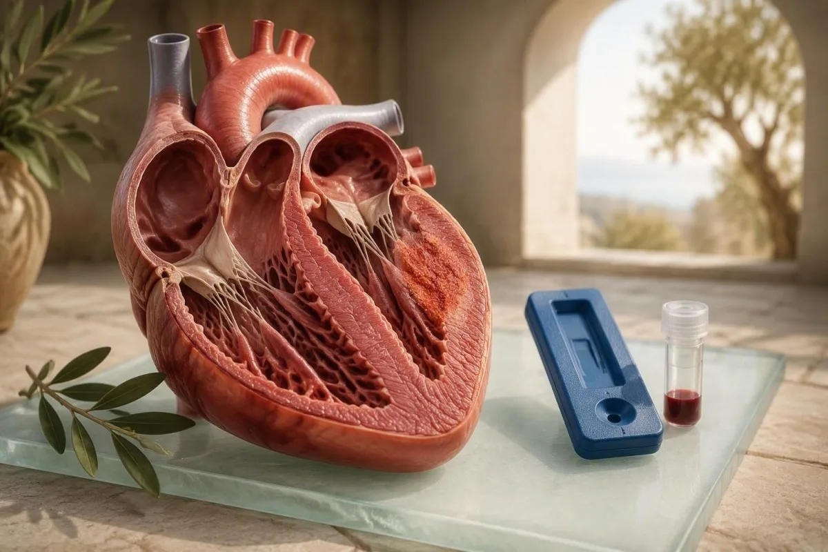
ትሮፖኒን የልብ ጡንቻ ጉዳትን ሊያሳይ ይችላል፣ ነገር ግን የልብ እብጠት (myocarditis) መንስኤ መሆኑን ማረጋገጥ አይችልም። ምርመራው የሚገኘው የደም ምርመራ ውጤቶችን ከምልክቶች፣ ከ ECG ግኝቶች፣ ከኤኮካርዲዮግራፊ እና በአብዛኛው ከልብ MRI ጋር በማዛመድ ነው።.


- ትሮፖኒን ከላቦራቶሪው 99ኛ-ፐርሰንታይል የላይኛው የማጣቀሻ ወሰን በላይ የሆነ የልብ ጡንቻ ጉዳት ያሳያል እንጂ የልብ እብጠት (myocarditis) የተለየ ምርመራ አይደለም።.
- ከፍተኛ-ስሜታዊ ትሮፖኒን በቀላል ወይም ዘግይቶ በሚመጣ የልብ እብጠት (myocarditis) መደበኛ ሊሆን ይችላል፣ ስለዚህ መደበኛ ውጤት ሙሉ በሙሉ አያገለለውም።.
- ሲአርፒ ከ 10 mg/L በላይ የሆነ ንባብ ንቁ የሆነ ስርአታዊ እብጠትን ይደግፋል ነገር ግን በልብ ውስጥ ያለውን እብጠት በተለይ መለየት አይችልም።.
- ተከታታይ ምርመራ (Serial testing) አስፈላጊ ነው፡ ከ1-3 ሰአታት በላይ ያለው የትሮፖኒን መጨመር ወይም መቀነስ ከነጠላ ውጤት የበለጠ መረጃ ሰጭ ነው።.
- የልብ MRI ምልክቶች፣ ECG እና ትሮፖኒን ሲጨነቁ የልብ እብጠት (myocarditis) ለመደገፍ ጥቅም ላይ የሚውለው ዋናው ወራሪ ያልሆነ ምርመራ ነው።.
- የልብ ወሳጅ ቧንቧ በሽታ በአዋቂዎች ላይ የደረት ሕመም እና የጨመረ ትሮፖኒን ታማሚዎች፣ በተለይም ከ40 ዓመት በኋላ ወይም የቫስኩላር ስጋት ምክንያቶች ባሉበት ጊዜ ግምት ውስጥ መግባት አለበት።.
- አስቸኳይ ግምገማ የደረት ግፊት፣ እረፍት ላይ የትንፋሽ ማጠር፣ ራስን መሳት፣ የቆየ የልብ ምት መዛባት፣ ወይም ከጭንቀት ምልክቶች ጋር የጨመረ ትሮፖኒን ያስፈልገዋል።.
የልብ እብጠት (myocarditis) የደም ምርመራ የልብ እብጠትን ሊመረምር ይችላል?
ምንም ነጠላ የ myocardiitis የደም ምርመራ myocardiitis ን ለመመርመር ወይም ለማግለል አይችልም።. ትሮፖኒን የልብ ጡንቻን ጉዳት ያሳያል፣ CRP እና ESR ደግሞ በሰውነት ውስጥ በየትኛውም ቦታ እብጠትን ያመለክታሉ፤ ሁለቱም መንስኤውን አያረጋግጡም። እንደ ዶክተር ቶማስ ክላይን፣ የደም ምርመራ ምርመራውን እንደሚጀምር ለታካሚዎች እገልጻለሁ፣ ነገር ግን የልብ MRI እና ክሊኒካዊው ምስል ብዙውን ጊዜ ያረጋግጡታል።.
A የ myocardiitis የደም ምርመራ ደጋፊ ማስረጃ እንጂ ፍርድ አይደለም። ትሮፖኒን በ myocardiitis፣ በልብ ድካም፣ ፈጣን arrhythmia፣ pulmonary embolism፣ ከባድ የደም ግፊት፣ የኩላሊት በሽታ፣ sepsis፣ ወይም ከፍተኛ የጽናት የአካል ብቃት እንቅስቃሴ ምክንያት ሊጨምር ይችላል። የ2013 የአውሮፓ የልብ ማህበር የሥልጣን መግለጫ myocardiitis ን ከመologiche እሴት ከሚገኝ ይልቅ እንደ ክሊኒኮፓቶሎጂካል ሁኔታ ይገልጸዋል። ለ assay ልዩነት ቀላል-ቋንቋ ግምገማ ይመልከቱ የ troponin I እና T ውጤቶች.
በእኔ ክሊኒካዊ ልምድ፣ በጣም አሳሳች ሁኔታው የቫይረስ በሽታ ካለበት በኋላ ከፍተኛ የደረት ህመም ያለበት ወጣት ጎልማሳ እና በተመጣጣኝ ሁኔታ የጨመረ ትሮፖኒን ነው። ያ ሁኔታ myocardiitis ን ሊያመለክት ይችላል፣ ነገር ግን pericarditis, coronary spasm, ወይም ከባድ የአካል ብቃት እንቅስቃሴ በኋላ ያልተገናኘ የ troponin መጨመር ሊሆን ይችላል። hs-cTnT 38 ng/L ያለው 52 ዓመት የሆነ ሯጭ ማራቶንን ተከትሎ ከ 38 ng/L ፣ ትኩሳት ፣ አዲስ የትንፋሽ እጥረት እና ECG ለውጦች ጋር 22 ዓመት የሆነ ጎልማሳ የተለየ ውይይት ይፈልጋል።.
ካንቴስቲ እ.ኤ.አ. AI የደም ምርመራ ተንታኝ ትሮፖኒንን ከኩላሊት ተግባር፣ ከእብጠት ጠቋሚዎች፣ ከጉበት ኢንዛይሞች እና ከቀዳሚ ውጤቶች ጋር የሚያነብ እንጂ አንድ የታወቀ እሴትን እንደ ምርመራ አድርጎ የማይቆጥር ነው። የእኛ ስርዓት ለክሊኒሽያን ንድፉን ለማደራጀት ሊረዳ ይችላል፣ ነገር ግን የአስቸኳይ ጊዜ ግምገማን፣ ECG ን፣ ወይም ምስልን መተካት አይችልም። ተግባራዊ ህግ፡ የደረት ምልክቶች ካሉ፣ የመስመር ላይ ትርጉምን አይጠብቁ።.
የልብ እብጠት (myocarditis) ትሮፖኒን መጠን ምን ማለት ነው?
ከ assay-specific የላይኛው የማጣቀሻ ወሰን በላይ ያለው ትሮፖኒን የልብ-ጡንቻ ጉዳት መኖሩን ያሳያል፣ ነገር ግን myocardiitis ን መንስኤውን አይለይም።. አብዛኛዎቹ ከፍተኛ-sensitive assays የጤናማ የማጣቀሻ ህዝብ የ99ኛውን ፐርሰንትይል ይጠቀማሉ፣ በተለምዶ ለ hs-cTnT ወደ 14 ng/L እና ለ hs-cTnI ደግሞ እንደ አምራቹ ከ4-20 ng/L አካባቢ ነው።.
ዋጋው ከ hs-cTnT 16 ng/L ከአንድ ላቦራቶሪ ገደብ ትንሽ በላይ ሊሆን ይችላል፣ በዚያን ጊዜ hs-cTnI 1,500 ng/L በግልጽ ከፍተኛ ነው፤ ሁለቱም ቁጥሮች ብቻውን የ myocardiitis ክብደትን አይመዘግቡም። ላቦራቶሪዎች የ troponin I እና troponin T እሴቶችን በደህና መለዋወጥ አይችሉም ምክንያቱም their assays የተለያዩ ፀረ እንግዳ አካላትን፣ ማስተካከያዎችን እና የማጣቀሻ ገደቦችን ይጠቀማሉ። የላቦራቶሪ ሪፖርቱ የራሱ የላይኛው የማጣቀሻ ገደብ አስፈላጊው ቁጥር ነው።.
የጊዜ አካሄድ ከከፍተኛው ይልቅ ብዙ ጊዜ የክሊኒካዊ መረጃን ይይዛል። ትሮፖኒን በተለምዶ ከ2-4 ሰዓታት በኋላ ከአጣዳፊ ጉዳት በኋላ መነሳት ይጀምራል፣ እና ክሊኒኮች በተደጋጋሚ ከ1-3 ሰዓታት በኋላ በአስቸኳይ መንገዶች ያድሱታል። ግልጽ የሆነ ጭማሪ ወይም ውድቀት የአጣዳፊ ሂደትን ይደግፋል፤ የተረጋጋ ቀላል ጭማሪ በከባድ የኩላሊት በሽታ፣ በመዋቅር የልብ ህመም፣ ወይም ለረጅም ጊዜ በልብ ላይ በሚደርስ ውጥረት ሊከሰት ይችላል። የ የደረት ህመም የደም ምርመራ መመሪያ ተከታታይ ናሙናዎች ለምን ጥቅም ላይ እንደሚውሉ ያብራራል።.
መደበኛ ትሮፖኒን myocardiitis ን በአስተማማኝ ሁኔታ አያግልም። ትናንሽ የቲሹ ምላሽ አካባቢዎች፣ ከከፍተኛው በኋላ የዘገየ ምርመራ፣ እና አነስተኛ-sensitive assays ሁሉም መደበኛ ውጤት ሊሰጡ ይችላሉ። በተቃራኒው፣ በጣም ከፍተኛ እሴቶች ወዲያውኑ ዘላቂ ጉዳትን አያመለክቱም—አጣዳፊ myocardiitis ያለባቸው አንዳንድ ሕመምተኞች አስደናቂ ጭማሪዎች ያሏቸው ቢሆንም መደበኛ የመpump ተግባር ያገግማሉ።.
ትሮፖኒን በልብ እብጠት (myocarditis) እና በሌሎች ሁኔታዎች ለምን ይነሳል
Troponin rises when cardiac muscle cells release intracellular proteins after injury or intense cellular stress. Myocarditis is one cause, but coronary blockage remains the diagnosis clinicians must urgently exclude when chest pain and raised troponin occur together.
In myocarditis, immune activity within the myocardium can disrupt cell membranes and release cardiac troponin I or T into circulation. In a heart attack, blocked coronary flow causes the same laboratory signal through a different mechanism. That overlap is why an elevated troponin should never be casually labelled “viral myocarditis” without considering coronary disease, particularly in people over 40 or those with diabetes, smoking exposure, or high ApoB.
Symptoms help but are imperfect. Myocarditis may cause central chest ache, breathlessness, reduced exercise capacity, skipped beats, or no symptoms at all. A person with reproducible pain when pressing the chest wall and a normal ECG is less likely to have myocardial injury, yet this cannot be confirmed remotely. New chest discomfort plus sweating, nausea, breathlessness, or fainting warrants emergency care.
Kantesti የ የደም ምርመራ ባዮማርከር መመሪያ places a troponin result within its assay units and reference interval, which helps avoid a common error: comparing a value in ng/L with an internet cutoff reported in ng/mL. One ng/mL equals 1,000 ng/L; unit confusion can make a result look a thousand-fold more alarming than it is.
CRP, ESR, or a CBC የልብ እብጠት (myocarditis) ያሳያሉ?
CRP, ESR, and white-cell counts can support an inflammatory illness, but none can locate inflammation in the heart. A CRP above 10 mg/L is often considered elevated in routine practice, although the result can rise from a cold, dental infection, autoimmune flare, obesity, or recent intense exercise.
Myocarditis CRP may be normal, mildly raised, or substantially elevated. CRP rises within about 6-12 hours after inflammatory signalling and usually falls faster than ESR when the trigger settles. A CRP of 48 mg/L with fever and chest symptoms adds weight to an inflammatory diagnosis, but it does not distinguish myocarditis from pneumonia, influenza, or bacterial infection. Read more about symptom context in high hs-CRP results.
ESR is slower and less specific. An ESR above 20-30 mm/hour may persist for days to weeks because it is influenced by fibrinogen, anaemia, age, pregnancy, and immunoglobulin levels. I rarely use ESR alone to make a decision about suspected myocarditis; it is more useful when checking whether a broad inflammatory pattern has persisted across time.
A CBC can show mild leukocytosis, lymphocyte changes, or normal findings. White cells above 11.0 × 10⁹/L can accompany infection or stress, yet many confirmed myocarditis cases have normal counts. The useful clinical question is whether CRP and WBC disagree—a pattern explored in our WBC versus CRP guide—rather than assuming either confirms cardiac involvement.
CK-MB, CK, LDH, and myoglobin አሁንም ይረዳሉ?
CK-MB, total CK, LDH, and myoglobin are less specific than troponin for heart-muscle injury and cannot diagnose myocarditis. They can occasionally clarify whether strenuous exercise or generalized skeletal-muscle injury is contributing to an abnormal result.
ሲኬ-ሜባ is found in cardiac tissue but also in skeletal muscle, so a raised result can occur after vigorous exercise, trauma, or muscle inflammation. Some laboratories report a CK-MB index, calculated relative to total CK, but high-sensitivity troponin has largely replaced it for acute myocardial injury assessment. Our explanation of what CK-MB means covers its remaining niche.
ጠቅላላ ክሬቲን ኪናዝ (creatine kinase) can rise dramatically after resistance training, statin-associated muscle injury, seizures, or rhabdomyolysis. A CK of 2,000 IU/L after an unfamiliar endurance event can coexist with a small troponin rise and may point toward skeletal-muscle stress, but clinicians still need to rule out cardiac injury if symptoms are present. CK values above 5,000 IU/L raise concern for significant muscle breakdown and kidney stress.
LDH and myoglobin are even broader markers of tissue turnover. In my practice, they are most helpful when the panel also includes AST, potassium, creatinine, and urine findings—especially after exertion. A high creatine kinase pattern should be interpreted as a muscle-safety issue, not casually attributed to myocarditis.
BNP and NT-proBNP በሚጠረጠረው የልብ እብጠት (myocarditis) ላይ ምን ይጨምራሉ
BNP and NT-proBNP indicate cardiac wall stress and possible heart failure; they do not prove inflammation or myocarditis. NT-proBNP below 125 ng/L is often used in stable outpatient adults to make chronic heart failure less likely, while acute-care thresholds are higher and age-dependent.
Myocarditis can impair the heart’s pumping or relaxation function, causing BNP ወይም NT-proBNP to rise as cardiac chambers stretch. A raised NT-proBNP alongside breathlessness, ankle swelling, a fast heart rate, and an abnormal echocardiogram is more concerning than the same value in isolation. Kidney impairment, atrial fibrillation, older age, and pulmonary embolism can also raise NT-proBNP.
A low natriuretic peptide is reassuring but not absolute. Very early myocarditis, focal inflammation, obesity, and preserved heart function can all yield a normal value. The numerical cutoff must match the setting: an outpatient screening threshold should not be used to dismiss someone with acute breathlessness in an emergency department.
Kantesti AI یو የAI ባዮማርከር ትርጓሜ መድረክ that can show how NT-proBNP, creatinine, sodium, and troponin move together across separate draws. That longitudinal view is useful because a falling NT-proBNP over days may reflect improving haemodynamics, whereas a rising trend merits prompt clinical review. See NT-proBNP cutoffs and symptoms ለውጤት-ተኮር አውድ (context)።.
የኩላሊት፣ የጉበት እና የኤሌክትሮላይት ውጤቶች ንባቡን ለምን ይቀይራሉ
Creatinine, eGFR, potassium, AST, ALT, and sodium can change how clinicians interpret a raised troponin. Kidney disease can produce chronic low-level troponin elevation, while potassium abnormalities can trigger rhythm problems that mimic or complicate myocarditis.
An eGFR below 60 mL/min/1.73 m² is associated with more frequent baseline hs-troponin elevation, especially for troponin T. This does not mean the result should be ignored; clinicians look harder at the delta, symptoms, ECG, and prior baseline. A dynamic increase remains clinically meaningful even when kidney function is reduced.
Potassium below 3.0 mmol/L or above 6.0 mmol/L can increase arrhythmia risk and needs prompt medical attention, especially with palpitations or weakness. Sodium below 130 mmol/L in a person with breathlessness may signal significant fluid imbalance or heart failure, though many non-cardiac illnesses can cause it. The መሠረታዊ የሜታቦሊክ ፓነል መመሪያ explains these tests in plain language.
AST can rise with cardiac or skeletal-muscle injury, whereas ALT is more liver-specific. A disproportionate AST rise after a race may be muscular; AST and ALT elevations with jaundice, bilirubin changes, or medication exposure need a broader liver work-up. Context is everything here—one isolated enzyme almost never tells the whole story.
የቫይረስ ፀረ እንግዳ አካላት (viral antibodies) ወይም የራስ-ሙን (autoimmune) የደም ምርመራዎች መታዘዝ አለባቸው?
Routine viral antibody panels rarely confirm the cause of myocarditis, and positive antibodies often show past exposure rather than active cardiac infection. Autoimmune testing is targeted when the history, examination, or other organs suggest systemic inflammatory disease.
Most adults have antibodies to common viruses such as Epstein-Barr virus, cytomegalovirus, and coxsackievirus. A positive IgG usually reflects previous exposure; it does not prove that a virus is affecting the heart now. Even IgM can be falsely positive or persist longer than expected. The EBV antibody pattern guide shows why antibody timing is tricky.
Clinicians may order ANA, ENA antibodies, ANCA, rheumatoid factor, complement levels, thyroid testing, or eosinophil counts when there are clues such as rash, joint swelling, asthma, sinus disease, kidney changes, recurrent inflammation, or drug exposure. Eosinophilic myocarditis is uncommon but clinically important because it may need urgent specialist-directed treatment.
Cardiac MRI and, in selected unstable or treatment-changing cases, endomyocardial tissue examination are better tools for characterising myocardial involvement than broad viral serology. The evidence is honestly mixed on exactly which ancillary tests every patient needs; the work-up should follow the phenotype, not a fixed shopping list.
የትኞቹ ምርመራዎች የልብ እብጠት (myocarditis) ሊያረጋግጡ ወይም በጠንካራ ሁኔታ ሊደግፉ ይችላሉ?
Cardiac MRI is the leading non-invasive test for supporting myocarditis because it can show oedema and non-ischaemic tissue injury patterns. ECG, echocardiography, coronary assessment when appropriate, and sometimes endomyocardial biopsy complete the diagnostic pathway.
The updated Lake Louise cardiac MRI criteria use at least one T2-based marker of oedema and one T1-based marker of non-ischaemic injury, such as late gadolinium enhancement or abnormal T1 mapping. Ferreira and colleagues reported that combining these tissue markers improves diagnostic assessment compared with older MRI approaches (Ferreira et al., 2018). MRI is strongest when performed close to the symptomatic period, although timing depends on stability and local access.
An ECG may show ST-T changes, PR depression, conduction delay, or rhythm disturbance, but a normal ECG does not exclude myocarditis. Echocardiography evaluates ejection fraction, chamber size, valve function, and pericardial fluid; it can be entirely normal in mild focal disease. A reduced left-ventricular ejection fraction below 50% changes urgency and follow-up planning.
Endomyocardial biopsy is not routine for everyone. It becomes more relevant with cardiogenic shock, rapidly worsening heart failure, high-grade heart block, sustained ventricular arrhythmia, or suspicion of a subtype that would alter treatment. This distinction matters: blood tests can raise suspicion, but only carefully selected tissue analysis can directly identify myocardial inflammatory cell patterns.
የልብ እብጠት (myocarditis) የደም ምርመራዎች መቼ መደገም አለባቸው?
Repeat troponin timing depends on symptoms and setting: emergency pathways commonly repeat high-sensitivity troponin after 1-3 hours, while outpatient follow-up may occur over days or weeks. Repeating a test without a clinical plan can create noise rather than clarity.
For acute chest symptoms, clinicians commonly use an immediate sample and a second sample at 1 or 2 hours, depending on the local validated algorithm. The absolute change in ng/L matters more than a percentage change when starting values are low. Do not use a home-timed repeat test to manage active chest pain—emergency teams need the ECG and vital signs at the same time.
After confirmed or probable myocarditis, troponin may normalise within days to weeks, but recovery is uneven. CRP often improves earlier than MRI abnormalities, and symptoms can improve before electrical irritability has fully settled. I advise patients to record the date, symptoms, exercise level, medications, and illness around each blood draw; our የላቦራቶሪ ጊዜ መመሪያ explains why those details matter.
Dr. Thomas Klein’s practical rule is that a repeat result should answer a specific question: “Is injury continuing?”, “Is this baseline elevation?”, or “Is recovery on track?” A troponin test repeated after heavy exercise, dehydration, or a different assay method may be difficult to compare. Ideally, follow-up uses the same laboratory and assay.
የአካል ብቃት እንቅስቃሴ ትሮፖኒን ያለ የልብ እብጠት (myocarditis) ሊያሳድግ ይችላል?
Yes, prolonged strenuous exercise can cause a temporary troponin rise without clinical myocarditis, particularly after marathons, ultradistance events, or intense cycling. Symptoms, ECG changes, recovery kinetics, and imaging determine whether the elevation is benign exertional release or true myocardial injury.
Post-exercise troponin elevations often peak within 2-6 hours and fall toward baseline within 24-48 hours, whereas sustained or rising values are more concerning. The overlap is not perfect, so symptoms matter enormously. Chest pain, collapse, unusual shortness of breath, or persistent palpitations after exercise should never be dismissed as “just training.”
During suspected myocarditis, vigorous exercise is usually restricted because inflamed myocardium may be more vulnerable to dangerous arrhythmias. Many sports-cardiology protocols reassess athletes after 3-6 ወራት with symptoms, biomarkers, ECG monitoring, ventricular function, and sometimes MRI guiding return. The exact timeline varies with severity and residual findings.
A high CK after exercise can muddy the picture, especially when AST rises too. Our የቆይታ አትሌት የደም ምርመራ መመሪያ helps distinguish training-related lab shifts from patterns that need medical review. Rest is not merely cautious advice here—it is part of risk reduction while the diagnosis is unresolved.
ከፍ ያለ ትሮፖኒን አስቸኳይ እንክብካቤ የሚያስፈልገው መቼ ነው
A raised troponin with active chest pressure, breathlessness at rest, fainting, confusion, sustained palpitations, or new weakness needs emergency assessment now. The result may reflect myocarditis, heart attack, pulmonary embolism, severe arrhythmia, or another condition that cannot be safely sorted out online.
Call emergency services rather than driving yourself if chest symptoms are severe, persistent beyond 10-15 minutes, accompanied by collapse, or associated with blue lips, severe breathlessness, or a racing irregular heartbeat. Troponin is not a measure of pain intensity; a person can have a dangerous condition with a modest elevation or little pain.
There are quieter warning patterns too: resting heart rate persistently above 120 beats per minute, new inability to climb one flight of stairs, swelling, reduced urine output, or palpitations with dizziness. A recent viral illness does not make a cardiac cause less urgent. In fact, it can increase suspicion for myocarditis while coronary and pulmonary causes still require consideration.
If you have a result but no current emergency symptoms, contact the clinician who ordered it the same day when possible. The high troponin urgent-care guide lists common non-heart-attack causes, but it should not be used to self-triage dangerous symptoms.
የልብ እብጠት (myocarditis) የደም ፓነልን ያለ ከመጠን በላይ መተርጎም እንዴት ማንበብ ይቻላል
The safest way to read blood tests for heart inflammation is to look for a pattern: troponin trajectory, CRP, kidney function, electrolytes, symptoms, timing, and prior values. A single red flag on a report is a prompt for clinical context, not a final diagnosis.
ካንቴስቲ እ.ኤ.አ. በ AI የተጎላበተ የደም ምርመራ ትንተና መሳሪያ designed to compare laboratory values across time and highlight combinations that deserve clinician follow-up. For suspected myocarditis, our AI does not label someone with the condition from troponin alone; it identifies myocardial-injury markers, inflammatory context, and potentially relevant kidney or electrolyte confounders. That is deliberately conservative.
Before reviewing any report, confirm four details: the test name, units, laboratory upper reference limit, and sampling date. Then note whether strenuous exercise, fever, dehydration, kidney disease, supplements, or a recent hospital visit could affect the result. The የደም ንጽጽር መሳሪያ is particularly useful when comparing values obtained by the same laboratory.
A healthy-looking CRP or normal CBC should not overrule concerning symptoms, and a flagged troponin should not be interpreted without an ECG. This is one of those areas where context matters more than the number. If you use Kantesti, treat the report as a structured conversation starter for your doctor, not clearance to continue normal activity.
ያልተለመደ የልብ እብጠት (myocarditis) የደም ምርመራ ከተደረገ በኋላ የሚጠየቁ ጥያቄዎች
After abnormal cardiac blood work, ask what the result means for you today, what dangerous alternatives have been excluded, and what test will change management next. A precise question often gets a more useful answer than asking whether the number is simply “bad.”
Useful questions include: “Which troponin assay was used?”, “What is my change between samples in ng/L?”, “Could my kidney function explain part of this?”, “Do I need ECG, echocardiography, coronary assessment, or cardiac MRI?”, and “What exercise restriction applies while we investigate?” These questions work because they focus on decisions rather than trying to force a diagnosis from a number.
Ask whether repeat testing should occur in the same laboratory and whether any medications or supplements need review. Non-steroidal anti-inflammatory drugs, stimulants, decongestants, bodybuilding products, cocaine, and some cancer therapies can be relevant in selected cases. Never stop prescribed medication without the clinician managing your care.
Kantesti’s clinical content is reviewed with physician oversight; you can read about those standards in our የሕክምና ማረጋገጫ አቀራረብ. As of September 22, 2026, the most reliable approach remains unchanged: troponin identifies injury, while expert clinical assessment and cardiac imaging establish whether myocarditis is the explanation.
ምርምር፣ የህክምና ግምገማ እና የመስመር ላይ ትርጓሜ ገደቦች
Online information can explain a myocarditis blood test, but it cannot assess your ECG, blood pressure, oxygen level, rhythm, or cardiac imaging. Those missing data are exactly why troponin should be interpreted as a clinical signal rather than a standalone diagnosis.
The core evidence base is clear on one point: myocardial injury and myocarditis are not synonyms. Caforio et al. (2013) emphasised the need for integrated clinical assessment, while Ferreira et al. (2018) established modern MRI tissue-characterisation criteria that improve non-invasive diagnosis. Neither paper supports diagnosing myocarditis from CRP, CK-MB, or troponin alone.
For related laboratory context, our research library includes the د سیرم پروټینونو د حوالې لارښود እና complement እና ANA መመሪያ. These tests may be relevant when clinicians suspect systemic immune disease, but they are not screening tests for every person with chest pain or an elevated troponin.
At Kantesti, we aim to make laboratory language less opaque while retaining the uncertainty that safe medicine requires. Our የሕክምና አማካሪ ቦርድ helps guide clinical review standards. If your symptoms are active or worsening, seek in-person care rather than relying on any article, app, or uploaded report.
በተደጋጋሚ የሚጠየቁ ጥያቄዎች
የደም ምርመራ የልብ ጡንቻ መቆጣትን (myocarditis) መለየት ይችላል?
Blood work cannot diagnose myocarditis by itself. Troponin above the assay-specific 99th-percentile upper reference limit shows myocardial injury, while CRP above about 10 mg/L supports systemic inflammation, but neither identifies the cause or location of inflammation. Clinicians combine symptoms, ECG, echocardiography, coronary assessment when appropriate, and cardiac MRI to support the diagnosis. Endomyocardial biopsy is reserved for selected severe or treatment-changing cases.
የ troponin መጠን myocarditis ን ያሳያል?
No troponin cutoff specifically indicates myocarditis because the same value can occur with heart attack, pulmonary embolism, rapid arrhythmia, kidney disease, or exercise. Many hs-cTnT assays use approximately 14 ng/L as the 99th-percentile upper reference limit, while hs-cTnI cutoffs vary widely by assay and sometimes by sex. A rise or fall over 1-3 hours is more informative than one isolated result. Chest symptoms and ECG findings determine urgency.
Myocarditis with normal troponin ሊኖርዎት ይችላል?
Yes, myocarditis can occur with normal troponin, particularly when inflammation is mild, focal, late in its course, or tested after the biomarker peak. A normal troponin makes substantial ongoing myocardial injury less likely at that sampling time, but it does not fully rule out myocarditis. Persistent chest pain, palpitations, fainting, or breathlessness still require medical assessment. Cardiac MRI can detect tissue changes that blood tests miss.
CRP በ myocarditis ከፍ ያለ ነው?
CRP can be high in myocarditis, but it is neither sensitive nor specific for cardiac inflammation. A CRP above 10 mg/L commonly reflects active inflammation and values of 30-100 mg/L can occur in many infections or autoimmune conditions. Some people with MRI-supported myocarditis have normal CRP. CRP is most useful when considered alongside symptoms, troponin, ECG, and changes over time.
ከሚያልፍ የልብ ጡንቻ እብጠት ጋር ትሮፖኒን ለምን ያህል ጊዜ ከፍ ይላል?
Troponin may remain elevated for several days and occasionally longer in myocarditis, depending on the degree and persistence of myocardial injury. High-sensitivity troponin often rises within hours of acute injury, but the exact timing differs between troponin I and T assays and between patients. A falling result usually suggests injury is settling, although it does not by itself confirm recovery of heart function. Follow-up may include ECG, echocardiography, rhythm monitoring, and cardiac MRI.
የትሮፖኒን መጠኔ ከቫይረስ ሕመም በኋላ ከፍ ካለ የአካል ብቃት እንቅስቃሴ ማድረግ አለብኝ?
You should avoid strenuous exercise and seek medical advice if troponin is elevated after a viral illness, especially with chest pain, palpitations, fainting, or breathlessness. Sports-cardiology practice commonly restricts competitive or vigorous exercise for 3-6 months after confirmed myocarditis, with return guided by symptoms, biomarkers, cardiac function, and rhythm assessment. Exercise can transiently raise troponin after endurance events, but that cannot be assumed without evaluation. Active chest symptoms require urgent in-person assessment.
ዛሬ የAI-የኃይል የደም ምርመራ ትንተና ያግኙ
በፍጥነት እና ትክክለኛ የላቦራቶሪ ምርመራ ትንተና ለማግኘት Kantestiን የሚያምኑ ከ2 ሚሊዮን በላይ ተጠቃሚዎችን ይቀላቀሉ። የደም ምርመራ ውጤትዎን ይስቀሉ እና በ15,000+ ባዮማርከሮች ላይ የተሟላ ትርጓሜን በሰከንዶች ውስጥ ይቀበሉ።.
📚 የተጠቀሱ የምርምር ህትመቶች
📖 ውጫዊ የሕክምና ማጣቀሻዎች
⚕️ የሕክምና ማስተባበያ
ይህ ጽሑፍ ለትምህርታዊ ዓላማ ብቻ ነው እና የሕክምና ምክር አይደለም። ለምርመራ እና ለሕክምና ውሳኔዎች ሁልጊዜ ብቁ የጤና ባለሙያን ያማክሩ።.
የE-E-A-T እምነት ምልክቶች
ልምድ
በሐኪም መሪነት የላቦራቶሪ ትርጓሜ የስራ ፍሰቶች ክሊኒካዊ ግምገማ።.
ባለሙያነት
በክሊኒካዊ አውድ ውስጥ ባዮማርከሮች እንዴት እንደሚሰሩ ላይ የላቦራቶሪ ሕክምና ትኩረት።.
ስልጣን ያለው
በዶክተር ቶማስ ክላይን የተፃፈ ከዶክተር ሳራ ሚቸል እና ፕሮፌሰር ዶክተር ሃንስ ዌበር ግምገማ ጋር።.
አስተማማኝነት
ለማስጠንቀቂያ ምላሽ መቀነስ ግልጽ የቀጣይ መንገዶች ያለው በማስረጃ የተደገፈ ትርጓሜ።.