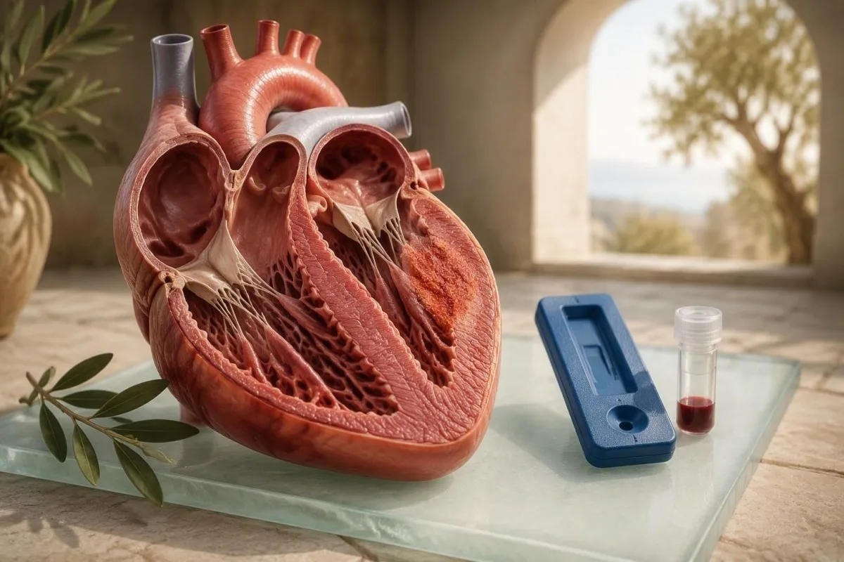
ට්රොපොනින් හෘද පේශි වලට වන හානිය හඳුනාගත හැකි නමුත්, එය මයෝකාඩයිටිස් (myocarditis) රෝගයට හේතුව බවට ඔප්පු කළ නොහැකිය. රෝග විනිශ්චය පැමිණෙන්නේ රුධිර පරීක්ෂණ ප්රතිඵල, රෝග ලක්ෂණ, ECG සොයාගැනීම්, echocardiography සහ සාමාන්යයෙන් cardiac MRI සමඟ සැසඳීමෙනි.


- ට්රොපොනින් රසායනාගාරයේ 99 වන ප්රතිශතයේ ඉහළ යොමු සීමාවට ඉහළින් හෘද පේශි වලට වන හානියක් පෙන්නුම් කරයි, මයෝකාඩයිටිස් රෝග විනිශ්චයක් නොවේ.
- අධි-සංවේදී troponin සැහැල්ලු හෝ ප්රමාද වී ඉදිරිපත් වන මයෝකාඩයිටිස් වලදී සාමාන්ය විය හැක, එබැවින් සාමාන්ය ප්රතිඵලයක් එය සම්පූර්ණයෙන්ම බැහැර නොකරයි.
- සී.ආර්.පී. 10 mg/L ට වඩා ඉහළින් ක්රියාකාරී පද්ධතිමය දැවිල්ලට සහාය දක්වයි නමුත් හදවතේ දැවිල්ල නිශ්චිතව හඳුනාගත නොහැකිය.
- අනුක්රමික පරීක්ෂණ (Serial testing) වැදගත් වේ: 1-3 පැය තුළ ඉහළ යන හෝ පහළ යන ට්රොපොනින් රටාවක් තනි ප්රතිඵලයකට වඩා තොරතුරු සපයයි.
- Cardiac MRI රෝග ලක්ෂණ, ECG, සහ ට්රොපොනින් කනස්සල්ලක් ඇති කරන විට මයෝකාඩයිටිස් වලට සහාය දැක්වීමට භාවිතා කරන ප්රධාන ආක්රමණශීලී නොවන පරීක්ෂණයයි.
- Coronary disease පපුවේ වේදනාව සහ ඉහළ ගිය ට්රොපොනින් සහිත වැඩිහිටියන් තුළ, විශේෂයෙන් වයස 40 න් පසු හෝ සනාල අවදානම් සාධක සහිතව සලකා බැලිය යුතුය.
- හදිසි ඇගයීමක් පපුවේ පීඩනය, විවේකයේදී හුස්ම හිරවීම, ක්ලාන්ත වීම, අඛණ්ඩ ස්පන්දන, හෝ ආබාධිත රෝග ලක්ෂණ සහිත ඉහළ ට්රොපොනින් සඳහා අවශ්ය වේ.
හෘදයේ දැවිල්ලට මයෝකාඩයිටිස් රුධිර පරීක්ෂණය මගින් රෝග විනිශ්චය කළ හැකිද?
Myocarditis රෝග විනිශ්චය කිරීමට හෝ බැහැර කිරීමට තනි රුධිර පරීක්ෂණයක් නොමැත. Troponin හෘද පේශිවලට සිදුවන හානිය හඳුනාගන්නා අතර, CRP සහ ESR ශරීරයේ කොතැනක හෝ දැවිල්ල හඳුනාගනී; කිසිවක් හේතුව තහවුරු නොකරයි. ආචාර්ය තෝමස් ක්ලයින් ලෙස, මම රෝගීන්ට පවසන්නේ රුධිර පරීක්ෂාවෙන් පරීක්ෂණය ආරම්භ වන බවයි, නමුත් හෘද MRI සහ සායනික පින්තූරය සාමාන්යයෙන් එය තහවුරු කරයි.
A myocarditis රුධිර පරීක්ෂාව සහායක සාක්ෂි මිස තීන්දුවක් නොවේ. Troponin myocarditis, හෘදයාබාධ, වේගවත් අරිද්මික්, පු pulmpmonary embolism, දැඩි අධි රුධිර පීඩනය, වකුගඩු රෝගය, සෙප්සිස්, හෝ දැඩි විඳදරාගැනීමේ අභ්යාස නිසා ඉහළ යා හැකිය. 2013 යුරෝපීය කාඩියෝලොජි සංගමයේ තත්ත්ව ප්රකාශය myocarditis විස්තර කරන්නේ එක් රසායනාගාර අගයකින් (Caforio et al., 2013) කළ රෝග විනිශ්චයක් වෙනුවට සායනික රෝග විද්යාත්මක තත්වයක් ලෙසය. Assay වෙනස්කම් වල භාෂා-සාමාන්ය සමාලෝචනයක් සඳහා, බලන්න troponin I සහ T ප්රතිඵල.
මගේ සායනික අත්දැකීම්වල, වඩාත් වැරදි ලෙස මෙහෙයවන අවස්ථාව වන්නේ, වෛරස් ආසාදනයකින් පසු තරුණ වැඩිහිටියෙකුට තියුණු පපුවේ අපහසුතාවයක් සහ මධ්යස්ථව ඉහළ ගිය troponin එකක් තිබීමයි. එම රටාව myocarditis සඳහා සුදුසු විය හැකිය, නමුත් එය pericarditis, coronary spasm, හෝ දෘඩ ව්යායාමයකින් පසු සම්බන්ධයක් නැති troponin ඉහළ යාමද විය හැකිය. 40 ත් 38 ත් අතර වයසේ පසුවන 52 හැවිරිදි ධාවකයෙකු, මැරතෝනයකින් පසු hs-cTnT 38 ng/L සමඟ, 22 හැවිරිදි 38 ng/L, උණ, නව හුස්ම හිරවීම සහ ECG වෙනස්වීම් ඇති අයෙකුගෙන් වෙනස් සංවාදයක් අවශ්ය වේ.
කන්ටෙස්ටි යනු AI රුධිර පරීක්ෂණ විශ්ලේෂකය එය රෝග විනිශ්චයක් ලෙස එක් ලකුණු කළ අගයක් සලකනවා වෙනුවට වකුගඩු ක්රියාකාරිත්වය, දැවිල්ල සලකුණු, අක්මා එන්සයිම සහ පෙර ප්රතිඵල සමඟ troponin කියවීම් සිදු කරයි. අපගේ පද්ධතිය වෛද්යවරයෙකු සඳහා රටාව සංවිධානය කිරීමට උපකාර කළ හැකිය, නමුත් එය හදිසි ඇගයීම, ECG, හෝ රූපවාහිනිය ප්රතිස්ථාපනය කළ නොහැක. ප්රායෝගික රීතිය: පපුවේ රෝග ලක්ෂණ පවතී නම්, මාර්ගගත අර්ථ නිරූපණය සඳහා රැඳී නොසිටින්න.
මයෝකාඩයිටිස් හි ට්රොපොනින් මට්ටම් ඇත්ත වශයෙන්ම අදහස් කරන්නේ කුමක්ද?
Assay-specific upper reference limit ට වඩා Troponin ඉහළින් තිබීම හෘද මාංශපේශී තුවාලයක් ඇති බවට ලකුණකි, නමුත් එය myocarditis හේතුව ලෙස හඳුනා නොගනී. බොහෝ අධි-සංවේදී Assay වල සෞඛ්ය සම්පන්න යොමු ජනගහනයක 99 වන ප්රතිශතය භාවිතා කරයි, සාමාන්යයෙන් hs-cTnT සඳහා 14 ng/L පමණ වන අතර, නිෂ්පාදකයා අනුව hs-cTnI සඳහා 4-20 ng/L පමණ වේ.
අගයක් hs-cTnT 16 ng/L එක රසායනාගාරයක කප්-ඕෆ් එකට වඩා තරමක් ඉහළින් තිබිය හැකිය, අතර hs-cTnI 1,500 ng/L පැහැදිලිවම ඉහළය; කිසිම අගයක් පමණක් myocarditis වල බරපතලකම ශ්රේණිගත නොකරයි. රසායනාගාර වෙනස් antibodies, calibrations, සහ යොමු සීමාවන් භාවිතා කරන නිසා රසායනාගාරවලට troponin I සහ troponin T අගයන් ආරක්ෂිතව හුවමාරු කළ නොහැක. රසායනාගාර වාර්තාවේ තමන්ගේම ඉහළ යොමු සීමාව අදාළ වන අගයයි.
peak එකට වඩා කාලගතිකය බොහෝ විට වැඩි සායනික තොරතුරු දරයි. Troponin සාමාන්යයෙන් උග්ර තුවාලයකින් පැය 2-4 කට පසුව ඉහළ යාමට පටන් ගනී, සහ වෛද්යවරුන් හදිසි මාර්ගවල පැය 1-3 කට පසුව එය නැවත නැවතත් කරයි. පැහැදිලි නැගීමක් හෝ පහත වැටීමක් උග්ර ක්රියාවලියකට සහාය වන අතර; නිදන්ගත වකුගඩු රෝගය, ආගන්තුක හෘද රෝග, හෝ දිගුකල් පැවතෙන හෘදයාබාධ සමඟ ස්ථායර මෘදු ඉහළ යාමක් සිදුවිය හැකිය. የደረት ህመም የደም ምርመራ መመሪያ මාලා සාම්පල භාවිතා කරන්නේ මන්දැයි පැහැදිලි කරයි.
සාමාන්ය troponin myocarditis විශ්වාසදායක ලෙස බැහැර නොකරයි. පටක ප්රතිචාරයේ කුඩා නාභිගත ප්රදේශ, peak එකෙන් පසුව ප්රමාද වූ පරීක්ෂණය, සහ අඩු සංවේදී Assay සියල්ල සාමාන්ය ප්රතිඵලයක් ලබා දිය හැකිය. ඊට වෙනස්ව, ඉතා ඉහළ අගයන් අනිවාර්යයෙන්ම ආපසු හැරවිය නොහැකි හානියක් අදහස් නොකරයි - උග්ර myocarditis ඇති සමහර රෝගීන් කැපී පෙනෙන ඉහළ යාමක් ඇති නමුත් සාමාන්ය පොම්ප කිරීමේ ක්රියාකාරිත්වය යථා තත්ත්වයට පත් කරයි.
මයෝකාඩයිටිස් සහ වෙනත් රෝග තත්ව වලදී ට්රොපොනින් ඉහළ යන්නේ ඇයි?
Troponin ඉහළ යන්නේ හෘද මාංශ පේශි සෛල තුවාල වීමෙන් හෝ දැඩි සෛලීය ආතතියෙන් පසු සෛලීය ප්රෝටීන නිකුත් කරන විටය. හෘද මාංශයෙහි දැවිල්ල එක් හේතුවක් වන නමුත්, පපුවේ වේදනාව සහ ඉහළ ගිය ට්රොපොනින් එකට ඇති වූ විට, ධමනි අවහිර වීම රෝග විනිශ්චය ලෙස වෛද්යවරු විසින් හදිසි ලෙස බැහැර කළ යුතුය.
හෘද මාංශයෙහි දැවිල්ලෙහිදී, හෘද මාංශ පේශි තුළ ප්රතිශක්තිකරණ ක්රියාකාරිත්වය සෛල පටල බාධා කර මුදා හැරිය හැක හෘද ට්රොපොනින් I හෝ T සංසරණයට. හෘදයාබාධයකදී, අවහිර වූ ධමනි ප්රවාහය විවිධ යාන්ත්රණයක් හරහා එකම රසායනාගාර සංඥාව ඇති කරයි. එම අතිච්ඡාදනය නිසා, කොරොනරි රෝගය, විශේෂයෙන් 40 ට වැඩි පුද්ගලයින් තුළ හෝ දියවැඩියාව, දුම්පානයට නිරාවරණය වීම හෝ ඉහළ ApoB ඇති අය තුළ නොසලකා හැරිය නොහැකිව ඉහළ ගිය ට්රොපොනින් “වෛරස් හෘද මාංශයෙහි දැවිල්ල” ලෙස කිසි විටෙකත් අහඹු ලෙස නම් කළ නොහැක.
රෝග ලක්ෂණ උපකාරී වන නමුත් පරිපූර්ණ නොවේ. හෘද මාංශයෙහි දැවිල්ල මධ්යම පපුවේ වේදනාව, හුස්ම හිරවීම, ව්යායාම ධාරිතාව අඩුවීම, ස්පන්දනය මග හැරීම හෝ කිසිදු රෝග ලක්ෂණයක් නොමැතිව සිදුවිය හැක. පපුවේ බිත්තියට තද කරන විට පුනරාවර්තන වේදනාව සහ සාමාන්ය ECG ඇති පුද්ගලයෙකුට හෘද මාංශ පේශි තුවාලයක් ඇති වීමට ඇති ඉඩකඩ අඩුය, එහෙත් මෙය දුරස්ථව තහවුරු කළ නොහැක. නව පපුවේ අපහසුතාවය, දහඩිය දැමීම, ඔක්කාරය, හුස්ම හිරවීම හෝ සිහිය නැතිවීම හදිසි ප්රතිකාර සඳහා සුදුසු වේ.
Kantesti’s රුධිර පරීක්ෂණ ජෛව සලකුණු මාර්ගෝපදේශය එහි ඇසසි යුනිට් සහ යොමු පරතරය තුළ ට්රොපොනින් ප්රතිඵලය ස්ථානගත කරයි, එය පොදු වැරදීමක් මඟවා ගැනීමට උපකාරී වේ: ng/L හි අගයක් ng/mL හි වාර්තා කරන ලද අන්තර්ජාල කප්-ඕෆ් එකක් සමඟ සංසන්දනය කිරීම. එක ng/mL යනු 1,000 ng/L ට සමාන වේ; ඒකක ව්යාකූලත්වය ප්රතිඵලය එයට වඩා දහස් ගුණයකින් භයානක ලෙස පෙනීමට හේතු විය හැක.
CRP, ESR, හෝ CBC මගින් මයෝකාඩයිටිස් පෙන්විය හැකිද?
CRP, ESR, සහ සුදු රුධිරාණු ගණන් දැවිල්ල ඇති කරන රෝගයකට සහාය විය හැකි නමුත්, කිසිවක් හදවතේ දැවිල්ල හඳුනාගත නොහැක. 10 mg/L ට වඩා වැඩි CRP සාමාන්ය පිළිවෙතේදී ඉහළ ගිය එකක් ලෙස සලකනු ලැබේ, කෙසේ වෙතත් ප්රතිඵලය සීතල, දන්ත ආසාදනය, ස්වයංක්රීය ප්රතිශක්තිකරණ ආබාධ, තරබාරුකම හෝ මෑතකදී දැඩි ව්යායාම වලින් ඉහළ යා හැක.
Myocarditis CRP සාමාන්ය, මදක් ඉහළ ගිය හෝ සැලකිය යුතු ලෙස ඉහළ ගිය එකක් විය හැක. CRP ආසන්න වශයෙන් 6-12 පැය තුළ දැවිල්ල සංඥා කිරීමෙන් පසු ඉහළ යන අතර, ට්රිගර් එක පදිංචි වූ විට ESR ට වඩා වේගයෙන් පහත වැටේ. 48 mg/L හි CRP උණ සහ පපුවේ රෝග ලක්ෂණ සමඟ දැවිල්ල ඇති රෝග විනිශ්චයට බරක් එක් කරයි, නමුත් එය pneumonia, influenza, හෝ bacterial infection වලින් myocarditis වෙන් කරන්නේ නැත. රෝග ලක්ෂණ සන්දර්භය පිළිබඳව වැඩිදුර කියවන්න ඉහළ hs-CRP ප්රතිඵල.
ESR මන්දගාමී හා අඩු නිශ්චිත ය. පැයකට 20-30 mm ට වඩා වැඩි ESR දින සිට සති දක්වා පැවතිය හැක, මන්ද එය ෆයිබ්රිනොජන්, රක්තහීනතාවය, වයස, ගැබ් ගැනීම සහ ඉමියුනොග්ලොබුලින් මට්ටම් වලින් බලපෑමට ලක් වේ. සැක සහිත හෘද මාංශයෙහි දැවිල්ල පිළිබඳ තීරණයක් ගැනීමට මම කලාතුරකින් ESR පමණක් භාවිතා කරමි; පුළුල් දැවිල්ල රටාවක් කාලය පුරා පැවතියේදැයි පරීක්ෂා කරන විට එය වඩාත් ප්රයෝජනවත් වේ.
CBC මගින් මදක් ලියුකෝසයිටෝසිස්, ලිම්ෆොසයිට් වෙනස්කම් හෝ සාමාන්ය සොයාගැනීම් පෙන්විය හැකිය. 11.0 × 10⁹/L ට වඩා වැඩි සුදු රුධිරාණු ආසාදනය හෝ ආතතිය සමඟ එක්විය හැකි නමුත්, තහවුරු කරන ලද හෘද මාංශයෙහි දැවිල්ලෙහි බොහෝ අවස්ථා වල සාමාන්ය ගණන් ඇත. ප්රයෝජනවත් සායනික ප්රශ්නය වන්නේ CRP සහ WBC එකඟ නොවේද—අපේ WBC එදිරිව CRP මාර්ගෝපදේශය— හි ගවේෂණය කරන ලද රටාවක්, මිස කිසිවක් හෘද සම්බන්ධතාවය තහවුරු කරන බවට උපකල්පනය කිරීම නොවේ.
CK-MB, CK, LDH, සහ myoglobin තවමත් උපකාරී වේද?
CK-MB, මුළු CK, LDH, සහ myoglobin හෘද මාංශ පේශි තුවාල සඳහා troponin ට වඩා අඩු නිශ්චිත වන අතර myocarditis රෝග විනිශ්චය කළ නොහැක. ඒවා ඉඳහිට අධික ව්යායාම හෝ සාමාන්යකරණය වූ අස්ථි මාංශ පේශි තුවාලයක් අසාමාන්ය ප්රතිඵලයකට දායක වන්නේද යන්න පැහැදිලි කළ හැකිය.
සීකේ-එම්බී හෘද පටක වල මෙන්ම අස්ථි මාංශ පේශි වලද දක්නට ලැබේ, එබැවින් දැඩි ව්යායාම, කම්පනය හෝ මාංශ පේශි දැවිල්ලෙන් පසු ඉහළ ගිය ප්රතිඵලයක් ඇතිවිය හැකිය. සමහර රසායනාගාර මුළු CK ට සාපේක්ෂව ගණනය කරන ලද CK-MB දර්ශකයක් වාර්තා කරයි, නමුත් ඉහළ සංවේදීතාවයකින් යුත් ට්රොපොනින් විසින් එය උග්ර හෘද තුවාල ඇගයීම සඳහා බොහෝ දුරට එය ප්රතිස්ථාපනය කර ඇත. අපගේ පැහැදිලි කිරීම CK-MB යනු කුමක්දැයි එහි ඉතිරි ස්ථානය ආවරණය කරයි.
Total ක්රියේටීන් කිනාස් (creatine kinase) ප්රතිරෝධක පුහුණුව, ස්ටැටින්-ශාරීරික මාංශ පේශි තුවාල, අල්ලා ගැනීම් හෝ රුධිරය කැටිගැසීමෙන් පසු නාටකාකාර ලෙස ඉහළ යා හැකිය. නොදන්නා විඳදරාගැනීමේ සිදුවීමකින් පසු 2,000 IU/L හි CK එකක් කුඩා ට්රොපොනින් ඉහළ යාමක් සමඟ එකට පැවතිය හැකි අතර, ඇටසැකිලි-මාංශ පේශි ආතතියක් පෙන්නුම් කළ හැකිය, නමුත් රෝග ලක්ෂණ තිබේ නම් හෘද තුවාලයක් බැහැර කිරීමට වෛද්යවරුන්ට තවමත් අවශ්ය වේ. 5,000 IU/L ට වඩා වැඩි CK අගයන් සැලකිය යුතු මාංශ පේශි බිඳවැටීම සහ වකුගඩු ආතතිය පිළිබඳ සැලකිල්ලක් දක්වයි.
LDH සහ මයෝග්ලොබින් පටක හැරවුම් වල තවත් පුළුල් සලකුණු වේ. මගේ පුරුද්දේදී, පැනලයට AST, පොටෑසියම්, ක්රියේටිනින් සහ මුත්රා සොයාගැනීම් - විශේෂයෙන් ශ්රමය පසු - අතිරේකව පවතින විට ඒවා වඩාත් ප්රයෝජනවත් වේ. A ඉහළ ක්රියේටීන් කිනේස් රටාවක් හෘද මාංශපේශි දැවිල්ලට හේතුවක් නොවන, මාංශ පේශි-ආරක්ෂිත ගැටලුවක් ලෙස අර්ථ දැක්විය යුතුය.
BNP සහ NT-proBNP සැක සහිත මයෝකාඩයිටිස් වලට එකතු කරන්නේ මොනවාද?
BNP සහ NT-proBNP හෘද බිත්ති ආතතිය සහ හෘදයාබාධයක් පෙන්නුම් කරයි; ඒවා දැවිල්ලක් හෝ හෘද මාංශපේශි දැවිල්ලක් ඔප්පු නොකරයි. NT-proBNP 125 ng/L ට වඩා අඩු අගය බොහෝ විට ස්ථාවර බාහිර රෝගීන් තුළ නිදන්ගත හෘදයාබාධයක් අඩු කිරීමට භාවිතා කරයි, අතර උග්ර-හෘද අරෝහණ ඉහළ සහ වයස්-ගත වේ.
හෘද මාංශපේශි දැවිල්ල හෘදයේ පොම්ප කිරීම හෝ ලිහිල් කිරීමේ කාර්යයට බාධා කළ හැකි අතර, එයට හේතු වේ BNP හෝ NT-proBNP හෘද කුටි දිගු වන විට ඉහළ යයි. හුස්ම හිරවීම, විලුඹ ඉදිමීම, වේගවත් හෘද ස්පන්දනය සහ අසාමාන්ය ඉකෝකාර්ඩියෝග්රෑම් සමඟ ඉහළ NT-proBNP එකක්, එය තනිව පැවතීමට වඩා සැලකිල්ලක් දක්වයි. වකුගඩු ආබාධ, කර්ණික ෆයිබ්රිලේෂන්, වයස්ගත වීම සහ පු pulmanary embolism ද NT-proBNP ඉහළ යාමට හේතු විය හැක.
අඩු නැටියුරෙටික් පෙප්ටයිඩයක් සැනසීමක් නමුත් සම්පූර්ණ නොවේ. ඉතා මුල් හෘද මාංශපේශි දැවිල්ල, නාභිගත දැවිල්ල, තරබාරුකම සහ සංරක්ෂිත හෘද ක්රියාකාරිත්වය සියල්ල සාමාන්ය අගයක් ලබා දිය හැකිය. සංඛ්යාත්මක කප්-ඕෆ් සැකසුම සමඟ ගැලපිය යුතුය: බාහිර තිරගත කිරීමේ සීමාවක් ගිලන්රථ දෙපාර්තමේන්තුවේ උග්ර හුස්ම හිරවීම ඇති අයව බැහැර කිරීමට භාවිතා නොකළ යුතුය.
Kantesti AI යනු AI biomarker interpretation platform වෙනත් අඳින ලද ස්ථාන හරහා NT-proBNP, ක්රියේටිනින්, සෝඩියම් සහ ට්රොපොනින් එකට ගමන් කරන ආකාරය පෙන්විය හැකිය. එම දිගු දර්ශනය ප්රයෝජනවත් වන්නේ දින ගණනක් පුරා NT-proBNP පහත වැටීම වැඩිදියුණු වූ රක්තහීනතාවයට පිළිබිඹු විය හැකි අතර, නැගී එන ප්රවණතාවක් ඉක්මන් වෛද්ය සමාලෝචනයක් ලැබිය යුතුය. බලන්න NT-proBNP කප්-ඕෆ් සහ රෝග ලක්ෂණ ප්රතිඵල-විශේෂ සන්දර්භය සඳහා.
වකුගඩු, අක්මාව සහ ඉලෙක්ට්රොලයිට් ප්රතිඵල කියවීම වෙනස් කරන්නේ ඇයි?
ක්රියේටිනින්, eGFR, පොටෑසියම්, AST, ALT, සහ සෝඩියම් වෛද්යවරුන් විසින් ඉහළ ට්රොපොනින් අර්ථකථනය කරන ආකාරය වෙනස් කළ හැකිය. වකුගඩු රෝගය නිදන්ගත අඩු-මට්ටමේ ට්රොපොනින් ඉහළ යාමක් ඇති කළ හැකිය, අතර පොටෑසියම් අසාමාන්යතා හෘද මාංශපේශි දැවිල්ලට සමාන හෝ සංකීර්ණ වන රිද්ම ගැටළු අවුලුවාලිය හැකිය.
60 mL/min/1.73 m² ට වඩා අඩු eGFR එකක්, විශේෂයෙන් ට්රොපොනින් T සඳහා, නිතර නිතර බේස්ලයින් hs-troponin ඉහළ යාමක් සමඟ සම්බන්ධ වේ. මෙයින් අදහස් කරන්නේ ප්රතිඵලය නොසලකා හැරිය යුතු බව නොවේ; වෛද්යවරුන් වැඩිපුර දෙස බලන්නේ ඩෙල්ටා, රෝග ලක්ෂණ, ECG, සහ පෙර බේස්ලයින්. වකුගඩු ක්රියාකාරිත්වය අඩු වූ විට පවා ගතික වැඩිවීමක් වෛද්යමය වශයෙන් අර්ථවත් වේ.
3.0 mmol/L ට වඩා අඩු හෝ 6.0 mmol/L ට වඩා වැඩි පොටෑසියම් ධමනි අවදානම වැඩි කළ හැකි අතර, විශේෂයෙන් පැල්පිටේෂන් හෝ දුර්වලතා සමඟ ඉක්මන් වෛද්ය අවධානය අවශ්ය වේ. හුස්ම හිරවීම ඇති පුද්ගලයෙකු තුළ 130 mmol/L ට වඩා අඩු සෝඩියම් සැලකිය යුතු තරල අසමතුලිතතාවයක් හෝ හෘදයාබාධයක් සංඥා කළ හැකිය, නමුත් බොහෝ හෘද නොවන රෝග එයට හේතු විය හැක. The basic metabolic panel මාර්ගෝපදේශය මෙම පරීක්ෂණ සරල භාෂාවෙන් පැහැදිලි කරයි.
AST හෘද හෝ ඇටසැකිලි-මාංශ පේශි තුවාල සමඟ ඉහළ යා හැකිය, අතර ALT වඩා අක්මාව-නිර්දිෂ්ට වේ. තරඟයකින් පසු අසමාන AST ඉහළ යාමක් මාංශපේශී විය හැකිය; කහට, බිලිරුබින් වෙනස්කම්, හෝ ඖෂධ නිරාවරණය සමඟ AST සහ ALT ඉහළ යාමක් පුළුල් අක්මා වැඩ-අප් එකක් අවශ්ය වේ. මෙහි සන්දර්භය සෑම දෙයක්ම වේ - එක් නාභිගත එන්සයිමයක් කලාතුරකින් කතාවක් නොකියයි.
වෛරස් ප්රතිදේහ හෝ ස්වයං ප්රතිශක්ති රුධිර පරීක්ෂණ ඇණවුම් කළ යුතුද?
සාමාන්ය වෛරස් ප්රතිදේහ පැනල් කලාතුරකින් හෘද මාංශපේශි දැවිල්ලට හේතුව තහවුරු කරයි, අතර ධනාත්මක ප්රතිදේහ බොහෝ විට සක්රිය හෘද ආසාදනයකට වඩා අතීත නිරාවරණය පෙන්වයි. ස්වයං-ප්රතිශක්තිකරණ පරීක්ෂාව ඉතිහාසය, පරීක්ෂාව, හෝ වෙනත් අවයව පද්ධතිමය දැවිල්ල රෝගයක් යෝජනා කරන විට ඉලක්ක කරනු ලැබේ.
Most adults have antibodies to common viruses such as Epstein-Barr virus, cytomegalovirus, and coxsackievirus. A positive IgG usually reflects previous exposure; it does not prove that a virus is affecting the heart now. Even IgM can be falsely positive or persist longer than expected. The EBV antibody pattern guide shows why antibody timing is tricky.
Clinicians may order ANA, ENA antibodies, ANCA, rheumatoid factor, complement levels, thyroid testing, or eosinophil counts when there are clues such as rash, joint swelling, asthma, sinus disease, kidney changes, recurrent inflammation, or drug exposure. Eosinophilic myocarditis is uncommon but clinically important because it may need urgent specialist-directed treatment.
Cardiac MRI and, in selected unstable or treatment-changing cases, endomyocardial tissue examination are better tools for characterising myocardial involvement than broad viral serology. The evidence is honestly mixed on exactly which ancillary tests every patient needs; the work-up should follow the phenotype, not a fixed shopping list.
කුමන පරීක්ෂණ මගින් මයෝකාඩයිටිස් තහවුරු කළ හැකිද හෝ ශක්තිමත්ව සහාය දැක්විය හැකිද?
Cardiac MRI is the leading non-invasive test for supporting myocarditis because it can show oedema and non-ischaemic tissue injury patterns. ECG, echocardiography, coronary assessment when appropriate, and sometimes endomyocardial biopsy complete the diagnostic pathway.
The updated Lake Louise cardiac MRI criteria use at least one T2-based marker of oedema and one T1-based marker of non-ischaemic injury, such as late gadolinium enhancement or abnormal T1 mapping. Ferreira and colleagues reported that combining these tissue markers improves diagnostic assessment compared with older MRI approaches (Ferreira et al., 2018). MRI is strongest when performed close to the symptomatic period, although timing depends on stability and local access.
An ECG may show ST-T changes, PR depression, conduction delay, or rhythm disturbance, but a normal ECG does not exclude myocarditis. Echocardiography evaluates ejection fraction, chamber size, valve function, and pericardial fluid; it can be entirely normal in mild focal disease. A reduced left-ventricular ejection fraction below 50% changes urgency and follow-up planning.
Endomyocardial biopsy is not routine for everyone. It becomes more relevant with cardiogenic shock, rapidly worsening heart failure, high-grade heart block, sustained ventricular arrhythmia, or suspicion of a subtype that would alter treatment. This distinction matters: blood tests can raise suspicion, but only carefully selected tissue analysis can directly identify myocardial inflammatory cell patterns.
මයෝකාඩයිටිස් රුධිර පරීක්ෂණ නැවත නැවත කළ යුත්තේ කවදාද?
Repeat troponin timing depends on symptoms and setting: emergency pathways commonly repeat high-sensitivity troponin after 1-3 hours, while outpatient follow-up may occur over days or weeks. Repeating a test without a clinical plan can create noise rather than clarity.
For acute chest symptoms, clinicians commonly use an immediate sample and a second sample at 1 or 2 hours, depending on the local validated algorithm. The absolute change in ng/L matters more than a percentage change when starting values are low. Do not use a home-timed repeat test to manage active chest pain—emergency teams need the ECG and vital signs at the same time.
After confirmed or probable myocarditis, troponin may normalise within days to weeks, but recovery is uneven. CRP often improves earlier than MRI abnormalities, and symptoms can improve before electrical irritability has fully settled. I advise patients to record the date, symptoms, exercise level, medications, and illness around each blood draw; our රසායනාගාර කාලරාමු මාර්ගෝපදේශය (lab timeline guide) explains why those details matter.
Dr. Thomas Klein’s practical rule is that a repeat result should answer a specific question: “Is injury continuing?”, “Is this baseline elevation?”, or “Is recovery on track?” A troponin test repeated after heavy exercise, dehydration, or a different assay method may be difficult to compare. Ideally, follow-up uses the same laboratory and assay.
මයෝකාඩයිටිස් නොමැතිව ව්යායාම ට්රොපොනින් ඉහළ නැංවිය හැකිද?
Yes, prolonged strenuous exercise can cause a temporary troponin rise without clinical myocarditis, particularly after marathons, ultradistance events, or intense cycling. Symptoms, ECG changes, recovery kinetics, and imaging determine whether the elevation is benign exertional release or true myocardial injury.
Post-exercise troponin elevations often peak within 2-6 hours and fall toward baseline within 24-48 hours, whereas sustained or rising values are more concerning. The overlap is not perfect, so symptoms matter enormously. Chest pain, collapse, unusual shortness of breath, or persistent palpitations after exercise should never be dismissed as “just training.”
During suspected myocarditis, vigorous exercise is usually restricted because inflamed myocardium may be more vulnerable to dangerous arrhythmias. Many sports-cardiology protocols reassess athletes after තුළ හදිසියේ වෙනස් වන ලිපිඩ් පැනලයක් with symptoms, biomarkers, ECG monitoring, ventricular function, and sometimes MRI guiding return. The exact timeline varies with severity and residual findings.
A high CK after exercise can muddy the picture, especially when AST rises too. Our දිගුකාලීන ක්රීඩකයාගේ රුධිර පරීක්ෂණ මාර්ගෝපදේශය helps distinguish training-related lab shifts from patterns that need medical review. Rest is not merely cautious advice here—it is part of risk reduction while the diagnosis is unresolved.
ඉහළ ගිය ට්රොපොනින් වලට හදිසි වෛද්ය ප්රතිකාර අවශ්ය වන්නේ කවදාද?
A raised troponin with active chest pressure, breathlessness at rest, fainting, confusion, sustained palpitations, or new weakness needs emergency assessment now. The result may reflect myocarditis, heart attack, pulmonary embolism, severe arrhythmia, or another condition that cannot be safely sorted out online.
Call emergency services rather than driving yourself if chest symptoms are severe, persistent beyond 10-15 minutes, accompanied by collapse, or associated with blue lips, severe breathlessness, or a racing irregular heartbeat. Troponin is not a measure of pain intensity; a person can have a dangerous condition with a modest elevation or little pain.
There are quieter warning patterns too: resting heart rate persistently above 120 beats per minute, new inability to climb one flight of stairs, swelling, reduced urine output, or palpitations with dizziness. A recent viral illness does not make a cardiac cause less urgent. In fact, it can increase suspicion for myocarditis while coronary and pulmonary causes still require consideration.
If you have a result but no current emergency symptoms, contact the clinician who ordered it the same day when possible. The high troponin urgent-care guide lists common non-heart-attack causes, but it should not be used to self-triage dangerous symptoms.
මයෝකාඩයිටිස් රුධිර පැනලය අධික ලෙස අර්ථ නිරූපණය නොකර කියවන්නේ කෙසේද?
The safest way to read blood tests for heart inflammation is to look for a pattern: troponin trajectory, CRP, kidney function, electrolytes, symptoms, timing, and prior values. A single red flag on a report is a prompt for clinical context, not a final diagnosis.
කන්ටෙස්ටි යනු AI මගින් බලගැන්වූ රුධිර පරීක්ෂණ විශ්ලේෂණ මෙවලම designed to compare laboratory values across time and highlight combinations that deserve clinician follow-up. For suspected myocarditis, our AI does not label someone with the condition from troponin alone; it identifies myocardial-injury markers, inflammatory context, and potentially relevant kidney or electrolyte confounders. That is deliberately conservative.
Before reviewing any report, confirm four details: the test name, units, laboratory upper reference limit, and sampling date. Then note whether strenuous exercise, fever, dehydration, kidney disease, supplements, or a recent hospital visit could affect the result. The රුධිර සංසන්දන මෙවලම is particularly useful when comparing values obtained by the same laboratory.
A healthy-looking CRP or normal CBC should not overrule concerning symptoms, and a flagged troponin should not be interpreted without an ECG. This is one of those areas where context matters more than the number. If you use Kantesti, treat the report as a structured conversation starter for your doctor, not clearance to continue normal activity.
අසාමාන්ය මයෝකාඩයිටිස් රුධිර පරීක්ෂණ වලින් පසු ඇසිය යුතු ප්රශ්න
After abnormal cardiac blood work, ask what the result means for you today, what dangerous alternatives have been excluded, and what test will change management next. A precise question often gets a more useful answer than asking whether the number is simply “bad.”
Useful questions include: “Which troponin assay was used?”, “What is my change between samples in ng/L?”, “Could my kidney function explain part of this?”, “Do I need ECG, echocardiography, coronary assessment, or cardiac MRI?”, and “What exercise restriction applies while we investigate?” These questions work because they focus on decisions rather than trying to force a diagnosis from a number.
Ask whether repeat testing should occur in the same laboratory and whether any medications or supplements need review. Non-steroidal anti-inflammatory drugs, stimulants, decongestants, bodybuilding products, cocaine, and some cancer therapies can be relevant in selected cases. Never stop prescribed medication without the clinician managing your care.
Kantesti’s clinical content is reviewed with physician oversight; you can read about those standards in our වෛද්ය සත්යාපන (medical validation) ප්රවේශය. As of September 22, 2026, the most reliable approach remains unchanged: troponin identifies injury, while expert clinical assessment and cardiac imaging establish whether myocarditis is the explanation.
පර්යේෂණ, වෛද්ය සමාලෝචනය සහ අන්තර්ජාල අර්ථකථනයේ සීමාවන්
Online information can explain a myocarditis blood test, but it cannot assess your ECG, blood pressure, oxygen level, rhythm, or cardiac imaging. Those missing data are exactly why troponin should be interpreted as a clinical signal rather than a standalone diagnosis.
The core evidence base is clear on one point: myocardial injury and myocarditis are not synonyms. Caforio et al. (2013) emphasised the need for integrated clinical assessment, while Ferreira et al. (2018) established modern MRI tissue-characterisation criteria that improve non-invasive diagnosis. Neither paper supports diagnosing myocarditis from CRP, CK-MB, or troponin alone.
For related laboratory context, our research library includes the සෙරුම් ප්රෝටීන සඳහා යොමු මාර්ගෝපදේශය සහ අනුපූරක සහ ANA මාර්ගෝපදේශය. These tests may be relevant when clinicians suspect systemic immune disease, but they are not screening tests for every person with chest pain or an elevated troponin.
At Kantesti, we aim to make laboratory language less opaque while retaining the uncertainty that safe medicine requires. Our වෛද්ය උපදේශක මණ්ඩලය helps guide clinical review standards. If your symptoms are active or worsening, seek in-person care rather than relying on any article, app, or uploaded report.
නිතර අසන ප්රශ්න
රුධිර පරීක්ෂාවකින් මයෝකාඩයිටිස් හඳුනාගත හැකිද?
Blood work cannot diagnose myocarditis by itself. Troponin above the assay-specific 99th-percentile upper reference limit shows myocardial injury, while CRP above about 10 mg/L supports systemic inflammation, but neither identifies the cause or location of inflammation. Clinicians combine symptoms, ECG, echocardiography, coronary assessment when appropriate, and cardiac MRI to support the diagnosis. Endomyocardial biopsy is reserved for selected severe or treatment-changing cases.
හෘද මාංශයෙහි දැවිල්ලක් (myocarditis) ඇති බවට යෝජනා කරන ට්රොපොනින් මට්ටම කුමක්ද?
No troponin cutoff specifically indicates myocarditis because the same value can occur with heart attack, pulmonary embolism, rapid arrhythmia, kidney disease, or exercise. Many hs-cTnT assays use approximately 14 ng/L as the 99th-percentile upper reference limit, while hs-cTnI cutoffs vary widely by assay and sometimes by sex. A rise or fall over 1-3 hours is more informative than one isolated result. Chest symptoms and ECG findings determine urgency.
සාමාන්ය ට්රොපොනින් සමඟ මයෝකාඩයිටිස් ඇති විය හැකිද?
Yes, myocarditis can occur with normal troponin, particularly when inflammation is mild, focal, late in its course, or tested after the biomarker peak. A normal troponin makes substantial ongoing myocardial injury less likely at that sampling time, but it does not fully rule out myocarditis. Persistent chest pain, palpitations, fainting, or breathlessness still require medical assessment. Cardiac MRI can detect tissue changes that blood tests miss.
myocardiitis හි CRP ඉහළද?
CRP can be high in myocarditis, but it is neither sensitive nor specific for cardiac inflammation. A CRP above 10 mg/L commonly reflects active inflammation and values of 30-100 mg/L can occur in many infections or autoimmune conditions. Some people with MRI-supported myocarditis have normal CRP. CRP is most useful when considered alongside symptoms, troponin, ECG, and changes over time.
myoarditis සමඟ troponin කොපමණ කලක් ඉහළ යනවාද?
Troponin may remain elevated for several days and occasionally longer in myocarditis, depending on the degree and persistence of myocardial injury. High-sensitivity troponin often rises within hours of acute injury, but the exact timing differs between troponin I and T assays and between patients. A falling result usually suggests injury is settling, although it does not by itself confirm recovery of heart function. Follow-up may include ECG, echocardiography, rhythm monitoring, and cardiac MRI.
වෛරස් ආසාදනයකින් පසු ට්රොපොනින් ඉහළ මට්ටමක පවතී නම් මම ව්යායාම කළ යුතුද?
You should avoid strenuous exercise and seek medical advice if troponin is elevated after a viral illness, especially with chest pain, palpitations, fainting, or breathlessness. Sports-cardiology practice commonly restricts competitive or vigorous exercise for 3-6 months after confirmed myocarditis, with return guided by symptoms, biomarkers, cardiac function, and rhythm assessment. Exercise can transiently raise troponin after endurance events, but that cannot be assumed without evaluation. Active chest symptoms require urgent in-person assessment.
අදම AI බලයෙන් රුධිර පරීක්ෂණ විශ්ලේෂණය ලබාගන්න
තත්පර කිහිපයකින් ක්ෂණික හා නිවැරදි රසායනාගාර පරීක්ෂණ විශ්ලේෂණය සඳහා Kantesti විශ්වාස කරන ලොව පුරා මිලියන 2කට වැඩි පරිශීලකයන්ට එක්වන්න. ඔබගේ රුධිර පරීක්ෂණ ප්රතිඵල උඩුගත කර, තත්පර කිහිපයකින් 15,000+ ජෛව සලකුණු පිළිබඳ සවිස්තර අර්ථකථනය ලබාගන්න.
📚 යොමු කර ඇති පර්යේෂණ ප්රකාශන
ක්ලයින්, ටී., මිචෙල්, එස්., සහ වෙබර්, එච්. (2026). සෙරුම් ප්රෝටීන් මාර්ගෝපදේශය: ග්ලෝබියුලින්, ඇල්බියුමින් සහ A/G අනුපාත රුධිර පරීක්ෂාව. Kantesti AI වෛද්ය පර්යේෂණ.
ක්ලයින්, ටී., මිචෙල්, එස්., සහ වෙබර්, එච්. (2026). C3 C4 අනුපූරක රුධිර පරීක්ෂණය සහ ANA ටයිටර් මාර්ගෝපදේශය. Kantesti AI වෛද්ය පර්යේෂණ.
📖 බාහිර වෛද්ය යොමු
⚕️ වෛද්ය වියාචනය
මෙම ලිපිය අධ්යාපනික අරමුණු සඳහා පමණක් වන අතර වෛද්ය උපදෙස් ලෙස නොසැලකේ. රෝග නිદાન සහ ප්රතිකාර තීරණ සඳහා සෑම විටම සුදුසුකම් ලත් සෞඛ්ය සේවා සපයන්නෙකුගෙන් උපදෙස් ලබාගන්න.
E-E-A-T විශ්වාස සංඥා
අත්දැකීම්
වෛද්යවරයා විසින් මෙහෙයවන ලද රසායනාගාර අර්ථකථන ක්රියාවලි පිළිබඳ සමාලෝචනය.
ප්රවීණතාව
සායනික සන්දර්භය තුළ ජෛව සලකුණු (biomarkers) හැසිරෙන ආකාරය පිළිබඳ රසායනාගාර වෛද්ය විද්යා අවධානය.
අධිකාරීත්වය
ආචාර්ය තෝමස් ක්ලයින් විසින් ලියන ලද අතර ආචාර්ය සාරා මිචෙල් සහ මහාචාර්ය ආචාර්ය හෑන්ස් වෙබර් විසින් සමාලෝචනය කරන ලදී.
විශ්වසනීයත්වය
අනතුරු ඇඟවීම් අඩු කිරීමට පැහැදිලි පසුකැඳවීම් මාර්ග සහිත සාක්ෂි-පාදක අර්ථකථනය.