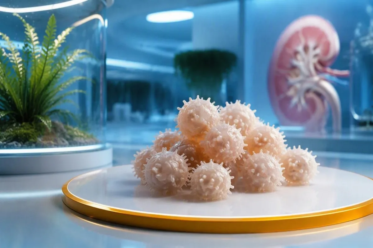
Burr cells, also called echinocytes, are often created after the sample leaves the body. Persistent, well-distributed findings can still provide a useful prompt to review renal function, electrolytes, and the wider clinical picture.


- Burr cells are red cells with many short, evenly spaced projections; the laboratory term is echinocytes.
- Artifact is common: delayed smear preparation, drying conditions, or a poor sample-to-EDTA ratio can create echinocytes after collection.
- A repeat smear matters: burr cells confined to the feathered edge or appearing only on one slide are more likely technical than biological.
- Kidney context: persistent echinocytes with reduced eGFR, rising creatinine, or high urea deserve clinician review, but they do not diagnose kidney disease alone.
- Electrolytes matter: potassium, bicarbonate or total CO2, sodium, calcium, magnesium, and phosphate can clarify whether metabolic stress is present.
- Liver distinction: advanced liver disease more classically produces irregular acanthocytes or spur cells, not uniform burr cells.
- No universal percentage cutoff exists: morphology reports are usually graded as few, moderate, or many rather than counted as a diagnostic percentage.
- Urgent symptoms override morphology: confusion, severe weakness, markedly reduced urine, chest symptoms, or palpitations need prompt clinical assessment.
What burr cells actually mean on a smear
Burr cells on a blood smear are echinocytes: red cells with numerous small, evenly distributed, blunt projections. In practice, a single report of “few” burr cells is more often a slide or specimen effect than a diagnosis, especially when the CBC is otherwise ordinary. I am Dr. Thomas Klein, and in 15 years of reviewing laboratory patterns, I have learned that the location and repeatability of the finding matter more than the word itself.
Echinocytes blood smear findings describe shape, not a disease. A mature red cell normally measures about 7 to 8 micrometres across and has a smooth disc-like outline; an echinocyte develops 10 to 30 short, similarly sized surface spicules. Dè tha CBC a’ toirt a-steach helps explain why this visual observation may appear beside perfectly normal haemoglobin, MCV, and reticulocyte results.
The practical question is whether the shape existed in circulation or formed on glass. Kantesti is an AI blood test analyzer that places a morphology comment beside creatinine, eGFR, bicarbonate, liver tests, and prior results rather than treating it as a stand-alone diagnosis. A morphology flag without symptoms or corroborating chemistry is usually a follow-up clue, not a verdict.
Most laboratories report burr cells semi-quantitatively as few, moderate, or many because a reproducible diagnostic percentage has never been standardized across smear methods. Bain’s review of diagnostic blood-film interpretation makes the same broader point: morphology gains meaning when it agrees with the patient’s history and other laboratory abnormalities (Bain, 2005).
Echinocytes versus acanthocytes, crenation, and fragments
Echinocytes have regular, evenly spaced spicules, whereas acanthocytes have fewer, uneven projections of variable length. This difference is clinically useful because acanthocytes raise a different set of concerns, including severe liver dysfunction, rare lipid disorders, or neuroacanthocytosis syndromes.
An echinocyte looks like a tidy cogwheel under the microscope. An acanthocyte, sometimes called a spur cell, has irregular thorn-like protrusions and a distorted central pallor; confusing the two can send an otherwise sensible work-up down the wrong path. Our guide to cruthachadh rouleaux shows another reason a direct visual review is different from automated CBC numbers.
Crenation is often used loosely for any scalloped red cell, but it commonly reflects drying or osmotic change on the slide. Schistocytes are different again: they are fragmented, helmet-like cells with sharp angles, and even a low but confirmed level can carry more urgency in the right clinical setting than numerous burr cells.
When I review a report that says both burr cells and fragments, I ask for the original morphology wording, platelet count, LDH, bilirubin, haptoglobin, and the clinical setting. A patient with haemoglobin 92 g/L, platelets 55 × 10^9/L, and confirmed schistocytes needs a very different response from someone with haemoglobin 142 g/L and “occasional echinocytes.”
Why burr cells are often a sample artifact
The most common cause of isolated burr cells is an artifact introduced during storage, slide preparation, or drying. Echinocytes that cluster at the thin feathered edge of a slide, rather than appearing across the monolayer, are particularly suspicious for a technical effect.
Red cells continue to exchange water and ions after collection. A sample held too long before film preparation, exposed to heat, or spread on a humid or contaminated slide can develop uniform surface spicules; the same phenomenon can occur when the anticoagulant ratio is relatively high because a tube was underfilled.
A fresh redraw can settle the issue quickly. For non-urgent morphology questions, clinicians commonly request a properly filled EDTA specimen and a promptly made film, ideally compared with the initial slide; our explanation of ath-dheuchainn às deidh sèididh covers why collection quality changes more than one result.
Do not try to “treat” a burr-cell comment by drinking extreme amounts of water or taking supplements. Overhydration can lower sodium, while a heavy workout, vomiting, diarrhoea, or fasting can independently alter creatinine, urea, and acid-base markers—then the redraw answers less than it should.
How laboratories decide whether echinocytes are persistent
Persistent echinocytes are supported when the same morphology appears in the central monolayer of a freshly prepared repeat smear, not merely at an edge or in one field. The laboratory should also check whether there is a specimen-quality comment and whether other red-cell changes travel with it.
The best viewing area is where cellular elements lie singly, with minimal overlap and a visible central pallor. An experienced morphologist scans several fields rather than selecting the most dramatic one; this is why a smartphone photograph of a single microscope field cannot reliably establish a percentage or a cause.
A meaningful report may pair echinocytes with anisopoikilocytosis, polychromasia, target cells, or evidence of fragmentation. A sudden mismatch between an unchanged CBC and a new morphology flag is also worth checking against prior specimens; delta checks on laboratory results are designed to identify implausible changes before they become a clinical story.
There is no validated rule that “more than 5% burr cells equals kidney disease.” That claim circulates online but ignores variation in stain quality, observer grading, analyser workflow, and the fact that a film is a two-dimensional sample of a dynamic circulation.
When burr cells should prompt a kidney review
Persistent burr cells warrant kidney context review when they occur with reduced eGFR, a rising creatinine, albuminuria, high urea, or symptoms of uraemia. They do not establish chronic kidney disease by themselves, and a normal eGFR does not become abnormal because a smear contains echinocytes.
In adults, chronic kidney disease requires an eGFR below 60 mL/min/1.73 m² for at least 3 months, or another marker of kidney damage such as persistent albuminuria. The 2024 KDIGO guideline emphasizes confirming chronicity rather than diagnosing CKD from one creatinine result (KDIGO, 2024). Basic metabolic panel results are often the sensible starting point after a persistent morphology finding.
Uraemia can alter red-cell membrane metabolism and has long been associated with echinocytes, but the association is neither sensitive nor specific. I have seen a person with eGFR 34 mL/min/1.73 m² and repeated burr cells, and another with eGFR 28 mL/min/1.73 m² whose films were unremarkable—clinical medicine is annoyingly like that.
Kantesti AI, an AI blood test interpretation platform, flags a morphology-plus-renal pattern for review when eGFR falls, creatinine rises, or urine protein is reported across time. It cannot inspect the physical slide, so the laboratory’s confirmed morphology remains the primary evidence.
Read creatinine, eGFR, and urine albumin together
Creatinine and eGFR estimate filtration, while urine albumin identifies kidney damage that creatinine can miss. A single elevated creatinine can reflect dehydration, recent muscle injury, creatine use, or cooked-meat intake, so its direction over days to weeks is often more useful than one number.
Creatinine reference intervals vary by laboratory, muscle mass, sex, and assay; many adult laboratories use roughly 60 to 110 µmol/L, but the eGFR reported beside it is usually more clinically interpretable. A urine albumin-to-creatinine ratio below 3 mg/mmol is generally A1, 3 to 30 mg/mmol is A2, and above 30 mg/mmol is A3 albuminuria.
The reason we worry about low eGFR plus albuminuria is that together they support intrinsic kidney disease, whereas a mildly raised creatinine after a long run may not. ìrean galar dubhaig leantainneach explain the G and A categories clinicians combine in routine renal risk assessment.
Cystatin C can be useful when creatinine seems misleading, particularly in very muscular people, frailty, restrictive diets, or major weight change. It is not a magic tie-breaker—thyroid disease, systemic steroid use, smoking, and inflammation can influence cystatin C too.
Electrolyte and acid-base results that add context
Potassium, bicarbonate or total CO2, sodium, calcium, magnesium, and phosphate are the most useful chemistry results to review alongside persistent echinocytes. Burr cells do not predict a particular electrolyte result, but renal retention, gastrointestinal loss, and acid-base disturbance can affect red-cell membrane behaviour and clinical risk.
A potassium result above 6.0 mmol/L, particularly with weakness, palpitations, a rapidly rising value, or reduced kidney function, needs same-day clinical assessment. In contrast, a potassium of 5.3 mmol/L from a difficult collection may be pseudohyperkalaemia; the sample haemolysis index and a prompt repeat are often decisive.
Total CO2 on a chemistry panel usually approximates bicarbonate. A value below 22 mmol/L can support metabolic acidosis in CKD or diarrhoeal illness, although respiratory disorders and sample handling also affect interpretation; electrolyte panel guidance details the combinations that should not wait.
Phosphate above 1.8 mmol/L is more concerning when eGFR is substantially reduced, while phosphate below 0.5 mmol/L can cause marked weakness and needs urgent assessment. The cell shape is never the emergency—the symptoms and the chemistry are.
Liver clues: burr cells are not classic spur cells
Liver disease can coexist with echinocytes, but advanced hepatic dysfunction more characteristically produces acanthocytes or spur cells with irregular projections. If a report uses the word “burr,” it is reasonable to confirm the morphology before assuming it indicates liver disease.
The liver-related pattern that catches my attention is irregular acanthocytosis plus rising bilirubin, low albumin, prolonged INR, thrombocytopenia, or clinical jaundice—not isolated echinocytes. Albumin below 30 g/L and INR above 1.5 can signal reduced hepatic synthetic function, although anticoagulant medication and nutritional status must be considered.
AST and ALT are markers of cellular injury, not direct measures of liver function. If alkaline phosphatase, GGT, direct bilirubin, and symptoms suggest a cholestatic pattern, liver test interpretation is more useful than trying to infer a diagnosis from the film.
Alcohol exposure, severe malnutrition, and end-stage liver disease can produce overlapping shape abnormalities, which is one reason morphology must be reviewed by someone who sees the actual slide. A normal ALT does not rule out cirrhosis, and a burr-cell comment does not prove it.
CBC patterns that make a smear finding more important
Burr cells become more clinically meaningful when anaemia, thrombocytopenia, a high reticulocyte count, or evidence of haemolysis appears alongside them. An isolated morphology comment with haemoglobin, platelets, bilirubin, and LDH all stable is usually lower priority.
Adult haemoglobin reference intervals vary, but many laboratories use approximately 120 to 160 g/L for women and 130 to 170 g/L for men. A fall of 20 g/L over a short interval matters more than being 2 g/L below a printed reference range, particularly if fatigue, breathlessness, dark urine, or jaundice is present.
LDH, indirect bilirubin, haptoglobin, and reticulocytes help separate increased red-cell turnover from underproduction. Kantesti is an AI-powered blood test analysis tool that compares these linked markers over time, but a clinician must decide whether a smear warrants urgent manual review. Hematocrit and haemoglobin differences can also expose dilution or index-related confusion.
Platelets below 100 × 10^9/L combined with new anaemia and confirmed fragments should be reviewed urgently, even when a report also mentions burr cells. This is where I prefer a direct conversation with the laboratory rather than relying on a portal flag.
Symptoms and urine findings that change the next step
Reduced urine output, swelling, breathlessness, persistent nausea, confusion, dark urine, or new high blood pressure make renal and metabolic review more urgent than the smear wording itself. A urine dipstick and albumin-to-creatinine ratio can add information that no peripheral smear can provide.
Dipstick protein can be transient after fever, strenuous exercise, or concentrated urine, which is why a first-morning urine ACR is often preferred for confirmation. Blood plus protein on urine testing, particularly with high blood pressure or falling eGFR, deserves prompt clinician review; urine dipstick results explain common false positives too.
Foamy urine alone is not a reliable test for proteinuria. Persistent foam together with ankle swelling, low serum albumin, or an elevated ACR is more informative, while visible red or cola-coloured urine should not be dismissed as a burr-cell issue.
Seek urgent care for fainting, severe shortness of breath, chest pain, new confusion, inability to keep fluids down, or very little urine for 12 hours. In these scenarios, the immediate priorities are vital signs, ECG when potassium is a concern, kidney function, and urine assessment.
Medicines, dehydration, and exposures to mention
Diuretics, lithium, NSAIDs, some antibiotics, contrast exposure, vomiting, diarrhoea, and prolonged fasting can change renal chemistry without being the direct cause of burr cells. Giving your clinician this timeline often explains a borderline result faster than another broad test panel.
Non-steroidal anti-inflammatory drugs can reduce kidney perfusion, especially during dehydration or when combined with diuretics and renin-angiotensin system medicines. Lithium requires regular creatinine, eGFR, thyroid, and calcium monitoring because reduced clearance can become clinically significant before a patient feels unwell; see our lithium monitoring guide.
Creatine supplements may raise measured creatinine without proportionate kidney injury, while heavy resistance training can transiently raise creatinine and CK. That does not mean every high result is harmless—new proteinuria, persistent eGFR decline, or potassium elevation changes the balance.
Bring a list of prescribed medicines, over-the-counter pain relievers, herbal preparations, supplements, recent imaging contrast, and illnesses from the preceding 14 days. “Natural” products are not automatically kidney-neutral, especially when ingredients or doses are uncertain.
A sensible repeat-testing plan after an isolated finding
For an asymptomatic person with isolated few burr cells and otherwise reassuring results, a fresh CBC with manual smear review in days to a few weeks is usually reasonable. The interval should shorten when creatinine is rising, potassium is high, haemoglobin is falling, or symptoms are present.
Before a planned redraw, maintain ordinary hydration, avoid an unusually punishing workout for 24 to 48 hours, and do not start or stop prescribed medicines without advice. A clinician may request CBC, creatinine with eGFR, urea or BUN, sodium, potassium, chloride, bicarbonate, calcium, phosphate, magnesium, liver panel, urinalysis, and urine ACR depending on context.
Repeat timing is not one-size-fits-all. An incidental report in a well person may wait 2 to 4 weeks, whereas potassium 6.1 mmol/L, new eGFR 24 mL/min/1.73 m², or a creatinine rise of 50% from baseline calls for same-day or urgent clinical direction.
Kantesti AI can organize previous results and identify whether a creatinine or bicarbonate change is likely real rather than a one-off fluctuation. For a deeper explanation of kidney chemistry, our Stiùireadh BUN-gu-creatinine is useful, but it cannot replace repeat sampling.
Common mistakes people make after seeing echinocytes
Echinocytes do not mean you have “toxic blood,” need a detox, or have kidney failure. They are a morphology clue with a high artifact rate, and the safest response is confirmation plus targeted clinical context.
The most common error is treating a report comment as a diagnosis. Another is searching for a universal echinocyte percentage threshold when laboratories intentionally use semi-quantitative reporting because staining, storage, and slide quality affect what is visible.
Do not take potassium, magnesium, bicarbonate, or phosphate supplements simply because burr cells appear on a report. Potassium supplements can be dangerous in CKD, and sodium bicarbonate can worsen fluid retention or blood pressure; stuthan cur-ris ann an tinneas dubhaig leantainneach explains why lab-guided choices matter.
The evidence around mild isolated echinocytosis is honestly limited. That uncertainty is not a failure of medicine—it is a reason to use the repeat film, urine testing, symptom history, and longitudinal results rather than over-reading an imperfect biological snapshot.
How Kantesti frames a smear comment alongside your results
Kantesti does not diagnose burr cells from a CBC alone; it interprets the laboratory-reported smear comment in relation to renal, electrolyte, liver, urine, and trend data. This approach reduces the temptation to label an artifact as disease while still surfacing combinations that merit human review.
Kantesti is an AI lab test interpretation service that reads reported echinocytes alongside eGFR, creatinine, potassium, total CO2, albumin, bilirubin, haemoglobin, and previous values. A one-time “few” result with stable renal markers is framed differently from repeat moderate echinocytes plus a 20 mL/min/1.73 m² eGFR decline.
Our clinical approach also records what the laboratory did not measure: symptoms, fasting, exercise, hydration, medicines, and whether the sample was difficult to collect. The technical principles behind this workflow are described in our stiùireadh teicneòlais AI, including why AI output should support—not replace—professional judgment.
As of September 18, 2026, the most reliable use of AI here is pattern recognition and preparation for a clinician conversation. It cannot establish slide quality, identify a rare red-cell disorder from a photograph, or perform a physical examination.
Questions to take to your clinician or laboratory team
Ask whether the burr cells were diffuse or edge-only, whether the film was freshly made, and whether a manual repeat is recommended. These three questions often provide more clarity than ordering an unstructured battery of tests.
Useful questions include: “Was this reported as few, moderate, or many?” “Were acanthocytes, schistocytes, or polychromasia also present?” and “Do my eGFR, urine ACR, potassium, bicarbonate, bilirubin, and platelet count change the interpretation?” Specific questions produce specific answers.
Ask whether your result needs a repeat smear, renal panel, urinalysis, liver panel, haemolysis tests, or no action beyond routine follow-up. Dr. Thomas Klein’s usual rule is simple: if the repeat slide is clean and the chemistry is stable, stop chasing the shape; if both persist, investigate the pattern.
Tha susbaint meidigeach Kantesti air a lèirmheas le stiùireadh lighiche, agus tha an Bòrd Comhairleachaidh Meidigeach reflects the clinical standards we apply when explaining uncertainty. Bring your complete report, not a cropped screenshot, because reference intervals and specimen comments change the interpretation.
Ceistean Bitheanta
A bheil ceallan burr air smear fala dona?
Chanadaichean chan eil iad gu fèin-ghluasadach dona o chionn gu bheil mòran air an cruthachadh le aois a’ chruinneachaidh, tiormachadh sleamhnachaidh, no suidheachadh cruinneachaidh an dèidh don chruinneachadh obair-lann a ghabhail. Aithisg air beagan echinocytes le haemoglobin àbhaisteach, truinnsearan, creatinine, eGFR, potasium, agus gun chomharran mar as trice cunnart ìosal. Tha feum air ath-sgrùdadh air eadar-mheadhanachaidh no iomadh echinocyte nuair a thachras iad le eGFR fo 60 mL/min/1.73 m², a’ togail creatinine, pròtain ann an fual, no neo-riaghailteachdan metabolach. Tha measadh èiginneach iomchaidh airson laigse mòr, palpitations, troimh-chèile, lùghdachadh mòr ann an fual, no toradh potasium os cionn 6.0 mmol/L.
A bheil ceallan burr a' ciallachadh tinneas dubhaig?
Faodaidh ceallan-ghaoil nochdadh ann an uraemia agus tinneas dubhaig leantainneach, ach chan eil iad a’ dearbhadh tinneas dubhaig leotha fhèin. Feumaidh tinneas dubhaig leantainneach eGFR fo 60 mL/min/1.73 m² airson co-dhiù 3 mìosan no comharra eile a mhaireas fada de mhilleadh dubhaig, leithid albuminuria. Tha co-mheas albuim-gu-creatinine ann an fual de 3 mg/mmol no barrachd a’ toirt taic do mhilleadh dubhaig nuair a thèid a dhearbhadh, fhad ‘s a tha sgrìobadh ath-aithris àbhaisteach agus comharran dubhaig seasmhach a’ dèanamh artifact nas dualtaiche. Bidh luchd-clionaigeach a’ mìneachadh echinocytes còmhla ri ceimigeachd nan dubhaig agus toraidhean fual, chan ann mar dheuchainn na sheasamh fhèin.
Dè an diofar eadar echinocytes agus acanthocytes?
Tha 10 gu 30 pròiseactan goirid aig echinocytes, le spàintean cothromach, agus tha spàintean neo-riaghailteach aig acanthocytes de dh'fhaid is de leud neo-chothromach. Gu tric is e rud a thachras a th' ann an echinocytes no comharra a dh'fhaodadh a bhith ann de dhroch dubhaig no metabolism. Tha ceangal nas làidire aig acanthocytes, ris an canar cuideachd ceallan spòraidh, ri droch dhroch obrachadh grùthan agus cuid de thinneasan tearc lipid no neurologic. Bu chòir do phroifeasanta obair-lann ionnsaichte an diofar a dhearbhadh oir faodaidh aithisg portal faclan neo-fhoirmeil a chleachdadh.
An urrainn do dìth uisgeachadh ceallan burr adhbhrachadh?
Faodaidh dìth uisge cur gu neo-dhìreach ri mìneachadh cheallan burr le bhith a’ dùnadh air comharran dubhaig agus a’ cruthachadh cuideam metabolach, ach chan e sin adhbhar sònraichte a chaidh a dhearbhadh airson echinocytes ann an a h-uile euslainteach. Nas trice, tha ullachadh slaodach smeur, cus anticoagulant an coimeas ri meud an sampall, no buairidhean tiormachaidh a’ toirt a-mach echinocytes an dèidh cruinneachadh. Faodaidh dìth uisge urea agus creatinine àrdachadh gu sealach, rud a dh’ fhaodadh beachd smeur mar a thachras a bhith a’ coimhead nas draghail na tha e. Faodaidh neach-clionaigeach deuchainnean dubhaig agus smeur ùr ath-aithris às deidh gabhail ri uisge àbhaisteach, mar as trice a’ seachnadh eacarsaich mòr airson 24 gu 48 uairean ro làimh.
An urrainn do thinneas ae a bhith ag adhbhrachadh cheallan burr?
Faodaidh tinneas grùthan a bhith ann còmhla ri echinocytes, ach tha acanthocytes neo-riaghailteach no spur cells nas sònraichte do dhroch dhroch ghnìomhachd hepatic na burr cells aon-ghnèitheach. Tha dragh grùthan a’ fàs nuair a nochdas cumadh neo-riaghailteach-spicule le àrdachadh bilirubin, albuim fo timcheall air 30 g/L, INR os cionn 1.5, truailleagan ìosal, buidheachas, no gleidheadh fluid. Chan eil ach AST agus ALT a’ tomhas milleadh cealla seach gnìomhachd synthesis grùthan. Tha ath-sgrùdadh obair-lann den fhìor sleamhnan ciallach nuair a chleachdas an aithisg burr cells ach is e droch thinneas grùthan a th’ anns a’ cheist clionaigeach.
Am bu chòir ceallan burr ath-aithris?
A repeat CBC with prompt manual smear preparation is often appropriate when burr cells are an unexpected isolated finding. For a well person with few echinocytes and stable laboratory results, repeat testing may occur in roughly 2 to 4 weeks; the exact timing depends on the clinician and laboratory context. Repeat testing should be faster when potassium is elevated, creatinine is rising, eGFR is below 30 mL/min/1.73 m², haemoglobin is falling, or symptoms are present. The repeat slide is most useful when it is prepared from a properly filled, fresh EDTA specimen.
Faigh Mion-sgrùdadh Deuchainn Fala le Cumhachd AI an-diugh
Thig còmhla ri còrr is 2 mhillean neach air feadh an t-saoghail a tha a’ earbsa Kantesti airson mion-sgrùdadh sa bhad, ceart air deuchainnean obair-lann. Luchdaich suas na toraidhean deuchainn fala agad agus faigh mìneachadh coileanta air biomarcair 15,000+ ann an diogan.
📚 Foillseachaidhean Rannsachaidh le Iomraidhean
Klein, T., Mitchell, S., & Weber, H. (2026). A' bhuinneach às dèidh trosgadh, breacan dubha anns an stòl & stiùireadh GI 2026. Rannsachadh Leigheis AI Kantesti.
Klein, T., Mitchell, S., & Weber, H. (2026). Stiùireadh Slàinte nam Ban: Ovulation, Menopause & Comharraidhean Hormonail. Rannsachadh Leigheis AI Kantesti.
📖 Iomraidhean Meidigeach Taobh a-muigh
⚕️ Àicheadh Meidigeach
Tha an artaigil seo dìreach airson adhbharan foghlaim agus chan eil e a’ dèanamh comhairle mheidigeach. Cuir fios an-còmhnaidh gu solaraiche cùram slàinte teisteanasach airson co-dhùnaidhean breithneachaidh is leigheis.
Comharran earbsa E-E-A-T
Eòlas
Lèirmheas clionaigeach air a stiùireadh le lighiche air sruthan-obrach mìneachaidh obair-lann.
Eòlas
Fòcas air leigheas obair-lann air mar a bhios bith-chomharraidhean (biomarkers) a’ giùlan ann an co-theacsa clionaigeach.
Ùghdarrasachd
Air a sgrìobhadh le Dr. Thomas Klein le ath-sgrùdadh le Dr. Sarah Mitchell agus Prof. Dr. Hans Weber.
Earbsachd
Mìneachadh stèidhichte air fianais le slighean leanmhainn soilleir gus dragh a lughdachadh.