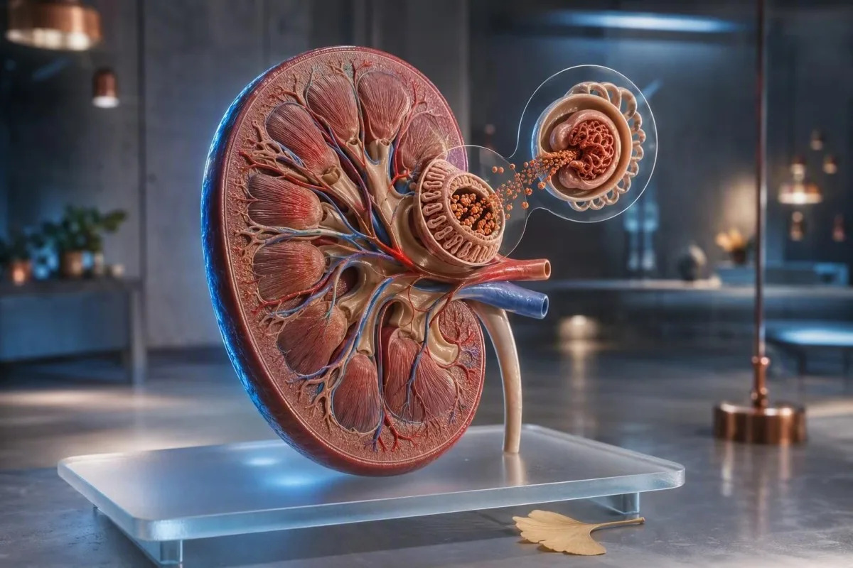
چند حباب پس از جریان سریع ادرار معمولاً ناشی از فیزیک است. کف ادراری که به طور مکرر کاسه توالت را میپوشاند، پس از سیفون ماندگار است، یا با تورم همراه است، مستلزم آزمایش آلبومین ادرار است و نه حدس و گمان.


- حبابهای کوتاه که در عرض چند ثانیه از بین میروند، معمولاً نشاندهنده سرعت ادرار، باقیمانده شوینده توالت، یا ادرار غلیظ هستند و نه بیماری کلیوی.
- ادرار کفآلود پایدار زمانی نگرانکنندهتر است که لایه متراکم سفید رنگی به مدت 1-2 دقیقه در دفعات مکرر باقی بماند.
- ACR ادرار کمتر از 3 میلیگرم در میلیمول (30 میلیگرم در گرم) دفع آلبومین طبیعی تا خفیف افزایش یافته در اکثر بزرگسالان است.
- آلبومینوری 3-30 میلیگرم در میلیمول (30-300 میلیگرم در گرم) باید با تکرار نمونههای اول صبح در طول حداقل 3 ماه تأیید شود.
- ACR بالاتر از 30 میلیگرم در میلیمول (300 میلیگرم در گرم) آلبومینوری به شدت افزایش یافته است و نیاز به ارزیابی بالینی فوری دارد.
- علائم هشداردهنده شامل تورم صورت یا پا، خون قابل مشاهده، تنگی نفس، کاهش حجم ادرار، تب، یا بارداری همراه با سردرد و فشار خون بالا است.
- جمعآوری مهم است: از ورزش سنگین تا ۲۴ ساعت قبل خودداری کنید، از نمونه تمیز وسط جریان ادرار اول صبح استفاده کنید و در مورد قاعدگی یا عفونت ادراری به پزشک اطلاع دهید.
- آزمایش خون کمک می کند: کراتینین، eGFR، آلبومین سرم، گلوکز و HbA1c مشخص می کنند که آیا پروتئین ادرار بازتابی از بیماری کلیوی، دیابت یا بیماری سیستمیک است یا خیر.
چگونه حبابهای بیضرر را از ادرار کفآلود پایدار تشخیص دهیم
علتهای کفآلود شدن ادرار از جریان سریع که به آب برخورد می کند تا نشت آلبومین از فیلتر کلیه متغیر است. حباب های بی ضرر بزرگ، شفاف و به سرعت ناپدید می شوند؛ کف نگران کننده ریزتر، سفیدتر، مکررتر است و علی رغم کاسه تمیز، حدود ۱-۲ دقیقه باقی می ماند.
کشش سطحی بیشتر حباب های یکباره را توضیح می دهد. جریان قوی اول صبح، کم آبی بدن، غلظت مواد حل شده، یا باقیمانده شوینده می تواند هوا را در سطح آب بدون هیچ گونه اتلاف پروتئین به دام بیندازد؛ این امر به ویژه پس از پرواز طولانی، تب یا ورزش شدید شایع است.
الگو بیش از یک مشاهده مهم است. در ۱۵ سال طبابت بالینی خود، از بیماران می خواهم که کف را در سه روز مجزا با استفاده از یک کاسه تمیز و بدون سفیدکننده مشاهده کنند؛ یک لایه کف ریز قابل تکرار دلیل بهتری برای آزمایش است تا یک اپیزود چشمگیر پس از ورزش.
کف به تنهایی نمی تواند پروتئینوری را تشخیص دهد زیرا فسفات، نمک های صفراوی، مایع منی و التهاب مجاری ادراری نیز می توانند کشش سطحی را تغییر دهند. اگر ادرار به جای کف آلوده باشد، تشخیص افتراقی به سمت کریستال ها، سلول ها یا عفونت شیفت می یابد؛ راهنمای ما به سرنخ های ادرار کدر به تفکیک این الگوها کمک می کند.
یک قانون ساده مشاهده خانگی
از کاسه توالت عکس نگیرید یا “تست تکان دادن” خانگی انجام ندهید. در عوض، توجه کنید که آیا کف در یک کاسه تمیز در حداقل ۳ روز از ۷ روز رخ می دهد، آیا بیش از ۱ دقیقه طول می کشد، و آیا تورم یا تغییر فشار خون وجود دارد. این جزئیات درخواست آزمایشگاهی را بسیار مفیدتر می کند.
چرا پروتئین در ادرار میتواند کف پایدار ایجاد کند
پروتئین ادرار کف آلود اغلب آلبومین است، پروتئینی با بار منفی که به طور معمول در جریان خون باقی می ماند. هنگامی که فیلتر گلومرولی نفوذپذیرتر می شود، آلبومین کشش سطحی را کاهش می دهد و می تواند حباب های کوچک و پایداری ایجاد کند.
یک بزرگسال سالم معمولاً کمتر از ۳۰ میلی گرم آلبومین در روز دفع می کند، اگرچه یک نوار تست معمولی ممکن است از دست دادن سطح پایین آلبومین را تشخیص ندهد. نسبت آلبومین به کراتینین ادرار حساس تر، یا ACR، آلبومین را برای غلظت ادرار تصحیح می کند و برای غربالگری اولیه ترجیح داده می شود.
آلبومین تنها پروتئینی نیست که مهم است. زنجیره های سبک از اختلالات پلاسما سل، پروتئین های توبولار پس از برخی داروها، و پروتئین های گذرا در طول تب ممکن است از نوار تست طراحی شده عمدتاً برای آلبومین فرار کنند؛ به همین دلیل است که یک نوار منفی هر مورد کف پایدار را حل نمی کند.
دکتر توماس کلین یافته های ادرار را در کنار کراتینین سرم، eGFR، آلبومین و گلوکز بررسی می کند تا اینکه یک علامت بصری را به عنوان تشخیص در نظر بگیرد. ما راهنمای پروتئین سرم ما توضیح می دهد که چرا نتیجه پایین آلبومین خون و ACR ادرار بالا با هم وزن بیشتری نسبت به هر یک به تنهایی دارند.
دلایل رایج کف آلود شدن ادرار غیر کلیوی
بیشتر ادرار کف آلود موقتی دارای توضیح غیر کلیوی است: ادرار غلیظ، تخلیه سریع، انزال اخیر، باقی مانده صابون، تب، یا ورزش شدید. این علل معمولاً ظرف ۲۴-۴۸ ساعت پس از هیدراتاسیون طبیعی و بهبودی برطرف می شوند.
کم آبی بدن ادرار را تیره تر و غلیظ تر می کند، بنابراین حباب ها ممکن است حتی زمانی که دفع آلبومین طبیعی است، متراکم تر به نظر برسند. به جای مصرف بیش از حد مایعات، ادرار به رنگ کاهی کمرنگ را در روز بعد هدف قرار دهید؛ نوشیدن چندین لیتر به سرعت می تواند خود باعث کاهش سدیم در افراد مستعد شود.
ورزش می تواند آلبومین ادرار را به طور موقت افزایش دهد. مسافت ۱۰ کیلومتر، جلسه تمرین مقاومتی با شدت بالا، یا قرار گرفتن طولانی مدت در معرض گرما ممکن است پروتئین را در نمونه ای که بلافاصله پس از آن جمع آوری شده است، تولید کند، به همین دلیل آزمایشگاه ها معمولاً توصیه می کنند از ورزش شدید تا ۲۴ ساعت قبل از آزمایش ACR خودداری شود.
مایع منی در مجرای ادرار پس از انزال می تواند حباب های موقتی و نوار پروتئین مثبت رقیق ایجاد کند. ادرار صورتی یا رنگ چای قابل مشاهده، پرسش را به طور کلی تغییر می دهد و باید با استفاده از این راهنما برای خون در ادرار شود, ارزیابی شود، به خصوص پس از ۴۵ سالگی یا در هر فردی که در معرض دود سیگار قرار گرفته است.
چه زمانی کف پایدار، از دست دادن پروتئین معنیدار بالینی را نشان میدهد
کف پایدار هنگام تکرار و به ویژه در صورت همراهی با دیابت، فشار خون بالا، تورم، یا کاهش eGFR، نیاز به آزمایش پروتئین ادرار را ایجاب می کند. آلبومینوری می تواند یک هشدار اولیه کلیوی باشد، حتی زمانی که کراتینین در محدوده آزمایشگاهی باقی می ماند.
KDIGO، ACR کمتر از 3 میلی گرم در میلی مول، یا 30 میلی گرم در گرم را A1؛ 3-30 میلی گرم در میلی مول را A2؛ و بالاتر از 30 میلی گرم در میلی مول را آلبومینوری A3 تعریف می کند. این دسته بندی ها، خطر کلیوی و قلبی عروقی را با همراهی eGFR، که به تنهایی استفاده نمی شود، به دقیق ترین شکل پیش بینی می کنند (KDIGO, 2024).
دیابت و پرفشاری خون شایع ترین عوامل محرک مزمن آلبومینوری در سراسر جهان هستند، اما تنها عوامل نیستند. گلومرولونفریت، گلومرولوپاتی مرتبط با چاقی، لوپوس، پره اکلامپسی، آپنه خواب و برخی داروهای ضد التهابی نیز ممکن است نقش داشته باشند؛ یافته های پایدار نیاز به بررسی مکانیسم توسط پزشک دارند.
کانتستی یک آنالایزر آزمایش خون هوش مصنوعی که کراتینین، eGFR، آلبومین سرم، گلوکز و HbA1c را در یک زمینه طولی قرار می دهد. این روش نمی تواند ACR ادرار را از روی نمونه خون اندازه گیری کند، اما می تواند به شناسایی الگوی متابولیک یا کلیوی که منجر به فوریت بیشتر در نتایج ادرار می شود، کمک کند.
کدام آزمایشهای پروتئین ادرار به بهترین نحو به این سوال پاسخ میدهند
یک نمونه ادرار اول صبح ACR بهترین آزمایش اولیه برای از دست دادن مشکوک آلبومین در اکثر بزرگسالان است. نسبت پروتئین به کراتینین، یا PCR، زمانی مفید است که پروتئین غیر آلبومینی مشکوک باشد یا زمانی که نیاز به اندازه گیری کل پروتئین باشد.
ACR در بریتانیا و بسیاری از کشورهای دیگر به صورت میلی گرم در میلی مول، یا در ایالات متحده به صورت میلی گرم در گرم گزارش می شود. ACR 3 میلی گرم در میلی مول معادل حدود 30 میلی گرم در گرم است؛ این تبدیل مهم است زیرا بیماران گاهی اوقات واحدهای مختلف را با تغییر بزرگ در نتیجه اشتباه می گیرند.
نتیجه مثبت نوار تست ادرار برای پروتئین در حد ردیابی یا 1+، یک سرنخ غربالگری است، نه یک تشخیص. ادرار قلیایی، ادرار غلیظ و آلودگی می توانند منجر به مثبت کاذب شوند، در حالی که ادرار رقیق و پروتئین های غیر آلبومینی می توانند اطمینان کاذب ایجاد کنند؛ آزمایش کمی این عدم قطعیت را برطرف می کند.
جمع آوری ادرار 24 ساعته اکنون برای موارد خاص، مانند دفع پروتئین بسیار بالا، ارزیابی بارداری، یا نمونه های نامتجانس در محل، محفوظ است. جمع آوری ناکافی آن آسان است، بنابراین دستورالعمل های عملی ما را بخوانید جمعآوری ادرار ۲۴ ساعته قبل از شروع آن.
چگونه آزمایش پروتئین ادرار را با دقت جمعآوری کنیم
آزمایش دقیق پروتئین ادرار با یک نمونه تمیز ادرار میانی اول صبح که پس از 24 ساعت بدون ورزش شدید جمع آوری می شود، آغاز می شود. اولین ادرار پس از بیدار شدن ترجیح داده می شود زیرا وضعیت بدنی، هیدراتاسیون و فعالیت روزانه تأثیر کمتری بر دفع آلبومین دارند.
دست ها را بشویید، ظرف استریل ارائه شده را استفاده کنید، ادرار کردن را در توالت شروع کنید، سپس قسمت میانی را بدون لمس داخل ظرف یا در آن بگیرید. نمونه را فوراً تحویل دهید؛ اگر تأخیر اجتناب ناپذیر است، به جای گذاشتن آن در ماشین گرم، دستورالعمل های برودتی آزمایشگاه را دنبال کنید.
آزمایش های غیر فوری را در طول عفونت مجاری ادراری علامت دار، تب، اسهال، قاعدگی، یا ظرف 24 ساعت پس از ورزش شدید، به تعویق بیندازید. این شرایط می توانند به طور موقت دفع آلبومین را افزایش دهند، در حالی که آلودگی قاعدگی نیز می تواند خون و پروتئین ظاهری ایجاد کند؛; نتایج استراز لکوسیت ممکن است الگوی عفونی احتمالی را روشن کند.
پروتئینوری ارتوستاتیک شایع است، به خصوص در نوجوانان و جوانان. پروتئین موجود در ادرار روزانه اما غایب در نمونه اول صبح، اغلب خوش خیم است، اگرچه پزشک باید الگو را تأیید کند نه اینکه فرض کند.
چگونه نتایج ACR و پروتئین-کراتینین را تفسیر کنیم
ACR کمتر از 3 میلی گرم در میلی مول معمولاً اطمینان بخش است، در حالی که ACR تایید شده 3 میلی گرم در میلی مول یا بالاتر نیاز به پیگیری مبتنی بر ریسک دارد. یک نمونه مثبت، بیماری مزمن کلیه را مشخص نمی کند زیرا ورزش، عفونت و افزایش قند خون می توانند باعث آلبومینوری کوتاه مدت شوند.
برای بزرگسالان بدون دیابت، ACR تازه افزایش یافته معمولاً در عرض چند هفته با استفاده از نمونه اول صبح تکرار می شود. بیماری مزمن کلیه نیازمند ناهنجاری های موجود برای حداقل 3 ماه است، مگر اینکه آسیب حاد کلیوی مشخص یا پروتئین به شدت افزایش یافته باشد که نیاز به بررسی سریع تر داشته باشد (KDIGO, 2024).
ACR بالاتر از 70 میلی گرم در میلی مول، تقریباً 700 میلی گرم در گرم، معمولاً حتی در صورت حفظ eGFR، به خصوص با وجود خون در ادرار، نیاز به بحث فوری تخصصی دارد. پروتئینوری در محدوده نفروتیک به طور کلی PCR بالاتر از 300 میلی گرم در میلی مول یا دفع پروتئین بالاتر از 3.5 گرم در روز است، که اغلب با ادم و آلبومین سرم پایین همراه است.
کانتستی یک سرویس تفسیر آزمایشهای آزمایشگاه AI منتشر میشوند که می تواند شاخص های خونی کلیوی را در طول تاریخ سازماندهی کرده و روند رو به کاهش eGFR را مشخص کند. تفسیر روند، میکروسکوپی ادرار پزشک، تأیید ACR و بررسی دارو را پشتیبانی می کند، اما هرگز جایگزین آن نمی شود.
آزمایشهای خونی که نتایج ادرار کفآلود را معنیدار میکنند
کراتینین، eGFR، آلبومین سرم، گلوکز، HbA1c، الکترولیت ها و چربی ها به توضیح این موضوع کمک می کنند که آیا پروتئین ادرار منعکس کننده آسیب فیلتر کننده کلیه است یا یک اختلال متابولیک گسترده تر. کراتینین طبیعی، آلبومینوری اولیه را رد نمی کند.
eGFR از کراتینین محاسبه می شود و در بسیاری از افراد دیابتی دیرتر از نشت خفیف آلبومین کاهش می یابد. eGFR کمتر از 60 میلی لیتر در دقیقه در 1.73 متر مربع به مدت 3 ماه یا بیشتر، یکی از معیارهای بیماری مزمن کلیه را برآورده می کند، اما مقادیر باید بر اساس سن، توده عضلانی و بیماری اخیر تفسیر شوند.
آلبومین سرم پایین، تقریباً زیر ۳۵ گرم در لیتر، همراه با پروتئین ادراری زیاد و تورم جدید مچ پا، باعث نگرانی در مورد دفع پروتئین قابل توجه کلیوی میشود. برعکس، آلبومین بالا با ادرار تیره، اغلب نشاندهنده کمآبی است؛ راهنمای eGFR پس از کم آبی دلیل اینکه آزمایش مجدد پس از بهبودی میتواند منطقی باشد را توضیح میدهد.
انجمن دیابت آمریکا حداقل سالیانه آزمایش UACR و eGFR را در دیابت نوع ۲ و در دیابت نوع ۱ که ۵ سال یا بیشتر طول کشیده است، توصیه میکند (کمیته راهبردی حرفهای ADA، ۲۰۲۴). HbA1c در مورد میانگین قند خون به ما میگوید، اما افزایش ناگهانی قند همچنان میتواند به طور موقت آلبومین ادرار را قبل از تغییر HbA1c افزایش دهد.
داروها، مکملها و بیماریهایی که میتوانند بر پروتئین ادرار تأثیر بگذارند
داروهای ضد التهاب مسکن، لیتیوم، برخی از درمانهای سرطان و عفونتهای شدید میتوانند تصفیه کلیه یا دفع پروتئین لولهای را تغییر دهند. به دلیل اینکه ادرار کفآلود به نظر میرسد، داروی تجویز شده را قطع نکنید؛ درخواست بررسی ادرار و آزمایش خون کلیه را بدهید.
داروهای ضد التهاب غیر استروئیدی مانند ایبوپروفن و ناپروکسن میتوانند جریان خون کلیه را کاهش دهند، به خصوص در هنگام کمآبی، نارسایی قلبی یا بیماری مزمن کلیه (CKD) موجود. خطر زمانی افزایش مییابد که یک داروی ضد التهاب غیر استروئیدی با یک دیورتیک و یک مهارکننده ACE یا ARB ترکیب شود، ترکیبی که پزشکان گاهی اوقات آن را “سه گانه کشنده” مینامند.”
داروهای SGLT2 ممکن است باعث کاهش جزئی اولیه eGFR شوند اما معمولاً در بیماران مناسب، آلبومینوری را در طول زمان کاهش میدهند. این تغییر همودینامیک مورد انتظار با آسیب پیشرونده متفاوت است؛ به تغییرات eGFR با درمان SGLT2 برای تمایزهای عملی مراجعه کنید.
ویتامین C با دوز بالا میتواند با برخی از نوارهای تست ادرار تداخل داشته باشد، در حالی که کراتین میتواند تفسیر کراتینین را بدون آسیب رساندن لزوماً به کلیهها، پیچیده کند. لیست کاملی از داروهای تجویزی، مسکنهای بدون نسخه، پودرهای پروتئین و محصولات گیاهی را به قرار ملاقات بیاورید - نامها و دوزها مهم هستند.
دیابت و فشار خون: بررسی ریسک با بالاترین بازده
دیابت و فشار خون بالا، کف پایدار را از نظر بالینی مهمتر میکنند زیرا هر دو میتوانند سالها قبل از ظاهر شدن علائم باعث آلبومینوری شوند. فشار خون در حدود ۱۴۰/۹۰ میلیمتر جیوه یا بالاتر و ACR در حدود ۳ میلیگرم در میلیمول یا بالاتر باید منجر به یک بررسی ساختاریافته مراقبت اولیه شود.
برای بسیاری از افراد مبتلا به CKD و آلبومینوری، پزشکان در صورت تحمل، فشار خون سیستولیک دفترچه استاندارد شده را زیر ۱۲۰ میلیمتر جیوه هدف قرار میدهند، اگرچه اهداف باید بر اساس سن، خطر سقوط، بارداری و روش اندازهگیری فردی شوند. قرائتهای خانگی که به اشتباه انجام شدهاند نباید به تنهایی باعث تغییر دارو شوند.
آلبومینوری همچنین یک نشانگر خطر عروقی است. دلیل نگرانی ما در مورد افزایش ACR همراه با فشار خون بالا این است که این دو با هم استرس اندوتلیال و گلومرولی را نشان میدهند، در حالی که هر یک از این یافتهها یک بار در طول بیماری حاد ممکن است موقتی باشد؛ راهنمای آزمایشگاهی فشار خون بالا ما ارزیابی گستردهتر را ترسیم میکند.
کانتستی یک پلتفرم تفسیر بیومارکرهای AI که در بیش از ۱۰۰ کشور برای مقایسه روند قند خون، HbA1c، نشانگرهای کلیوی و لیپید استفاده میشود. برای ادرار کف آلود پایدار، تست بعدی مرتبط، آزمایش ACR آزمایشگاهی ادرار است، که در حالت ایدهآل باید با همان تیم بالینی که فشار خون یا دیابت را مدیریت میکند، بررسی شود.
ادرار کفآلود در بارداری و در کودکان به قوانین متفاوتی نیاز دارد
ادرار کف آلود در دوران بارداری، در صورت بروز همراه با سردرد، تغییر دید، درد بالای شکم، تورم ناگهانی، یا افزایش فشار خون، نیاز به ارزیابی در همان روز دارد. پروتئینوری پس از ۲۰ هفته میتواند بخشی از پرهاکلامپسی باشد، اما صرف ظاهر آن هرگز تشخیصی نیست.
در بارداری، نسبت پروتئین به کراتینین ۳۰ میلیگرم در میلیمول یا بیشتر، یا ۳۰۰ میلیگرم پروتئین در ۲۴ ساعت، در زمینه بالینی مناسب، پروتئینوری قابل توجهی را تأیید میکند. نمونه ادرار با ظاهر طبیعی نمیتواند پرهاکلامپسی را در صورتی که فشار خون ۱۴۰/۹۰ میلیمتر جیوه یا بالاتر همراه با علائم نگران کننده باشد، رد کند.
کودکان اغلب پس از تب، ورزش یا کمآبی، پروتئینوری موقتی دارند. پروتئین پایدار در سه نمونه، فشار خون بالا، ادم، یا خون در ادرار، نیاز به ارزیابی اطفال دارد؛ آزمایش صبحگاهی اول به ویژه برای رد پروتئینوری وضعیتی در نوجوانان مفید است.
بارداری تصفیه را تغییر میدهد، بنابراین مقادیر کراتینین که برای بزرگسالان غیر باردار “طبیعی” به نظر میرسند، ممکن است اطمینانبخش نباشند. راهنمای ما به مقادیر GFR در بارداری زمینه را فراهم میکند، اما علائم فوری باید مستقیماً به اورژانس زایمان مراجعه کنند تا تفسیر آنلاین.
علائم هشدار دهنده: چه زمانی ادرار کفآلود نیاز به مراقبت فوری پزشکی دارد
در صورت بروز ادرار کف آلود همراه با تنگی نفس، افزایش سریع تورم پا یا صورت، کاهش قابل توجه ادرار، خون قابل مشاهده، تب همراه با درد پهلو، یا علائم هشدار دهنده بارداری، به دنبال ارزیابی فوری پزشکی باشید. این ترکیبات میتوانند نشاندهنده بیماری قابل توجه کلیوی، قلبی، عفونی، یا مرتبط با بارداری باشند.
تورم جدید صورت هنگام بیدار شدن، تنگ شدن کفشها در عصر و افزایش وزن بیش از ۲ کیلوگرم در ۲-۳ روز میتواند نشاندهنده احتباس مایعات باشد. هنگامی که این علائم همراه با کف پایدار همراه باشد، پزشکان فوراً پروتئین ادرار، کراتینین، آلبومین، فشار خون و گاهی یافتههای قفسه سینه را بررسی میکنند.
ادرار رنگ چای پس از ورزش شدید با درد شدید عضلانی، کف خوشخیم معمولی نیست؛ این میتواند نشاندهنده آزادسازی میوگلوبین باشد و نیاز به آزمایش فوری خون و ادرار دارد. همین امر در مورد ادرار رنگ کولا با کاهش خروجی صدق میکند که ممکن است نشاندهنده خونریزی گلومرولی یا آسیب عضلانی باشد.
دکتر توماس کلین توصیه میکند که بیماران در صورت وجود علائم هشدار دهنده، منتظر نتیجه تست نواری خانگی نباشند. برای تغییرات رنگی مربوط به فعالیت بدنی، مقاله ما در مورد هماچوری در دوندگان طیف خوشخیم و ویژگیهای غیرخوشخیم آن را توضیح میدهد.
پزشکان پس از نتیجه مثبت پروتئین ادرار چه کاری انجام میدهند
پس از مثبت شدن ACR یا PCR، پزشکان معمولاً نتیجه را تأیید میکنند، فشار خون و eGFR را اندازهگیری میکنند، رسوب ادرار را بررسی میکنند و دیابت، داروها، علائم خودایمنی و سابقه خانوادگی را مرور میکنند. مرحله بعدی به کمیت، تداوم و وجود خون همراه یا کاهش فیلتراسیون بستگی دارد.
میکروسکوپی ادرار میتواند سلولهای قرمز خون، سلولهای سفید خون و سیلندرهایی را که تنها با عدد پروتئین قابل توضیح نیستند، شناسایی کند. سیلندرهای گلبول قرمز یا گلبولهای قرمز دیسمورفیک نشاندهنده منشأ گلومرولی هستند و ارجاع فوری به نفرولوژی را تسریع میکنند؛ سیلندرهای هیالین ساده میتوانند پس از کمآبی یا ورزش رخ دهند، همانطور که در راهنمای سیلندرهای هیالین.
ما پوشش داده شده است. اگر آلبومینوری تأیید شد، درمان معمولاً عامل ایجاد کننده را هدف قرار میدهد: کنترل فشار خون، مدیریت قند خون، حمایت برای وزن و ترک سیگار، و داروهای محافظ کلیه در صورت لزوم. سونوگرافی کلیه، تستهای ایمنی، الکتروفورز سرم یا بیوپسی کلیه، تستهای انتخابی هستند - نه عواقب خودکار یک تست نواری ۱+.
متدولوژی بالینی Kantesti در برابر استانداردهای ایمنی ساختاریافته بررسی میشود و خوانندگان میتوانند اعتبارسنجی پزشکی ما. ما را بررسی کنند. نقش ما این است که نتایج آزمایش خون موجود را برای بحث آسانتر کنیم، نه اینکه یک اختلال گلومرولی را از روی یک تصویر یا یک مقدار آزمایشگاهی منفرد تشخیص دهیم.
یک برنامه عملی هفت روزه برای کف پایدار
برای ادرار کفدار پایدار بدون علائم هشدار دهنده، یک تست ACR ادرار در مراقبتهای اولیه و آنالیز ادرار را در عرض ۱-۲ هفته ترتیب دهید، ترجیحاً با استفاده از نمونه صبحگاهی. فشار خون، هیدراتاسیون، ورزش، داروها و اینکه آیا کف بیش از ۱ دقیقه باقی میماند را برای هفت روز ثبت کنید.
روز ۱-۳: به طور معمول مایعات مصرف کنید، از ورزش شدید خودداری کنید و فقط در یک کاسه توالت تمیز بدون مواد شوینده اضافه شده مشاهده کنید. سعی نکنید با آب زیاد “مشکل را از بین ببرید”؛ هدف نمونه نماینده است، نه نمونه رقیق شده مصنوعی.
روز ۴-۷: درخواست آنالیز ادرار به همراه ACR را بدهید و بپرسید که آیا کراتینین، eGFR، پتاسیم، آلبومین سرم، گلوکز و HbA1c برای شرایط شما مناسب هستند. نتایج قبلی را بیاورید زیرا تغییر ACR از ۲ به ۸ میلیگرم بر میلیمولد معنای بیشتری نسبت به هر یک از اعداد به تنهایی دارد؛; پیگیری طولیِ نتایج میتواند به سازماندهی تاریخها کمک کند.
از ۴ سپتامبر ۲۰۲۶، پیام منطقی اطمینانبخش اما قاطع باقی میماند: اکثر حبابهای منفرد بیضرر هستند، در حالی که کف ریز تکراری شایسته یک تست کمی ادرار است. اگر برای آماده کردن سؤالات برای پزشک به کمک نیاز دارید، بهعنوان یک منبع شفافیت استفاده کنند و گزارش را بهعنوان یک نقطه شروع ساختاریافته برای مراقبت در نظر بگیرند، نه بهعنوان مجوزی برای شروع، توقف یا تغییر درمانِ تجویزی. ما استانداردهای بالینی پشت محتوای آموزشی ما را تعیین میکند.
سوالات متداول
آیا ادرار کفآلود همیشه نشانهی پروتئین در ادرار است؟
خیر، ادرار کفآلود همیشه نشانه از دست دادن پروتئین نیست. جریان سریع، کمآبی بدن، باقیمانده شوینده توالت، تب، ورزش و مایع منی میتواند باعث ایجاد حبابهایی شود که در عرض چند ثانیه از بین میروند. کف سفید ریز و مداومی که در چندین روز تکرار میشود، بیشتر نشانه پروتئینوری است و باید با نسبت آلبومین به کراتینین ادرار بررسی شود. نسبت ACR 3 میلیگرم بر میلیمول، معادل 30 میلیگرم بر گرم، یا بالاتر، به اندازهای غیرطبیعی تلقی میشود که نیاز به زمینه بالینی و اغلب تکرار آزمایش دارد.
کف ادرار چقدر باید طول بکشد تا نگران شوم؟
کف غلیظی که در دفعات مکرر حدود ۱ تا ۲ دقیقه باقی میماند، نگرانکنندهتر از حبابهای بزرگ است که به سرعت از بین میروند. مدت زمان پایدار ماندن کف، یک تست تشخیصی نیست زیرا مواد شیمیایی توالت و غلظت ادرار میتوانند پایداری حبابها را تغییر دهند. اگر این الگو در ۳ روز یا بیشتر تکرار شود، ظرف ۱ تا ۲ هفته آزمایش ادرار (urinalysis) و ACR ادرار را ترتیب دهید. در صورت بروز کف همراه با تورم، خون قابل مشاهده، تنگی نفس، تب، یا کاهش حجم ادرار، زودتر به پزشک مراجعه کنید.
برای چه آزمون پروتئین ادرار باید درخواست بدهم؟
بهترین تست اولیه برای اکثر بزرگسالان، نسبت آلبومین به کراتینین ادرار صبحگاهی است که به آن ACR یا UACR گفته میشود. ACR کمتر از 3 میلیگرم در میلیمول یا 30 میلیگرم در گرم، به طور کلی طبیعی تا کمی افزایش یافته است، در حالی که 3-30 میلیگرم در میلیمول نشاندهنده افزایش متوسط آلبومینوری است. در صورت مشکوک بودن به از دست دادن کلی یا غیرآلبومینی پروتئین، ممکن است نسبت پروتئین به کراتینین اضافه شود. تست نواری معمول برای غربالگری مفید است اما برای از دست دادن آلبومین در سطح پایین، دقت کمتری نسبت به ACR دارد.
آیا کمآبی بدن باعث کفآلود شدن ادرار میشود؟
بله، کمآبی بدن میتواند ادرار را غلیظتر کند و باعث ایجاد حباب یا کف قابل مشاهده بیشتری شود، بدون اینکه اتلاف پروتئین مداوم کلیوی وجود داشته باشد. این وضعیت اغلب همراه با ادرار زرد تیرهتر، تشنگی، قرار گرفتن در معرض گرما، استفراغ، اسهال یا عدم نوشیدن مایعات برای مدت طولانی رخ میدهد. قبل از انجام آزمایش غیر اورژانسی، ۲۴ تا ۴۸ ساعت به مصرف معمول مایعات خود بازگردید، اما از نوشیدن بیش از حد مایعات صرفاً برای رقیق کردن نتیجه خودداری کنید. اگر کف پس از هیدراتاسیون ادامه یافت، درخواست آزمایش ACR ادرار را بدهید.
آیا ورزش میتواند باعث ایجاد پروتئین و کف در ادرار شود؟
ورزش شدید میتواند باعث آلبومینوری موقت و ادرار کفآلود شود، به خصوص پس از دویدن، تمرینات مقاومتی سنگین، رویدادهای استقامتی یا ورزش در هوای گرم. این اثر معمولاً در عرض 24 تا 48 ساعت برطرف میشود، به همین دلیل نمونه ACR باید به طور ایدهآل پس از اجتناب از ورزش شدید به مدت 24 ساعت جمعآوری شود. پروتئینوری مداوم در نمونه صبحگاهی پس از بهبودی، کمتر با ورزش به تنهایی قابل توجیه است. ادرار تیره یا به رنگ کولا همراه با درد شدید عضلانی نیاز به ارزیابی فوری پزشکی دارد.
سطح ACR خطرناک چیست؟
آلبومینوری شدید با ACR بالاتر از ۳۰ میلیگرم در میلیمول (یا ۳۰۰ میلیگرم در گرم) شناخته میشود و نیاز به ارزیابی فوری پزشکی دارد، به ویژه اگر eGFR کاهش یافته باشد یا ادرار حاوی خون باشد. ACR بالاتر از ۷۰ میلیگرم در میلیمول (حدود ۷۰۰ میلیگرم در گرم) معمولاً حتی در صورت نرمال بودن کراتینین، بحث فوری تخصصی را ایجاب میکند. ریسک با ترکیبی از ACR، eGFR، فشار خون، وضعیت دیابت، علائم و اندازهگیریهای تکراری تعیین میشود. افزایش ACR تکمرحلهای معمولاً باید تأیید شود مگر اینکه وضعیت بالینی اورژانسی باشد.
همین امروز آنالیز آزمایش خون با هوش مصنوعی را دریافت کنید
به بیش از 2 میلیون کاربر در سراسر جهان بپیوندید که Kantesti را برای تحلیل فوری و دقیق آزمایشهای آزمایشگاهی مورد اعتماد قرار میدهند. نتایج آزمایش خون خود را بارگذاری کنید و در عرض چند ثانیه، تفسیر جامع 15,000+ از نشانگرهای زیستی را دریافت کنید.
📚 انتشارات پژوهشی ارجاعشده
کلاین، تی.، میچل، اس.، و وبر، اچ. (۲۰۲۶). چارچوب اعتبارسنجی بالینی v2.0 (صفحه اعتبارسنجی پزشکی). پژوهش پزشکی مبتنی بر هوش مصنوعی Kantesti.
کلاین، تی.، میچل، اس.، و وبر، اچ. (۲۰۲۶). آنالیزگر آزمایش خون با هوش مصنوعی: ۲.۵M آزمایش تحلیلشده | گزارش جهانی سلامت ۲۰۲۶. پژوهش پزشکی مبتنی بر هوش مصنوعی Kantesti.
📖 منابع پزشکی خارجی
⚕️ سلب مسئولیت پزشکی
این مقاله فقط برای اهداف آموزشی است و توصیه پزشکی محسوب نمیشود. برای تصمیمهای مربوط به تشخیص و درمان، همیشه با یک ارائهدهنده مراقبتهای بهداشتی واجد شرایط مشورت کنید.
سیگنالهای اعتماد E-E-A-T
تجربه
بازبینی بالینی مبتنی بر نظر پزشک از فرایندهای تفسیر آزمایشگاه.
تخصص
تمرکز بر پزشکی آزمایشگاهی و اینکه نشانگرهای زیستی در زمینه بالینی چگونه رفتار میکنند.
اقتدارگرایی
نوشتهشده توسط دکتر توماس کلاین، با بازبینی توسط دکتر سارا میچل و پروفسور دکتر هانس وبر.
قابل اعتماد بودن
تفسیر مبتنی بر شواهد با مسیرهای پیگیری روشن برای کاهش هشدارها.