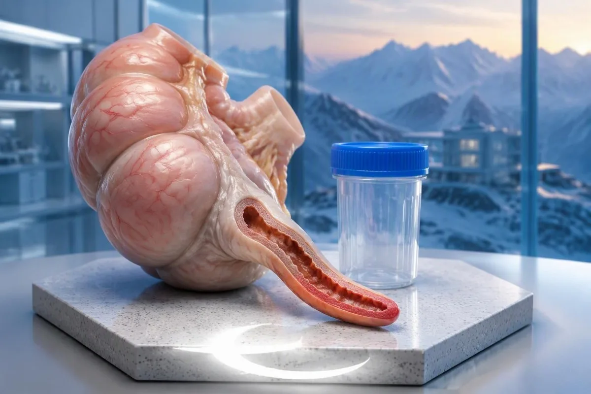
Nej – normale hvide blodlegemer og CRP kan ikke pålideligt udelukke blindtarmsbetændelse. Tidlige symptomer kan udvikle sig, før der ses laboratorieændringer, så forværring af mavesmerter skal vurderes, selv når alle resultater ser betryggende ud.


- Normal WBC betyder almindeligvis omkring 4,0–11,0 × 10⁹/L hos voksne; blindtarmsbetændelse kan stadig forekomme inden for dette interval.
- Normal CRP betyder ofte under 5 mg/L, selvom nogle laboratorier bruger under 10 mg/L; ingen af disse grænseværdier udelukker uafhængigt blindtarmsbetændelse.
- Tidlig testning kan overse den inflammatoriske respons, fordi CRP generelt først stiger flere timer efter den inflammatoriske stimulus, ofte omkring 6-8 timer.
- Pædiatrisk evidens fandt en følsomhed på ca. 85% for WBC ved 10,0 × 10⁹/L, hvilket betyder, at cirka 15% af de ramte børn faldt under denne specifikke grænse.
- Kombinerede resultater er mere informative end én markør, men normal WBC plus normal CRP tilsidesætter ikke vedvarende lokal ømhed eller forværrede smerter.
- Akut vurdering er nødvendig ved forværrede smerter i højre nedre del af maven, gentagen opkastning, stivhed i maven eller smerter forværret ved bevægelse – selv uden feber på 38°C.
- Usikkerhed ved billeddiagnostik er vigtig: en blindtarm, der ikke visualiseres på ultralyd, er ikke det samme som en blindtarm, der med sikkerhed vises at være normal.
- Sikkerhedsnet kræver en specifik revurderingsplan; vent ikke på endnu en blodprøve eller en AI-fortolkning, hvis symptomerne forværres.
Kan en normal blodprøve for blindtarmsbetændelse udelukke det?
A en normal blodprøve for blindtarmsbetændelse kan ikke pålideligt udelukke blindtarmsbetændelse, især i sygdommens første timer. Hvide blodlegemer og CRP måler kroppens reaktion, ikke selve blindtarmen; klinikere skal fortolke dem sammen med smerteudvikling, undersøgelsesfund og, når det er relevant, billeddiagnostik.
Kantesti er en AI blodprøveanalysator der kan forklare WBC- og CRP-resultater, men den kan ikke udelukke blindtarmsbetændelse ud fra en laboratorierapport. Vores organisation og kliniske fokus centrerer sig om fortolkning af resultater; en abdominal undersøgelse og en akut diagnostisk beslutning er forskellige opgaver.
To normale resultater reducerer normalt mistanken mere end ét normalt resultat, men omfanget af denne reduktion afhænger af den oprindelige risiko. En person med milde, aftagende ubehag er ikke ækvivalent med en person med støt forværret smerte i højre nedre del af maven, selvom begge har en WBC på 7,2 × 10⁹/L.
Et normalt laboratorieinterval er ikke en tærskel for udelukkelse af sygdom. Udtrykket inden for normalområdet beskriver sammenligning med en referencepopulation; det betyder ikke, at alle organer er blevet undersøgt, eller at en udviklende sygdom er blevet udelukket.
Min tilgang, som Dr. Thomas Klein, er at spørge, hvad resultatet rent faktisk besvarer: er der en målbar systemisk reaktion i øjeblikket? En CRP på 3 mg/L besvarer det snævre spørgsmål. Det forklarer ikke, hvorfor nogen nu har lokaliseret smerte, ikke kan tåle bevægelse eller er vendt tilbage efter en tidligere vurdering.
Hvilke blodprøver hjælper klinikere med at opdage blindtarmsbetændelse?
Blodprøver kan understøtte en diagnose af blindtarmsbetændelse, men ingen rutinemæssig blodmarkør identificerer specifikt blindtarmsbetændelse. En fuldstændig blodtælling med differentialtælling og CRP bruges almindeligvis; nyrefunktion, elektrolytter og andre tests hjælper med at vurdere dehydrering, alternative diagnoser og parathed til behandling eller billeddiagnostik.
De leukocyttallet, eller WBC, måler det samlede antal cirkulerende hvide celler. Differentialtællingen adskiller neutrofiler fra andre celletyper, hvilket er vigtigt, fordi et totalantal på 9,0 × 10⁹/L kan dække over et relativt højt neutrofilantal og et lavere lymfocyttal.
C-reaktivt protein, eller CRP, produceres primært af leveren som reaktion på inflammatorisk signalering. Et resultat på 25 mg/L indikerer en inflammatorisk reaktion, men det lokaliserer ikke reaktionen: blindtarmsbetændelse, en urinvejsinfektion, lungebetændelse og flere ikke-infektiøse tilstande kan alle øge CRP.
Rutinemæssige laboratorieintervaller varierer efter instrument, population og rapporteringspraksis. WBC på 10,5 × 10⁹/L kan blive markeret af et laboratorium og efterladt umarkeret af et andet; den kliniske betydning ændrer sig ikke brat, når en farvet advarsel forsvinder fra rapporten.
Uoverensstemmende resultater er ikke nødvendigvis laboratoriefejl. En øget WBC med en lav CRP kan afspejle forskellige responstidspunkter, mens en forhøjet CRP med en normal WBC kan afspejle en senere eller anderledes udtrykt reaktion; vores vejledning til WBC og CRP uoverensstemmelse forklarer den forskel.
Hvordan kan man have normal WBC ved blindtarmsbetændelse?
Normal WBC with appendicitis is possible because local inflammation does not always produce immediate or substantial leukocytosis. Symptom duration, baseline cell counts, medicines and immune function influence the result; a normal total count may also coexist with a concerning neutrophil pattern.
A total WBC should be read with the absolut neutrofiltal, not only the neutrophil percentage. For example, WBC of 8.0 × 10⁹/L with 80% neutrophils gives an absolute neutrophil count of 6.4 × 10⁹/L; the percentage alone can look more alarming than the absolute result.
Fawkner-Corbett et al. (2022) pooled 67 studies involving 34,839 children and found approximately 85% sensitivity for WBC at a cutoff of 10.0 × 10⁹/L. Roughly 15% of children with appendicitis therefore fell below that study cutoff—not necessarily below every laboratory's age-specific upper limit.
Baseline counts change the meaning of a single result. A rise from a person's usual WBC of 3.5 to 8.5 × 10⁹/L may represent a substantial response despite remaining unflagged; neutrophil reference ranges also differ by age and pregnancy status.
Some people have a naturally lower neutrophil baseline, including those with a Duffy-null associeret neutrofiltal. That finding is not itself evidence of impaired immunity, but it illustrates why a universal WBC cutoff cannot replace symptoms, examination or comparison with previous results.
Hvorfor kan CRP-niveauer ved blindtarmsbetændelse forblive normale tidligt?
CRP levels in appendicitis may remain normal early because inflammatory signalling must reach the liver before circulating CRP increases. CRP generally begins rising several hours after an inflammatory stimulus, often around 6–8 hours, but symptom onset and the biological start of inflammation are not identical timestamps.
A CRP of 2 mg/L shortly after pain begins is less reassuring than the same value after a longer, clearly improving illness. There is no reliable clock that lets a clinician say, 'At exactly eight hours, a normal CRP excludes appendicitis.' Biological responses vary, and patients may first notice symptoms at different stages.
Higher CRP can increase concern for more extensive inflammation, particularly when the history and imaging agree. However, CRP of 50 or 100 mg/L does not independently diagnose perforation; duration, another inflammatory condition and the patient's overall physiological state can influence the number.
Standard CRP and high-sensitivity CRP, or hs-CRP, measure the same protein using different analytical sensitivity. Cardiovascular hs-CRP categories such as below 1, 1–3 and above 3 mg/L are not appendicitis decision thresholds; fortolkning af CRP requires a different clinical question.
A mildly elevated baseline also complicates interpretation. Some medicines, including certain hormonal contraceptives, can influence CRP; our discussion of contraception and CRP results explains why a small elevation should not automatically be attributed to an acute abdominal cause—or used to dismiss that cause.
Er normal WBC og CRP sammen mere betryggende?
Normal WBC and CRP together are more reassuring than either alone, but they are not a universal rule-out test. Their value depends on symptom duration, examination findings, the population being assessed and the exact cutoffs used; persistent focal pain can still justify imaging or surgical assessment.
In the paediatric meta-analysis by Fawkner-Corbett et al. (2022), combining WBC ≥10.0 × 10⁹/L or CRP ≥10 mg/L produced approximately 97% sensitivity in the studies reporting that combination. This was an 'either marker positive' strategy, not a requirement that both markers be elevated.
A sensitivity of 97% does not mean that a child with two low results has a 3% chance of appendicitis. Sensitivity describes affected patients; the chance of disease after a negative result also depends on how likely appendicitis was before testing and on the specificity of the test strategy.
Thresholds deserve scrutiny. CRP below 10 mg/L counted as negative in that analysis, but a value of 8 mg/L is above the reference limit in many laboratories; calling every study-negative result 'completely normal' would therefore overstate what the evidence actually evaluated.
A clinician may repeat the examination and selected tests during supervised observation, but worsening pain should not wait for a laboratory rise. The timing of blood test changes is useful context, not permission to stay home until WBC or CRP crosses a particular line.
Hvordan ændrer symptomer og undersøgelse diagnosen?
Symptom progression and abdominal examination determine how much weight normal blood tests deserve. Pain migrating toward the right lower abdomen, focal tenderness, guarding and pain with movement increase concern; clinicians may use validated scores to organise these findings, but scores do not replace clinical judgement.
The classic sequence is central abdominal discomfort followed by more localised right-lower pain, but not every patient follows that pattern. An appendix behind the caecum may produce less obvious front-of-abdomen tenderness, while a pelvic appendix can cause urinary or bowel symptoms without the expected textbook presentation.
Consider a hypothetical 24-year-old with WBC of 7.8 × 10⁹/L and CRP of 4 mg/L whose discomfort becomes focal over several hours. Those results may justify caution rather than reassurance if the second examination reveals guarding; the change in the abdomen is new evidence, not a contradiction of the first test.
De Alvarado score has a maximum of 10 points, and the Appendicitis Inflammatory Response score has a maximum of 12. Both incorporate clinical features and laboratory information, so a low inflammatory response can lower the score even when an early examination remains concerning.
Di Saverio et al. (2020), in the WSES Jerusalem guidelines, recommend risk-stratified assessment and identify the AIR and Adult Appendicitis Score as useful adult tools. Our guide to unexplained pain testing makes the same practical distinction: a normal inflammatory marker does not explain away a focal symptom.
Hvilke symptomer kræver akut vurdering trods normale laboratorietal?
Worsening or persistent right-lower abdominal pain needs urgent assessment even with normal WBC and CRP. Seek emergency care for severe or spreading pain, a rigid abdomen, repeated vomiting, fainting, confusion or marked weakness; a fever is not required for appendicitis or another abdominal emergency.
Pain aggravated by walking, coughing or ordinary movement can suggest irritation of the abdominal lining. Do not repeatedly press your abdomen or try internet 'jump tests' to prove appendicitis; a negative home manoeuvre cannot exclude it, and a positive one does not identify the cause.
A temperature below 38°C does not exclude appendicitis. Children, older adults and immunosuppressed people may have less typical signs; a child who refuses to walk, has worsening abdominal pain or repeatedly vomits deserves prompt assessment rather than reassurance from an earlier normal panel.
Severe illness requires attention to circulation and breathing as well as abdominal findings. Clinicians may order lactate when shock or sepsis is suspected, but lactate testing limitations mean that neither a normal result nor a mildly elevated result independently settles the appendicitis question.
Call your local emergency number for collapse, confusion, difficulty breathing or severe rapidly worsening pain; in the UK, use 999 eller 112. For less severe but persistent or worsening symptoms, seek urgent same-day medical assessment, and do not delay because you have eaten, taken pain relief or are waiting for an online interpretation.
Hvad bidrager urin-, graviditets- og kemiske tests med?
Urine, pregnancy and chemistry tests help identify alternatives and treatment needs, not just appendicitis. Urinalysis can reveal urinary abnormalities, pregnancy testing changes the differential diagnosis and imaging plan, and kidney function or electrolyte tests assess dehydration and other consequences of vomiting.
Urinary white cells or microscopic blood can occur when an inflamed appendix irritates nearby urinary structures. A mildly abnormal urine result therefore does not automatically prove a urinary infection; the komplet guide til urinanalyse explains why collection quality and the entire urinary pattern matter.
Pregnancy testing is often appropriate for a person who could be pregnant and has acute lower abdominal pain. A negative urine test obtained very early can be misleading; our guide to early pregnancy test limitations explains when serum testing or repeat assessment may be necessary.
An ectopic pregnancy is a separate emergency that must be considered when pregnancy is possible, especially with one-sided pain, vaginal bleeding, dizziness or fainting. WBC of 6.0 × 10⁹/L and CRP of 3 mg/L do not exclude that diagnosis either; laboratory reassurance must not narrow the assessment prematurely.
Kantesti er en AI blodprøvefortolkningsplatform that can explain reported kidney markers and electrolytes alongside WBC and CRP. Its interpretation cannot determine whether urinary abnormalities arise from the appendix, a stone or infection; that distinction requires the patient's history, examination and sometimes imaging.
Hvornår er ultralyd, CT eller MR nødvendige?
Imaging is considered when appendicitis remains possible after clinical assessment, including when blood tests are normal. Ultrasound is commonly preferred initially in children and pregnancy, CT is frequently used in non-pregnant adults, and MRI can clarify selected cases without ionising radiation.
Ultrasound findings include appendix size, compressibility, surrounding fat changes and fluid. An outer diameter above approximately 6 mm can support appendicitis, but size alone is not definitive; a mildly enlarged appendix without supporting features is different from a non-compressible appendix with surrounding inflammatory changes.
'Appendix not visualised' is not synonymous with 'normal appendix'. Bowel gas, body habitus, appendix position and operator experience affect visibility; clinicians interpret a non-diagnostic scan with the examination and may choose observation, repeat ultrasound, MRI or CT rather than treating it as a negative answer.
CT can evaluate the appendix and several alternative causes of abdominal pain in one examination. Di Saverio et al. (2020) support selected low-dose CT strategies after inconclusive ultrasound, particularly in adolescents and young adults, while local pathways differ according to expertise, availability and the patient's circumstances.
Imaging choice also depends on where the pain is and what else is plausible. Right-upper abdominal pain may require a different assessment; our article on gallbladder blood test limitations illustrates another condition in which apparently reassuring inflammatory markers cannot substitute for examination and organ-specific imaging.
Hvem har brug for ekstra forsigtighed med normale inflammatoriske markører?
Children, pregnant patients, older adults and immunosuppressed people need particularly careful interpretation of normal results. Symptoms may be atypical, reference intervals may differ and some patients cannot mount a typical inflammatory response; clinical deterioration should carry more weight than an unflagged laboratory panel.
Children need age-specific WBC intervals, not automatic application of an adult 4.0–11.0 × 10⁹/L range. Younger children may struggle to describe migration or localisation of pain, so behaviour, hydration, movement and repeated examination can become more informative than an apparently ordinary laboratory result.
Pregnancy can increase the normal WBC baseline, making mild leukocytosis less specific for appendicitis. Pregnancy also changes the consequences of missed alternative diagnoses and the imaging discussion; ultrasound and MRI are useful options, but necessary diagnostic assessment should not be delayed solely because radiation is a concern.
Older adults may present with less fever or less obvious local tenderness, and delayed recognition can have serious consequences. New persistent abdominal pain in a 78-year-old with WBC of 8.0 × 10⁹/L still needs a broad assessment that considers appendicitis, obstruction, vascular disease and other urgent causes.
Immune-suppressing medicines can alter laboratory responses, while corticosteroids can themselves increase circulating neutrophils. These mixed effects make a single result difficult to interpret; our discussion of children's AI interpretation safety explains why age, symptoms and direct clinical assessment must remain central.
Hvad skal du gøre efter udskrivelse med normale blodprøver?
Discharge after normal blood tests should include clear return precautions and, when needed, a reassessment plan. Return promptly if pain worsens, localises, spreads, or is accompanied by repeated vomiting, fainting or increasing illness; an earlier reassuring assessment does not guarantee that an evolving condition remains low risk.
Ask what was actually established before discharge: was appendicitis considered unlikely, was a normal appendix demonstrated on imaging, or was ultrasound inconclusive? Those are three different conclusions, and each deserves a different explanation rather than a vague statement that 'the bloods were fine'.
A useful safety plan names the warning signs, where to return and when persistent symptoms should be reviewed. If a clinician recommends reassessment within 6–12 hours, follow that individual plan; it is not a universal instruction to wait that long when pain is getting worse.
Bring the original report, imaging summary, medicine list and a simple symptom timeline. Record when the pain began and how its location changed, not just its severity; our tjekliste til lægebesøg helps organise information without replacing the consultation.
At Kantesti AI, we distinguish an explanation of an old result from a decision about new symptoms. A WBC of 7.0 × 10⁹/L from yesterday remains yesterday's measurement; if today's abdomen is more tender or vomiting has developed, another online reading of the same report cannot answer today's clinical question.
Kan gentagne blodprøver eller tendenser afklare spørgsmålet?
Repeat blood tests can add information, but trends cannot independently confirm or exclude appendicitis. The direction of symptoms and examination findings matters more than a small isolated laboratory change; repeat testing should be part of a clinician-led plan, not a substitute for reassessment or indicated imaging.
A rise in CRP from 3 to 18 mg/L over several hours supports a developing inflammatory response. It still does not identify the appendix as the source; clinicians ask whether the pain, examination and imaging now fit appendicitis or point toward a different abdominal or extra-abdominal diagnosis.
CRP has a circulating half-life of roughly 19 timer, so its concentration does not track symptom changes minute by minute. After treatment, CRP may remain elevated despite early clinical improvement; conversely, temporary improvement in discomfort does not prove that an untreated abdominal problem has resolved.
Small WBC differences can reflect hydration, stress, medicines and measurement variation. A change from 8.1 to 8.6 × 10⁹/L should not be portrayed as proof of progression on its own, particularly if different laboratories were used or the patient's fluid status changed between samples.
After hospital treatment, follow the discharge team's instructions rather than chasing a normal CRP independently. Our guide to post-hospital blood test changes explains why recovery curves vary; recurrent fever, worsening abdominal pain or persistent vomiting require assessment even if a previous result was improving.
Hvad kan AI-tolkning hjælpe med – og hvad kan den ikke?
Kantesti er en AI-drevet værktøj til analyse af blodprøver that can explain WBC, neutrophil and CRP results, but it cannot rule out appendicitis or provide an abdominal examination. With worsening abdominal symptoms, seek urgent assessment rather than waiting for a report upload, automated answer or trend comparison.
A useful interpretation checks the reported units before discussing a threshold. WBC of 10 × 10⁹/L equals 10,000 cells/µL, while CRP of 1 mg/dL equals 10 mg/L; confusing either pair can make the same result appear tenfold different or incorrectly classified.
AI should preserve the distinction between a laboratory flag and a diagnostic conclusion. Our forklaring af AI-teknologi describes the interpretation workflow, but no laboratory-only workflow can reproduce guarding, percussion tenderness, changing vital signs or the findings of a properly performed imaging study.
Kantesti's report tools can help organise laboratory information for a clinician, including reported reference intervals and previous measurements when available. They should not assign an 'all clear' from WBC of 6.8 × 10⁹/L and CRP of 2 mg/L when the patient describes worsening focal abdominal pain.
If uploading results for a non-urgent explanation, check that the file belongs to the correct person and that dates, units and values are legible. Our tjekliste for privatliv ved upload af resultater covers sensible preparation; during an urgent abdominal episode, obtaining clinical care comes before uploading any document.
Forskningspublikationer: diagnostisk evidens og AI's begrænsninger
Appendicitis diagnostic evidence and AI technical benchmarks answer different questions. As of October 4, 2026, this article uses established appendicitis guidelines and diagnostic research; Kantesti's repository publications below should not be treated as proof that an AI system can exclude appendicitis from normal laboratory results.
Andersson's 2004 meta-analysis in the British Journal of Surgery evaluated clinical and laboratory diagnosis and supports combining findings rather than relying on one marker. WSES guidance from 2020 and the 2022 paediatric meta-analysis address different populations and methods; none supports a blanket claim that normal WBC and CRP exclude every case.
A pre-registered, rubric-based automated technical benchmark of the Kantesti blood-test interpretation engine on 100,000 synthetic test cases. (n.d.). Figshare. The DOI-linked record is listed below, with Søgning i ResearchGate-optegnelse og Søgning i publikationer på Academia links; these are searches, not claims of verified duplicate deposits or peer review.
Clinical validation framework v2.0 (Medical validation page). (n.d.). Zenodo. The DOI-linked record is listed below, with ResearchGate framework search og Academia framework search links; a framework describes assessment methods, not an independently established appendicitis rule-out sensitivity.
As Dr. Thomas Klein, my editorial standard is to keep those boundaries explicit: 100,000 synthetic cases are not 100,000 patients prospectively assessed for appendicitis. Our technical validation standards og medical advisory structure explain the wider context; neither replaces timely assessment of a person with worsening abdominal pain.
Ofte stillede spørgsmål
Kan blodprøver afsløre blindtarmsbetændelse?
Blodprøver kan støtte en diagnose af blindtarmsbetændelse, men kan ikke påvise den alene. En forhøjet WBC eller CRP indikerer en inflammatorisk respons uden at identificere dens placering. En voksen WBC på omkring 4,0–11,0 × 10⁹/L kan stadig forekomme ved blindtarmsbetændelse, især tidligt i sygdommen. Klinikere kombinerer laboratorieresultater med symptomudvikling, abdominal undersøgelse og billeddiagnostik, når det er nødvendigt.
Kan man have blindtarmsbetændelse med normal WBC og CRP?
Ja, blindtarmsbetændelse kan forekomme med både normal WBC og normal CRP, især før en betydelig systemisk inflammatorisk respons udvikles. En WBC på 7,0 × 10⁹/L og CRP på 3 mg/L udelukker ikke uafhængigt blindtarmsbetændelse. To lave markører kan reducere mistanken i en ellers lavrisikovurdering, men vedvarende fokale ømhed eller forværring af smerter kan stadig retfærdiggøre billeddiagnostik eller genvurdering. Søg akut lægehjælp, hvis symptomerne forværres, snarere end at vente på, at markørerne stiger.
Hvor lang tid tager CRP at stige ved blindtarmsbetændelse?
CRP begynder generelt at stige flere timer efter en inflammatorisk stimulus, ofte omkring 6-8 timer, men blindtarmsbetændelse følger ikke en pålidelig laboratorietidsplan. Den biologiske start af inflammation kan forekomme før eller efter, at patienten først genkender mavesymptomer. En CRP under 5 mg/L tidligt i sygdomsforløbet kan derfor ikke udelukke blindtarmsbetændelse. Forværrede symptomer kræver vurdering, uanset hvor mange timer der er gået.
Hvilken WBC-værdi eller CRP-niveau bekræfter blindtarmsbetændelse?
Ingen enkelt WBC-tælling eller CRP-niveau bekræfter blindtarmsbetændelse. WBC over cirka 11,0 × 10⁹/L er over mange voksne referenceintervaller, og CRP over 5 eller 10 mg/L er forhøjet afhængigt af laboratoriet, men mange andre tilstande fremkalder disse ændringer. Selv CRP på 100 mg/L fastslår ikke uafhængigt blindtarmsbetændelse eller perforation. Bekræftelse afhænger af den samlede kliniske vurdering, ofte understøttet af billeddiagnostik.
Udelukker en normal ultralyd blindtarmsbetændelse?
En betryggende ultralyd reducerer bekymring mest, når blindtarmen tydeligt visualiseres og fremstår normal i en ellers lavrisiko klinisk vurdering. En rapport, der angiver, at blindtarmen ikke blev visualiseret, svarer ikke til en normal blindtarm. En blindtarmdiameter over ca. 6 mm kan understøtte blindtarmsbetændelse, men størrelsen skal fortolkes med kompressionsevne og omgivende fund. Vedvarende eller forværret smerte kan kræve yderligere vurdering på trods af en indledende betryggende scanning.
Hvornår skal jeg vende tilbage til hospitalet efter normale blodprøver for blindtarmsbetændelse?
Vend straks tilbage, hvis mavesmerter forværres, flytter sig til nederste højre del af maven, spreder sig eller ledsages af gentagne opkastninger, besvimelse, krampetrækninger eller tiltagende sygdom. En temperatur under 38°C udelukker ikke blindtarmsbetændelse, og gårsdagens normale blodprøver fastslår ikke dagens diagnose. Følg enhver specifik genvurderingsinstruktion givet ved udskrivelsen, men vent ikke på den aftale, hvis symptomerne forværres. Ring til dit lokale alarmnummer ved kollaps, forvirring, åndedrætsbesvær eller stærke, hurtigt forværrede smerter.
Få AI-drevet blodprøveanalyse i dag
Bliv en del af over 2 millioner brugere på verdensplan, som har tillid til Kantesti for øjeblikkelig og præcis analyse af laboratorieprøver. Upload dine blodprøveresultater, og få en omfattende forstå blodprøveresultater af 15,000+-biomarkører på få sekunder.
📚 Refererede forskningspublikationer
Klein, T., Mitchell, S., & Weber, H. (2026). En præregistreret, rubric-baseret automatisk teknisk benchmark af Kantesti Blood-Test Interpretation Engine på 100.000 syntetiske testcases. Kantesti AI Medical Research.
Klein, T., Mitchell, S., & Weber, H. (2026). Klinisk valideringsramme v2.0 (Medical Validation Page). Kantesti AI Medical Research.
📖 Eksterne medicinske referencer
Fawkner-Corbett D et al. (2022). Diagnostic accuracy of blood tests of inflammation in paediatric appendicitis: a systematic review and meta-analysis. BMJ Open.
⚕️ Medicinsk ansvarsfraskrivelse
Denne artikel er kun til undervisningsformål og udgør ikke lægelig rådgivning. Rådfør dig altid med en kvalificeret sundhedsprofessionel for beslutninger om diagnose og behandling.
E-E-A-T Trust Signals
Erfaring
Lægefagligt ledet klinisk gennemgang af arbejdsgange til laboratorietolkning.
Ekspertise
Fokus på laboratoriemedicin på, hvordan biomarkører opfører sig i klinisk kontekst.
Autoritet
Skrevet af Dr. Thomas Klein med gennemgang af Dr. Sarah Mitchell og Prof. Dr. Hans Weber.
Troværdighed
Evidensbaseret fortolkning med klare opfølgningsspor for at reducere alarm.