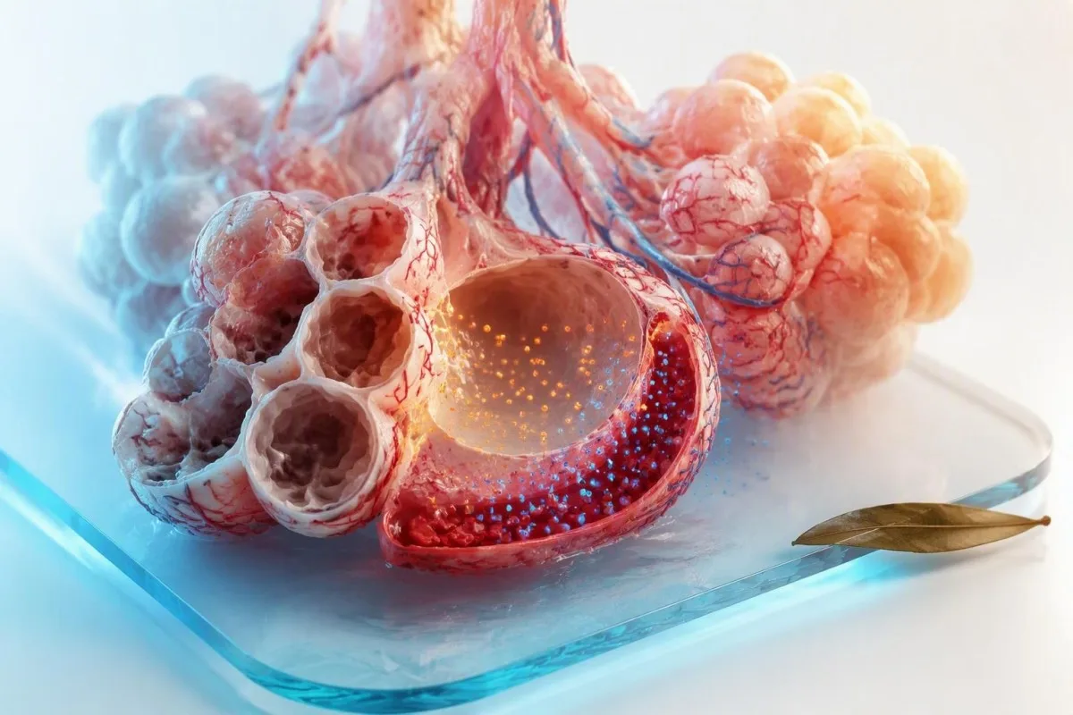
An ABG is a rapid snapshot of oxygen delivery, ventilation and acid-base balance. The safest way to read it is to identify the pH direction first, then decide whether carbon dioxide or bicarbonate best explains it.


- Blood gas pH is normally 7.35-7.45; pH below 7.20 or above 7.60 usually needs urgent clinical assessment.
- PaCO2 meaning is ventilation: 35-45 mmHg is typical, while a rising PaCO2 signals inadequate removal of carbon dioxide.
- PaO2 below 60 mmHg on room air indicates clinically significant hypoxaemia and needs prompt assessment in the right setting.
- Metabolic acidosis usually combines low bicarbonate below 22 mmol/L with low pH; diabetes, kidney failure, diarrhoea and lactic acidosis are common causes.
- Respiratory acidosis combines high PaCO2 above 45 mmHg with low pH and can occur with COPD flare-ups, sedatives or respiratory muscle fatigue.
- Winter’s formula estimates expected PaCO2 in metabolic acidosis: 1.5 × bicarbonate + 8, plus or minus 2 mmHg.
- Oxygen saturation can mislead when carbon monoxide exposure, poor circulation or an incorrect sample affects the result.
- Do not self-treat an ABG with oxygen, bicarbonate or breathing exercises without a clinician who knows your symptoms, medication list and sample conditions.
What arterial blood gas results show in minutes
Arterial blood gas results show whether the body is too acidic or alkaline, whether the lungs are clearing carbon dioxide, and whether oxygen is reaching arterial circulation. A typical adult ABG includes pH, PaCO2, PaO2, bicarbonate, base excess and measured oxygen saturation; the pattern matters far more than one flagged result.
An ABG is taken from an artery because arterial values best reflect gas exchange after blood has passed through the lungs. A venous sample can be useful for pH and bicarbonate trends, but its oxygen value cannot substitute for PaO2; see our explanation of serum, plasma and whole blood.
In practice, I read pH first, then PaCO2, then bicarbonate. Kantesti is an AI blood test analyzer that reads ABG values as a physiological pattern rather than presenting each number as an isolated red flag.
An ABG can change within 10-20 minutes after oxygen therapy, bronchodilators, vomiting, a seizure or a change in breathing support. That speed is why clinicians use it in acute care, but it also means a result from last week may no longer describe today’s physiology.
Why an artery rather than a vein?
Arterial circulation carries oxygenated blood from the lungs to organs, making PaO2 interpretable against the inspired oxygen concentration. Venous pH is commonly about 0.03 units lower and venous PCO2 about 4-6 mmHg higher than arterial values, although shock and poor circulation widen those gaps.
Normal ABG ranges and why your lab may differ
In adults breathing room air at sea level, pH is usually 7.35-7.45, PaCO2 35-45 mmHg, bicarbonate 22-26 mmol/L and PaO2 roughly 75-100 mmHg. PaO2 normally falls with age, and every oxygen result must be read alongside the FiO2, or percentage of oxygen being breathed.
A PaO2 of 68 mmHg may be meaningful in a 25-year-old breathing room air, yet can be close to expected in an older adult without breathlessness. Laboratories may use 80 mmHg as the lower reference limit, while acute-care teams often use 60 mmHg as the practical threshold for significant hypoxaemia.
Bicarbonate on an ABG is generally calculated from pH and PaCO2 rather than measured directly. If ABG bicarbonate and the total CO2 on a basic metabolic panel differ by more than about 2-3 mmol/L, clinicians consider timing, sample handling or a mixed disorder.
Base excess is usually -2 to +2 mmol/L. A base excess of -10 points toward a substantial metabolic acid load, whereas +10 supports metabolic alkalosis or renal compensation for longstanding CO2 retention.
Blood gas pH: the first result that sets the direction
A blood gas pH below 7.35 is acidaemia, while a pH above 7.45 is alkalaemia. The pH names the direction of the current problem; it does not by itself reveal whether the lungs, kidneys, gut, medicines or circulation caused it.
pH is logarithmic: a fall from 7.40 to 7.10 represents roughly twice the hydrogen ion concentration, not a small numerical drift. pH at or below 7.20, especially with confusion, low blood pressure or rapid breathing, is a clinical urgency rather than a result to watch at home.
Dr. Thomas Klein’s practical rule is simple: identify whether PaCO2 and bicarbonate push pH in the same direction or oppose it. High PaCO2 is acidic; low bicarbonate is acidic; their combination can produce severe acidaemia even when each abnormality seems only moderate.
A normal pH does not exclude danger. pH 7.40 with PaCO2 60 mmHg and bicarbonate 36 mmol/L may indicate compensated chronic respiratory acidosis, while pH 7.40 with PaCO2 20 and bicarbonate 12 can conceal two opposing acute disorders; electrolyte patterns help separate them.
PaCO2 meaning: how ABGs measure ventilation
PaCO2 measures carbon dioxide pressure in arterial blood and is the most direct ABG marker of effective ventilation. A PaCO2 above 45 mmHg means hypoventilation relative to metabolic CO2 production, while a level below 35 mmHg means hyperventilation.
PaCO2 rises when breathing becomes too shallow, too slow or mechanically ineffective. Opioids, benzodiazepines, severe asthma, COPD exacerbations, obesity hypoventilation, neuromuscular weakness and exhaustion can all produce values above 50-60 mmHg.
A PaCO2 of 70 mmHg is not automatically an emergency in someone with stable, known chronic CO2 retention; their pH and usual baseline matter. A sudden rise from 40 to 60 mmHg, however, can cause headache, drowsiness and confusion well before the number looks dramatic.
Low PaCO2 often reflects pain, fever, panic, pregnancy, pulmonary embolism or early sepsis, but it can also be appropriate compensation for metabolic acidosis. For symptom-led testing, our guide to mga pagsulay sa dugo sa kakulang sa gininhawa explains why an ABG is only one part of the work-up.
PaO2 and oxygen saturation: related but not interchangeable
PaO2 is dissolved oxygen pressure, whereas SaO2 is the percentage of haemoglobin carrying oxygen. PaO2 below 60 mmHg on room air generally corresponds to an oxygen saturation near 90%, where the oxygen-haemoglobin curve becomes steep and small declines matter more.
A pulse oximeter estimates saturation at the fingertip; an ABG directly measures PaO2 and may measure or calculate saturation depending on the analyser. Dark nail products, cold hands, movement, low perfusion and carbon monoxide exposure can make pulse oximetry less reliable, which is why clinicians compare symptoms, waveform quality and ABG data.
Giving supplemental oxygen can raise PaO2 substantially without fixing inadequate ventilation. A person receiving 40% oxygen with PaO2 70 mmHg may have more impaired gas exchange than someone breathing room air with the same PaO2.
The British Thoracic Society oxygen guideline recommends a target saturation of 94-98% for most acutely ill adults and 88-92% for patients at risk of hypercapnic respiratory failure (O’Driscoll et al., 2017). Chest pressure, blue-grey lips, collapse or new confusion requires emergency care; our chest pain testing guide covers associated urgent testing.
Metabolic acidosis patterns: bicarbonate, anion gap and causes
Metabolic acidosis is defined by low bicarbonate, usually below 22 mmol/L, driving pH downward. The next clinical question is whether the anion gap is high, which points toward unmeasured acids such as lactate, ketones, toxins or retained acids in kidney failure.
The common anion-gap calculation is sodium minus chloride minus bicarbonate; a typical value without potassium is about 8-12 mmol/L, although each laboratory sets its own range. Correct it for low albumin by adding roughly 2.5 mmol/L for every 1 g/dL albumin below 4.0.
Diarrhoea and renal tubular acidosis typically cause normal-gap, often chloride-rich metabolic acidosis. Ketoacidosis, lactic acidosis and advanced kidney failure more often raise the gap, but a normal gap does not make acidosis harmless; see our detailed renal tubular acidosis patterns.
Diabetic ketoacidosis can present with abdominal pain, thirst, vomiting and deep rapid breathing before glucose is known. Kitabchi et al. (2009) defined DKA by glucose above 250 mg/dL, arterial pH at or below 7.30 and bicarbonate at or below 18 mmol/L; ketones and clinical status determine urgency, not glucose alone.
Metabolic alkalosis: why vomiting and diuretics change ABGs
Metabolic alkalosis usually features bicarbonate above 26 mmol/L with a pH above 7.45. Vomiting, gastric drainage, loop or thiazide diuretics, low potassium and mineralocorticoid excess are frequent causes.
Chloride depletion is a surprisingly useful clue. When urine chloride is below about 20 mmol/L, vomiting or remote diuretic exposure is more likely to be chloride-responsive; higher urine chloride suggests ongoing diuretics or mineralocorticoid-driven alkalosis.
Compensatory breathing slows as bicarbonate rises, so PaCO2 often increases by about 0.7 mmHg for each 1 mmol/L bicarbonate above 24. PaCO2 above 55-60 mmHg is less easily explained by compensation alone and should prompt a search for additional respiratory failure.
Alkalosis can lower ionised calcium even when total calcium is normal, contributing to tingling, cramps or palpitations. Do not start salt or potassium products from an ABG alone, particularly with kidney or heart disease; our guide to ubos nga chloride nga mga resulta explains the medication clues.
Respiratory acidosis: high CO2 from reduced ventilation
Respiratory acidosis occurs when PaCO2 rises above 45 mmHg and lowers pH. Acute cases are often caused by sedating medicines, airway obstruction, severe lung disease or respiratory muscle fatigue, while chronic cases allow the kidneys time to retain bicarbonate.
For every 10 mmHg acute PaCO2 rise above 40, bicarbonate should increase by only about 1 mmol/L. In chronic respiratory acidosis, bicarbonate rises roughly 3.5-4 mmol/L per 10 mmHg, which is why a longstanding PaCO2 of 60 may coexist with bicarbonate near 32.
Kantesti is an AI blood test interpretation platform that compares PaCO2, pH and bicarbonate against expected compensation, helping identify when a supposed chronic pattern may actually be mixed. A result still needs clinician review, particularly if there is sleepiness, a new medication or worsening breathlessness.
In my experience, the most dangerous error is treating every high CO2 result as “just COPD.” New drowsiness, inability to speak full sentences, oxygen saturation falling below the prescribed target or pH below 7.30 needs same-day urgent assessment; persistent changes also benefit from medication safety trend review.
Respiratory alkalosis: low CO2 is not always anxiety
Respiratory alkalosis occurs when PaCO2 falls below 35 mmHg and pH rises above 7.45. Anxiety can cause it, but clinicians must first consider hypoxaemia, pulmonary embolism, pneumonia, fever, pregnancy, liver failure, salicylate exposure and sepsis.
In acute respiratory alkalosis, bicarbonate falls about 2 mmol/L for each 10 mmHg PaCO2 decrease below 40. Over 2-3 days, renal compensation lowers bicarbonate by about 4-5 mmol/L per 10 mmHg, so very low bicarbonate suggests chronicity or an added metabolic acidosis.
A patient with PaCO2 24 mmHg, bicarbonate 12 mmol/L and pH 7.35 does not have a reassuring “normal pH.” That combination strongly suggests respiratory alkalosis plus metabolic acidosis, a pattern seen in salicylate poisoning and severe systemic illness.
Tingling around the mouth or hands can occur because alkalosis temporarily reduces ionised calcium, but symptoms alone cannot identify the cause. If dizziness accompanies fast breathing, chest symptoms, fainting or unilateral leg swelling, seek urgent care rather than assuming panic; our giya sa blood test para sa pagkalipong nagpatin-aw sa mas lapad nga kalainan.
Mixed acid-base disorders: when two processes occur together
A mixed acid-base disorder exists when measured compensation is outside the expected range or when pH appears normal despite substantially abnormal PaCO2 and bicarbonate. Mixed patterns are common in emergency care because vomiting, infection, kidney dysfunction and lung disease often overlap.
Use the story before the equation. A person with septic shock may have lactic metabolic acidosis and respiratory alkalosis from increased respiratory drive, while a person with COPD who is vomiting may have respiratory acidosis plus metabolic alkalosis.
In high-gap metabolic acidosis, the rise in anion gap should roughly match the bicarbonate fall. If the gap rises by 20 mmol/L but bicarbonate falls by only 8 mmol/L, concurrent metabolic alkalosis is plausible; this “delta” reasoning is useful but not exact enough to diagnose without clinical context.
Lactate above 4 mmol/L in a clinically unwell patient is a high-risk finding that needs rapid assessment, while lower values can rise after seizures, beta-agonists or a difficult sample collection. Read our discussion of high lactate beyond sepsis before treating an isolated value as a diagnosis.
Compensation formulas that make ABG patterns clearer
Compensation reduces a pH disturbance but rarely restores pH completely to normal in a single acute disorder. The most useful bedside check in metabolic acidosis is Winter’s formula: expected PaCO2 equals 1.5 × bicarbonate + 8, plus or minus 2 mmHg.
If bicarbonate is 12 mmol/L, expected PaCO2 is about 26 mmHg. A measured PaCO2 of 40 indicates inadequate respiratory compensation and an added respiratory acidosis; a measured PaCO2 of 18 indicates an additional respiratory alkalosis.
For metabolic alkalosis, expected PaCO2 is approximately 0.7 × (bicarbonate minus 24) + 40, plus or minus 5. These equations are screening tools, not substitutes for examining the patient or checking inspired oxygen, respiratory rate and medication timing.
Kantesti AI interprets ABG test results by pairing compensation mathematics with related electrolytes, kidney markers and prior results when they are available. Ang giya sa teknolohiya sa AI describes why source values, units and collection time must be verified before any automated interpretation.
ABG sample problems that can change pH, CO2 or oxygen
Delayed processing, air exposure, excess liquid heparin and an unintended venous sample can distort ABG results. A result that conflicts sharply with the patient’s appearance, pulse oximeter or prior values should be repeated before major treatment decisions when it is safe to do so.
Air bubbles tend to drive PaO2 toward room-air levels of roughly 150 mmHg, lower PaCO2 and raise pH, particularly if a sample sits before analysis. White-cell or platelet counts that are extremely high can consume oxygen in the syringe, producing spuriously low PaO2—sometimes called leukocyte larceny.
Liquid heparin can dilute electrolytes and bicarbonate if the syringe is not properly prepared. Prolonged tourniquet time does not affect an arterial sample in the same way as a routine venous draw, but delay at room temperature still allows cell metabolism to change gases.
A radial arterial puncture can be uncomfortable, and I never dismiss a patient who says the sample felt unusual or was difficult to obtain. Before uploading a report, use our PDF ug photo accuracy checklist to confirm that pH, PaCO2 units, FiO2 and collection time were captured correctly.
Which ABG results need urgent clinical assessment
Urgent assessment is generally needed for pH at or below 7.20, pH at or above 7.60, PaO2 below 60 mmHg on room air, or a rapidly rising PaCO2 with drowsiness or respiratory distress. Symptoms and speed of change can make a less extreme value equally urgent.
Call emergency services now for severe breathlessness, blue-grey colour, chest pain, fainting, seizure, confusion, inability to stay awake or a rapidly deteriorating condition. An ABG is never a home triage tool when those symptoms are present, regardless of whether a portal flags the result.
A pH of 7.25 may be tolerated temporarily in chronic kidney disease under close supervision, but the same value during sudden diabetic ketoacidosis or opioid-related hypoventilation needs very different and often immediate treatment. Dr. Thomas Klein advises patients to record oxygen flow rate, recent medicines, vomiting or diarrhoea, and whether the sample was arterial when speaking to the care team.
The AARC blood gas analysis guideline stresses quality control, timely analysis and clinical correlation rather than interpretation from a printed number alone (AARC, 2013). Our Lupon sa Medikal nga Magtatambag reviews the clinical safeguards that guide Kantesti’s result explanations.
What to ask after receiving ABG test results
After an ABG, ask what oxygen concentration you were breathing, whether the result is acute or chronic, and whether the compensation fits a single disorder. Those three questions often clarify more than asking whether one individual number is “high” or “low.”
Ask for the exact pH, PaCO2, PaO2, bicarbonate, base excess, saturation and lactate, plus the FiO2 or oxygen flow at collection. If you have kidney disease, diabetes, COPD, sleep apnoea or use sedatives, bring a current medication list and previous results; trends can be more informative than one ABG.
As of September 17, 2026, Kantesti supports multilingual interpretation across 75+ languages, but it does not replace urgent examination, imaging, ECG assessment or emergency treatment. Kantesti is an AI-powered blood test analysis tool that highlights patterns and follow-up questions while keeping clinical diagnosis with the treating professional.
Keep a copy of the original report, not just a screenshot of flags, and ask when a repeat gas or metabolic panel is planned. Our mga sumbanan sa medikal nga pag-validate explain the clinical oversight and data-quality boundaries we use when interpreting complex laboratory reports.
Kanunay nga Gipangutana nga mga Pangutana
Unsa ang normal nga pH sa arterial blood gas?
Ang normal nga arterial blood gas pH sa kadaghanan sa mga hamtong mao ang 7.35-7.45. Ang pH nga ubos sa 7.35 acidaemia, ug ang pH nga labaw sa 7.45 alkalaemia. Ang pH nga 7.20 o ubos pa, o 7.60 o labaw pa sagad nagkinahanglan og dinalian nga clinical assessment, ilabi na kung adunay kalibog, ubos nga presyon sa dugo, mga sintomas sa dughan o kalisud sa pagginhawa. Ang hinungdan nagdepende sa PaCO2, bicarbonate, mga sintomas ug ang katulin sa kausaban.
Unsay buot ipasabot sa taas nga PaCO2 sa resulta sa ABG?
Ang PaCO2 nga labaw sa 45 mmHg nagpasabut nga ang pagtangtang sa carbon dioxide nabawasan kumpara sa produksyon, nga gitawag ug hypoventilation. Ang mga kantidad nga labaw sa 50-60 mmHg mahimong mahitabo sa COPD exacerbation, mga tambal nga makapahinanok, grabe nga hika, obesity hypoventilation o kahuyang sa kaunoran sa respiratoryo. Ang taas nga PaCO2 nga adunay ubos nga pH nagpaila sa acute o dili igo nga nabayran nga respiratory acidosis. Ang usa ka lig-on nga tawo nga adunay chronic CO2 retention mahimong adunay mas taas nga baseline, busa ang miaging mga resulta ug karon nga pagkaalerto hinungdanon.
Delikado ba ang PaO2 nga 60?
Ang PaO2 nga ubos sa 60 mmHg samtang nagginhawa og room air nagpakita og clinically significant hypoxaemia ug kasagaran nagkinahanglan og dinalian nga pagsusi. Sa hapit niining ang-ang, ang oxygen saturation kasagaran hapit na sa 90% tungod kay ang oxygen-haemoglobin dissociation curve mahimong titip. Ang sama nga PaO2 mahimong mas makapaalarma kon ang usa ka tawo nagdawat na og supplemental oxygen, adunay sakit sa dughan, morag asul-abohon, nalibog, o naningkamot pag-ayo sa pagginhawa. Ang edad, altitude, ug paghatud sa oxygen sa panahon sa sampling makaapekto usab sa interpretasyon.
Unsaon nako pagkahibalo kung ang ABG respiratory o metabolic?
Start with pH, then identify which value explains its direction. Low pH with high PaCO2 indicates respiratory acidosis, while low pH with bicarbonate below 22 mmol/L indicates metabolic acidosis. High pH with low PaCO2 indicates respiratory alkalosis, while high pH with bicarbonate above 26 mmol/L indicates metabolic alkalosis. Compensation calculations then help determine whether a second process is present.
Can anxiety cause abnormal ABG test results?
Anxiety can cause rapid breathing that lowers PaCO2 below 35 mmHg and produces respiratory alkalosis. However, low PaCO2 also occurs with pulmonary embolism, pneumonia, fever, pregnancy, sepsis, liver disease and salicylate exposure. Anxiety should not be assumed when rapid breathing comes with chest pain, fainting, low oxygen saturation, fever or a new medical illness. A clinician should interpret the ABG alongside vital signs and examination findings.
Why can my ABG oxygen result be different from my pulse oximeter?
An ABG measures PaO2 in arterial plasma, while a pulse oximeter estimates haemoglobin oxygen saturation through the skin. Cold hands, low circulation, movement, nail products and poor signal quality can alter pulse oximeter readings, while air exposure or delayed ABG processing can alter PaO2. Carbon monoxide exposure can also create misleading saturation readings unless co-oximetry is performed. A discrepancy should be checked against symptoms, oxygen delivery and repeat measurement rather than ignored.
Karon na ang AI-Powered Blood Test Analysis
Apil sa kapin sa 2 milyon nga mga user sa tibuok kalibutan nga nagsalig sa Kantesti para sa dayon ug tukma nga pag-analisa sa lab test. I-upload ang imong resulta sa blood test ug makadawat og komprehensibong pagsabot sa 15,000+ nga mga biomarker sulod sa mga segundo.
📚 Mga Napangalan nga Research Publications
Klein, T., Mitchell, S., & Weber, H. (2026). Normal nga Sakop sa aPTT: D-Dimer, Giya sa Pag-ihap sa Dugo gamit ang Protina C. Kantesti AI Medical Research.
Klein, T., Mitchell, S., & Weber, H. (2026). Giya sa mga Protina sa Serum: Pagsulay sa Dugo sa mga Globulin, Albumin ug A/G Ratio. Kantesti AI Medical Research.
📖 Mga Panlabas nga Sanggunian sa Medisina
American Association for Respiratory Care (2013). AARC clinical practice guideline: blood gas analysis and hemoximetry: 2013. Respiratory Care.
⚕️ Pagpasabot sa Medikal
Kini nga artikulo para sa katuyoan sa edukasyon ra ug dili kini mosangpot sa medikal nga tambag. Kanunay mokonsulta sa usa ka kwalipikado nga healthcare provider alang sa mga desisyon sa diagnosis ug pagtambal.
Mga E-E-A-T Trust Signals
Kasinatian
Pagsusi sa klinika nga gipangulohan sa doktor sa mga workflow sa interpretasyon sa lab.
Kahanas
Pokus sa medisina sa laboratoryo kung giunsa paglihok ang mga biomarker sa konteksto sa klinika.
Pagka-awtorisado
Gisulat ni Dr. Thomas Klein ug gisusi ni Dr. Sarah Mitchell ug Prof. Dr. Hans Weber.
Kasaligan
Interpretasyon nga base sa ebidensya, nga adunay klaro nga mga agianan sa sunod nga buhat aron makunhuran ang kabalaka.