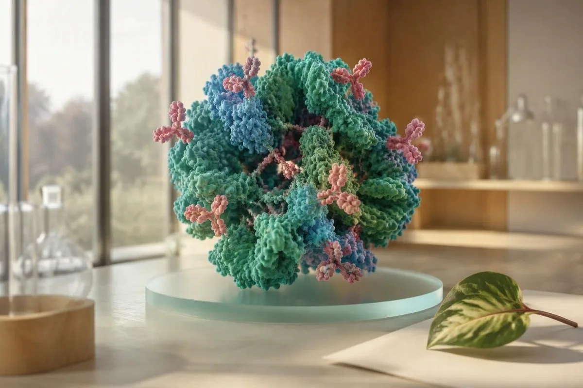
Anti-Sm is one of the most lupus-specific antibodies, but a laboratory result never replaces a clinical diagnosis. The useful question is whether the result fits your symptoms and the rest of your testing.


- Spesifisiteit of a confirmed anti-Sm result is usually about 95–99% for systemic lupus erythematosus.
- Sensitiwiteit is low: only about 5–30% of people with lupus have anti-Sm antibodies.
- ANA threshold of at least 1:80 on HEp-2 cells is the EULAR/ACR classification entry criterion.
- Six points are assigned to anti-Sm antibodies in the 2019 EULAR/ACR lupus classification criteria.
- No universal titre defines severity because anti-Sm assays use different antigens and reporting units.
- Nierkontroles include urinalysis, urine protein-to-creatinine ratio, creatinine/eGFR, C3, C4, CBC, and anti-dsDNA.
- Dringende simptome include chest pain with breathlessness, seizure, confusion, visible blood in urine, or rapidly reduced urine output.
What a Positive Anti-Sm Result Actually Means
'n anti-Smith antibody positive result means your immune system has formed antibodies against Sm nuclear proteins. A confirmed result is highly specific for systemic lupus erythematosus, often 95–99%, but it does not diagnose lupus by itself; symptoms, examination findings, urine testing, blood counts, complement, and other antibodies determine whether lupus is the best explanation.
I use anti-Sm as a diagnostic signpost, not a verdict. A patient with inflammatory joint swelling, rash, low C3, and protein in urine has a very different likelihood of lupus than a person with fatigue alone and one low-positive antibody. The ANA resultaat gids explains the usual starting point.
The name Smith came from an early patient, not a gene or family condition. Anti-Sm targets spliceosome proteins that process RNA inside cells; it does not directly measure current disease activity. That distinction matters when people assume a stronger-looking laboratory flag means more severe lupus.
As of September 28, 2026, an isolated anti-Sm result calls for careful review, not immediate treatment. In my clinical experience, a quiet urine abnormality can matter far more than the antibody number itself. Dr. Thomas Klein has also seen confirmed anti-Sm positivity in people who never developed classifiable SLE.
Why Anti-Sm Is Specific but Not Sensitive for Lupus
Anti-Sm antibodies are highly specific but poorly sensitive for lupus. Roughly 5–30% of people with established SLE test positive, depending on ancestry, cohort selection, and assay, which means a negative anti-Sm result cannot rule lupus out.
Specificity asks how rarely a test is positive without disease; sensitivity asks how often it detects disease when present. Anti-Sm performs very well on specificity and modestly on sensitivity. This is why the same antibody can be a persuasive clue when present yet unhelpful when absent.
Reported prevalence differs across populations. Some Black, Asian, and Hispanic lupus cohorts show higher anti-Sm prevalence than White European cohorts, although referral patterns and laboratory methods contribute. A group percentage cannot predict an individual's future kidney, skin, or joint involvement.
The 2019 EULAR/ACR criteria award anti-Sm antibodies 6 points, reflecting their discriminatory value, but antibodies alone do not establish classification (Aringer et al., 2019). Anti-dsDNA results may provide a more useful activity trend in some patients, particularly when renal disease is being assessed.
How an Anti-Sm Antibody Test Is Measured
Anti-Sm results have no universal normal range or severity titre. Laboratories use ELISA, multiplex bead assays, line immunoassays, or immunoprecipitation, and each method has its own negative, equivocal, and positive cutoffs.
One laboratory may report an index, another arbitrary units such as U/mL, and another only positive or negative. A value of 25 U/mL at one laboratory is not interchangeable with 25 U/mL elsewhere. The laboratory name, reference interval, and assay method belong in any specialist referral.
Low positives deserve scrutiny if the ANA is negative or the clinical picture is incompatible with lupus. Non-specific binding and assay discordance are uncommon but genuine issues, especially in broad automated panels. Read more about kwalitatiewe en kwantitatiewe toetse before comparing results across reports.
Kantesti is 'n KI-bloedtoetsontleder that reads the reporting laboratory, date, companion markers, and result language rather than treating one flag as a diagnosis. An unexpected anti-Sm result is best viewed as a reason for clinician-led confirmation and context review.
How Anti-Sm Fits the Lupus Classification Criteria
Anti-Sm contributes 6 points in the 2019 EULAR/ACR SLE classification criteria, but 10 points and at least one clinical criterion are required. An ANA of at least 1:80 on HEp-2 cells is the entry criterion before any points are counted.
Classification criteria make research cohorts consistent; they do not replace a physician's diagnostic judgment. A patient may have lupus before meeting every classification feature, or may meet a numerical threshold while another illness better explains the presentation. This nuance prevents both delayed care and overdiagnosis.
Anti-Sm sits in the SLE-specific antibody domain with anti-dsDNA; only the highest item within a domain counts. Kidney biopsy class III or IV lupus nephritis scores 10 points, while fever scores 2 and acute cutaneous lupus scores 6. The numbers describe a framework, not a disease meter.
The earlier SLICC system also required immunologic and clinical evidence, reinforcing the same core principle: laboratory autoimmunity needs clinical correlation (Petri et al., 2012). Our C3, C4 en ANA lei shows the tests commonly read alongside anti-Sm.
Symptoms That Make an Anti-Sm Result More Meaningful
Anti-Sm is more clinically meaningful when it accompanies objective lupus features. These include inflammatory arthritis, photosensitive or acute rashes, mouth ulcers, serositis, cytopenias, neurologic features, proteinuria, or active urinary sediment.
Inflammatory arthritis generally causes swollen or tender joints with morning stiffness lasting 30–60 minutes or longer, rather than pain only after a busy day. Lupus rashes commonly spare the nasolabial folds, but skin findings are variable enough that photographs and dermatology review can be genuinely useful.
A history of recurrent painless mouth ulcers, sunlight-triggered rash, pleuritic chest pain, unexplained low white cells, and Raynaud-type color changes raises the pre-test probability. No single symptom is unique to SLE. Fibromyalgia, viral illness, thyroid disease, medication effects, and other autoimmune conditions can overlap.
Document patterns rather than trying to self-score lupus. A dated symptom diary, photographs of transient rash, medication list, and prior results help a rheumatologist substantially. People with cold-sensitive color changes may find our Raynaud blood-test overview useful while awaiting review.
Why Urine and Kidney Tests Matter After Anti-Sm Positivity
Urinalysis and urine protein measurement are essential after a concerning lupus antibody result because kidney inflammation can be silent. Persistent proteinuria of 500 mg per 24 hours, or a urine protein-to-creatinine ratio of 500 mg/g or higher, warrants prompt specialist assessment in suspected SLE.
A normal creatinine does not fully exclude early lupus nephritis. Creatinine rises late in some people because kidney reserve is substantial, while urine protein, red-cell casts, dysmorphic red cells, or declining complement may appear earlier. First-morning urine reduces dilution variability when a ratio is being checked.
Protein can arise from fever, strenuous exercise, diabetes, urinary infection, or contamination, so persistence matters. I usually want a repeat clean-catch urine sample and microscopy when a dipstick is positive. The practical details in our urine sediment guide can help readers understand the report.
New ankle swelling, foamy urine that persists, high blood pressure, or reduced urine should not wait for a routine annual review. Kidney involvement may require immunosuppressive treatment, but treatment choice depends on biopsy class, pregnancy plans, infection risk, and kidney function—not anti-Sm status alone.
The Companion Results Rheumatologists Check
The most useful companion tests are CBC with differential, creatinine/eGFR, urinalysis, urine protein ratio, C3, C4, anti-dsDNA, ESR, and CRP. Low C3 or C4 can support immune-complex activity, but normal complement does not exclude lupus.
Complement reference intervals vary, but many laboratories list C3 around 90–180 mg/dL and C4 around 10–40 mg/dL. A low result is more persuasive when it is newly reduced from that person's own baseline and coincides with proteinuria or rising anti-dsDNA. Low complement results need pattern-based interpretation.
CBC changes may include leukopenia below 4.0 × 10⁹/L, lymphopenia below 1.0 × 10⁹/L, thrombocytopenia below 100 × 10⁹/L, or hemolytic anemia. A single borderline count after a viral illness is not equivalent to persistent immune cytopenia. Timing and repeat samples matter.
CRP is often normal or only modestly raised during uncomplicated lupus activity, whereas a markedly high CRP should widen the search for infection, serositis, or another inflammatory process. That is a clinical rule of thumb, not an absolute rule; WBC and CRP patterns can be especially helpful.
Does Anti-Sm Predict Severe Lupus or a Flare?
Anti-Sm positivity does not reliably measure lupus severity, flare intensity, or treatment response. It often persists for years, so clinicians do not usually repeat it to decide whether inflammation is improving.
Some observational studies link anti-Sm with particular manifestations, including renal or neurologic disease in selected populations, but the associations are inconsistent after accounting for ancestry and coexisting antibodies. I would never forecast an individual's organ risk from anti-Sm alone. That approach creates anxiety without improving decisions.
For established lupus, trends in symptoms, examination, blood pressure, urine protein, urine sediment, creatinine, C3, C4, and anti-dsDNA are usually more actionable. A urine protein ratio that rises from 150 to 900 mg/g tells a more urgent story than a stable anti-Sm result.
Dr. Thomas Klein advises patients to ask which result will actually change the treatment plan. If the answer is anti-Sm alone, that deserves a conversation. A bloedtoets-tydlyn can help separate biological change from a single noisy measurement.
Can Anti-Sm Be Positive Without Lupus?
Anti-Sm positivity outside lupus is unusual but possible, especially with low-level assay signals or discordant testing. A result that conflicts with a negative ANA, absent clinical features, or a different laboratory method should be reviewed before a lifelong diagnosis is considered.
Technical differences are the commonest reason two reports disagree. Solid-phase assays may detect limited protein fragments, while other methods identify different epitopes or use different thresholds. Repeating the identical panel without a clinical question can add confusion; a rheumatologist may instead request a methodologically different confirmation.
Other systemic autoimmune diseases can occasionally produce broad autoantibody reactivity, but true isolated anti-Sm positivity remains far more associated with SLE than most antibodies on an ENA panel. A positive test does not explain every symptom: thyroid disease, anemia, sleep disorders, and medication effects still deserve ordinary medical care.
Do not stop or start medication solely because of an antibody portal result. The broader principles in our positive antibody guide apply here: confirm identity, establish clinical fit, then decide whether monitoring is needed.
When to Repeat or Confirm an Anti-Sm Test
Repeat anti-Sm testing is most useful when the original result is low-positive, unexpected, or technically discordant. Repeating a stable, clearly positive anti-Sm result every 3–6 months rarely adds useful information in established lupus.
Confirmation is reasonable when ANA is negative, symptoms are absent, the sample was tested as part of a large screen, or the report uses an equivocal category. Ask whether the laboratory can perform an ENA confirmation or whether a reference laboratory uses a different platform. Keep the original report; the assay name is useful data.
If symptoms are evolving, the repeat tests that usually matter more are urinalysis, urine protein ratio, CBC, creatinine, C3, C4, and anti-dsDNA. The right interval depends on findings: a patient with new proteinuria needs faster review than one with a normal examination and stable tests.
Kantesti is 'n AI bloedtoets interpretasie platform that can place serial laboratory reports side by side and flag meaningful context changes for discussion with a clinician. It cannot diagnose lupus, but organized trends can prevent an old antibody result from overshadowing newer kidney or blood-count changes.
Medicines, Pregnancy, and Anti-Sm Antibodies
Anti-Sm antibodies do not cause drug-induced lupus and are not the main antibody used to estimate pregnancy-specific neonatal lupus risk. Anti-Ro/SSA and anti-La/SSB, rather than anti-Sm, guide fetal heart-block surveillance discussions.
Classic drug-induced lupus more often involves antihistone antibodies and usually has less kidney or central nervous system involvement than idiopathic SLE. Hydralazine, procainamide, isoniazid, minocycline, and some biologic therapies are among medicines clinicians review when symptoms emerge. Never discontinue a prescribed medicine without a prescriber.
Pregnancy is safest when lupus has been clinically quiet for at least 6 months before conception, particularly after nephritis. Anti-Sm positivity itself does not determine delivery timing, anticoagulation, or treatment. Blood pressure, urine protein, kidney function, anti-Ro/La, and antiphospholipid antibodies are usually more decision-relevant.
People planning pregnancy should also understand a positive lupus antikoagulant resultaat, which is a clotting-risk test despite its confusing name. It is biologically different from anti-Sm and does not diagnose lupus by itself.
What to Expect at a Rheumatology Review
A rheumatology assessment after anti-Sm positivity usually combines a detailed symptom timeline, examination, repeat targeted laboratories, and urine testing. The appointment is not simply a yes-or-no antibody interpretation; it is an assessment of current organ involvement and alternative explanations.
Bring laboratory reports rather than screenshots alone, including dates and reference ranges. Mention rashes, fevers, mouth sores, joint stiffness duration, miscarriages, clots, neurologic symptoms, medication exposures, and family history. A normal exam between episodes does not invalidate a useful photograph of a transient rash.
The clinician may request ANA by indirect immunofluorescence, ENA profile, anti-dsDNA, complement, CBC, metabolic panel, urinalysis, urine protein ratio, antiphospholipid antibodies, and selected infection or thyroid tests. Not every person needs every test; focused testing lowers incidental findings.
Kantesti is 'n KI-aangedrewe bloedtoets-analisehulpmiddel used across 127+ countries to organize multi-laboratory reports before appointments. Readers can also review our 15,000-plus biomarker-gids to understand which results belong to kidney, immune, and blood-count domains.
When an Anti-Sm Positive Result Needs Urgent Care
Seek urgent medical assessment for chest pain with breathlessness, coughing blood, new seizure, new confusion, severe headache with neurologic symptoms, rapidly worsening swelling, or visibly bloody urine. These symptoms may reflect serious complications but are not caused by anti-Sm itself.
A sudden severe headache, weakness on one side, speech difficulty, or seizure needs emergency assessment regardless of whether lupus is confirmed. Similarly, pleuritic chest pain and shortness of breath can result from infection, pulmonary embolism, pericarditis, or other causes that cannot be distinguished safely at home.
Visible blood in urine, markedly reduced urine output, blood pressure of 180/120 mmHg or higher with symptoms, or rapidly increasing leg swelling should prompt same-day medical care. A dipstick alone cannot diagnose nephritis, but it can reveal why waiting is unwise. See our blood-in-urine guide for practical triage context.
For non-urgent situations, schedule primary-care or rheumatology follow-up rather than repeatedly searching isolated test names. The clinical standards behind our interpretation workflows are described in mediese validering, and human assessment remains essential when autoimmune disease is possible.
A Practical Plan After an Anti-Sm Positive Test
The next step after anti-Sm positivity is to verify the assay, screen for organ involvement, and arrange clinician review based on symptoms. There is no evidence-based diet, supplement, or detox that can erase anti-Sm antibodies or substitute for lupus assessment.
Start by obtaining the full report: ANA method and titre, anti-Sm result and units, reference range, and all ENA antibodies. Then collect CBC, creatinine/eGFR, urinalysis, urine protein ratio, C3, C4, and anti-dsDNA if they have not been checked. One well-organized assessment is better than scattered repeated panels.
Do not assume joint pain means lupus or that normal routine blood work excludes it. Symptoms lasting more than 6 weeks, objective swelling, recurrent rash, cytopenias, proteinuria, or low complement justify specialist input. Conversely, an asymptomatic person with reassuring repeat evaluation may need observation rather than medication.
Ons Mediese Adviesraad reviews the clinical guardrails used in Kantesti's educational content. The honest bottom line is reassuringly simple: anti-Sm is a strong clue, not a diagnosis, and the rest of the clinical picture tells you what it means.
Gereelde vrae
Beteken 'n anti-Smith-teenliggaam positiewe toets beslis dat ek lupus het?
Nee. 'n Bevestigde anti-Sm-teenliggaam positiewe resultaat is hoogs spesifiek vir sistemiese lupus erythematosus, algemeen rondom 95–99%, maar diagnose vereis steeds versoenbare kliniese bevindinge en uitsluiting van alternatiewe. Die 2019 EULAR/ACR-klassifikasiestelsel gee anti-Sm 6 punte, tog vereis dit ten minste 10 punte en een kliniese kriterium na 'n ANA-inskrywingskriterium van ten minste 1:80. 'n Reumatoloog moet simptome, urine, CBC, nierfunksie, komplement en anti-dsDNA assesseer eerder as om lupus slegs uit anti-Sm te diagnoseer.
Kan jy lupus hê met 'n negatiewe anti-Sm-teenliggaamtoets?
Ja. Slegs ongeveer 5–30% van mense met gevestigde sistemiese lupus erythematosus het anti-Sm-teenliggaampies, so 'n negatiewe anti-Sm-resultaat sluit nie lupus uit nie. ANA, anti-dsDNA, komplementvlakke, urienproteïen, bloedtelling, simptome en ondersoekbevindinge is meer insiggewend as 'n gekombineerde patroon. Die sensitiwiteit wissel volgens afkoms en toetsmetode, wat 'n ander rede is waarom 'n enkele negatiewe resultaat nie deurslaggewend is nie.
Watter anti-Sm vlak word as hoog beskou?
Daar is geen universele anti-Sm-vlak wat hoë siekteaktiwiteit of ernstige lupus definieer nie. Laboratoriums mag arbitrêre eenhede, indekswaardes, fluoressensiepatrone rapporteer, of bloot positiewe en negatiewe resultate, en hul afsnypunte is nie uitruilbaar nie. 'n Resultaat bo 'n laboratoriumafsnypunt is slegs klinies betekenisvol wanneer dit bevestig en geïnterpreteer word met simptome en metgesel toetsing. Anders as urienproteïen of kreatinien, word anti-Sm-titers nie roetine gebruik om 'n lupus-vlam op te spoor nie.
Watter toetse moet volg op 'n positiewe anti-Sm-teenliggaam?
Opvolging na anti-Sm-positiviteit sluit gewoonlik ANA deur indirekte immunofluoresensie, anti-dsDNA, C3, C4, CBC met differensiaal, kreatinien/eGFR, urinaleuse, en 'n urine-proteïen-tot-kreatinien-verhouding in. Aanhoudende proteinuria van 500 mg per 24 uur of 'n verhouding van 500 mg/g of hoër in vermoedelike SLE regverdig dringende spesialis evaluasie. Die presiese paneel hang af van simptome, swangerskapplanne, medikasieblootstelling, en die oorspronklike toets.
Kan anti-Sm-teenliggame negatief word?
Anti-Sm-teenstowwe kan wissel per toets en soms negatief word, maar dit bly dikwels jare lank opspoorbaar. 'n Verandering van positief na negatief bewys nie dat lupus opgelos het nie, en 'n stabiele positiewe resultaat bewys nie aktiewe inflammasie nie. Klinici volg gewoonlik simptome, proteïen in urine, kreatinien, komplement, CBC, en anti-dsDNA noukeuriger as herhaalde anti-Sm-metings. Herhaalde anti-Sm is die nuttigste wanneer die eerste resultaat grensgeval of klinies onverwags was.
Is anti-Sm the same as lupus anticoagulant?
No. Anti-Sm is an antinuclear antibody associated with systemic lupus erythematosus, while lupus anticoagulant is an antiphospholipid antibody test associated with clotting and pregnancy complications. Lupus anticoagulant can occur with or without lupus, and anti-Sm does not by itself predict a clot. Both tests may appear in an autoimmune work-up, but they answer different clinical questions and require separate interpretation.
Kry vandag KI-aangedrewe bloedtoets-analise
Sluit aan by meer as 2 miljoen gebruikers wêreldwyd wat Kantesti vertrou vir onmiddellike, akkurate laboratoriumtoetsanalise. Laai jou bloedtoetsresultate op en ontvang omvattende interpretasie van 15,000+-biomerkers binne sekondes.
📚 Verwysde navorsingspublikasies
Klein, T., Mitchell, S., & Weber, H. (2026). Nipah-virusbloedtoets: Vroeë opsporing en diagnosegids 2026. Kantesti KI Mediese Navorsing.
Klein, T., Mitchell, S., & Weber, H. (2026). B Negatiewe Bloedgroep, LDH-bloedtoets en retikulosiettelling-gids. Kantesti KI Mediese Navorsing.
📖 Eksterne mediese verwysings
⚕️ Mediese Vrywaring
Hierdie artikel is slegs vir opvoedkundige doeleindes en vorm nie mediese advies nie. Raadpleeg altyd ’n gekwalifiseerde gesondheidsorgverskaffer vir besluite oor diagnose en behandeling.
E-E-A-T Vertrouenseine
Ervaring
Kliniese oorsig gelei deur ’n geneesheer van laboratorium-interpretasie-werksvloei.
Kundigheid
Laboratoriumgeneeskunde fokus op hoe biomerkers in ’n kliniese konteks optree.
Gesagsvermoë
Geskryf deur dr. Thomas Klein met hersiening deur dr. Sarah Mitchell en prof. dr. Hans Weber.
Betroubaarheid
Bewysgebaseerde interpretasie met duidelike opvolgpaaie om alarm te verminder.