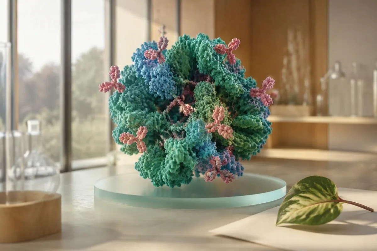
Anti-Sm ເປັນໜຶ່ງໃນພູມຕ້ານທານສະເພາະຂອງພະຍາດ lupus, ແຕ່ຜົນການທົດລອງໃນຫ້ອງທົດລອງບໍ່ສາມາດແທນການວິນິດໄສທາງດ້ານການແພດໄດ້. ຄຳຖາມທີ່ມີປະໂຫຍດແມ່ນວ່າຜົນໄດ້ຮັບນັ້ນກົງກັບອາການຂອງເຈົ້າ ແລະຜົນການທົດສອບອື່ນໆຂອງເຈົ້າຫຼືບໍ່.


- ສະເພາະ ຂອງຜົນ anti-Sm ທີ່ຢືນຢັນແລ້ວ ໂດຍປົກກະຕິແລ້ວແມ່ນປະມານ 95–99% ສຳລັບພະຍາດ systemic lupus erythematosus.
- ຄວາມອ່ອນໄຫວ ຕ່ຳ: ມີພຽງແຕ່ປະມານ 5–30% ຂອງຜູ້ທີ່ເປັນພະຍາດ lupus ທີ່ມີພູມຕ້ານທານ anti-Sm.
- ເງື່ອນໄຂ ANA ຂອງຢ່າງໜ້ອຍ 1:80 ໃນຈຸລັງ HEp-2 ແມ່ນເງື່ອນໄຂການເຂົ້າໃນການຈັດປະເພດ EULAR/ACR.
- ຫົກຄະແນນ ຖືກມອບໃຫ້ແກ່ພູມຕ້ານທານ anti-Sm ໃນເງື່ອນໄຂການຈັດປະເພດ lupus ຂອງ EULAR/ACR ປີ 2019.
- ບໍ່ມີຄ່າ titration ສາກົນ ກຳນົດຄວາມຮຸນແຮງ ເພາະວ່າການທົດສອບ anti-Sm ໃຊ້ antigens ແລະໜ່ວຍລາຍງານທີ່ແຕກຕ່າງກັນ.
- ການກວດໄຕ ລວມມີການວິເຄາະນໍ້າປັດສະວະ, ອັດຕາສ່ວນ protein-to-creatinine ຂອງນໍ້າປັດສະວະ, creatinine/eGFR, C3, C4, CBC, ແລະ anti-dsDNA.
- ອາການດ່ວນ ລວມມີອາການເຈັບໜ້າເອິກພ້ອມກັບຫາຍໃຈຍາກ, ຊັກ, ສະຕິສັບສົນ, ມີເລືອດອອກໃນນໍ້າປັດສະວະທີ່ເຫັນໄດ້, ຫຼືການຜະລິດປັດສະວະຫຼຸດລົງຢ່າງໄວ.
ໝາຍຄວາມວ່າແນວໃດທີ່ຜົນ Anti-Sm ເປັນບວກ
ອັນ ผลบวกของแอนติบอดีต่อต้านสม ผลลัพธ์หมายความว่าระบบภูมิคุ้มกันของคุณได้สร้างแอนติบอดีต่อโปรตีนนิวเคลียร์ Sm การยืนยันผลลัพธ์มีความเฉพาะเจาะจงสูงสำหรับโรค SLE เป็นระบบ ซึ่งมักจะ 95–99% แต่ก็ไม่สามารถวินิจฉัยโรค SLE ได้ด้วยตัวเอง อาการ การตรวจร่างกาย การตรวจปัสสาวะ การนับเม็ดเลือด คอมพลีเมนต์ และแอนติบอดีอื่นๆ จะเป็นตัวกำหนดว่าโรค SLE เป็นคำอธิบายที่ดีที่สุดหรือไม่.
ฉันใช้ anti-Sm เป็นเครื่องบ่งชี้ในการวินิจฉัย ไม่ใช่คำตัดสิน ผู้ป่วยที่มีอาการข้อบวมอักเสบ มีผื่น มีค่า C3 ต่ำ และมีโปรตีนในปัสสาวะ มีโอกาสเป็นโรค SLE แตกต่างจากผู้ที่มีอาการอ่อนเพลียเพียงอย่างเดียวและมีแอนติบอดีบวกเล็กน้อย ຄູ່ມືຜົນການທົດສອບ ANA ຂອງພວກເຮົາ อธิบายจุดเริ่มต้นปกติ.
ชื่อ Smith มาจากผู้ป่วยรายแรก ไม่ใช่จากยีนหรือโรคทางครอบครัว Anti-Sm มีเป้าหมายที่โปรตีน spliceosome ซึ่งประมวลผล RNA ภายในเซลล์ ไม่ได้วัดกิจกรรมของโรคในปัจจุบันโดยตรง ความแตกต่างนี้มีความสำคัญเมื่อผู้คนสันนิษฐานว่าผลการตรวจทางห้องปฏิบัติการที่ดูเหมือนแรงกว่าหมายถึงโรค SLE ที่รุนแรงกว่า.
ณ วันที่ 28 กันยายน 2026 ผล anti-Sm ที่แยกเดี่ยวเรียกร้องให้มีการตรวจสอบอย่างละเอียด ไม่ใช่การรักษาทันที จากประสบการณ์ทางคลินิกของฉัน ความผิดปกติของปัสสาวะที่เงียบสามารถมีความสำคัญมากกว่าตัวเลขของแอนติบอดีเอง. ດຣ. Thomas Klein ยังพบผลบวกของ anti-Sm ที่ยืนยันแล้วในผู้ที่ไม่เคยเป็นโรค SLE ที่สามารถจัดประเภทได้.
ເປັນຫຍັງ Anti-Sm ຈຶ່ງສະເພາະແຕ່ບໍ່ມີຄວາມອ່ອນໄຫວຕໍ່ພະຍາດ lupus
แอนติบอดี Anti-Sm มีความเฉพาะเจาะจงสูง แต่มีความไวต่ำสำหรับโรค SLE. ประมาณ 5–30% ของผู้ที่เป็นโรค SLE ที่ได้รับการยืนยันจะให้ผลบวก ขึ้นอยู่กับเชื้อชาติ การเลือกกลุ่มตัวอย่าง และวิธีการตรวจ ซึ่งหมายความว่าผล anti-Sm ที่เป็นลบไม่สามารถตัดโรค SLE ออกได้.
ความจำเพาะเจาะจงถามว่าการทดสอบมักจะให้ผลบวกโดยไม่มีโรคบ่อยแค่ไหน ความไวถามว่ามันตรวจพบโรคเมื่อมีอยู่บ่อยแค่ไหน Anti-Sm มีประสิทธิภาพที่ดีมากในด้านความจำเพาะเจาะจงและประสิทธิภาพปานกลางในด้านความไว นี่คือเหตุผลว่าทำไมแอนติบอดีชนิดเดียวกันจึงเป็นเบาะแสที่น่าเชื่อถือเมื่อมีอยู่ แต่ไม่มีประโยชน์เมื่อไม่มี.
ความชุกที่รายงานแตกต่างกันไปในแต่ละประชากร กลุ่มตัวอย่างโรค SLE ในคนผิวดำ เอเชีย และฮิสแปนิกบางกลุ่มแสดงความชุกของ anti-Sm สูงกว่ากลุ่มตัวอย่างคนผิวขาวในยุโรป แม้ว่ารูปแบบการส่งต่อและวิธีการทางห้องปฏิบัติการจะมีส่วนเกี่ยวข้องก็ตาม เปอร์เซ็นต์ของกลุ่มไม่สามารถทำนายการมีส่วนร่วมของไต ผิวหนัง หรือข้อต่อในอนาคตของแต่ละบุคคลได้.
เกณฑ์ EULAR/ACR ปี 2019 ให้คะแนนแอนติบอดี anti-Sm 6 คะแนน ซึ่งสะท้อนถึงคุณค่าในการจำแนกประเภท แต่แอนติบอดีเพียงอย่างเดียวไม่สามารถจัดประเภทได้ (Aringer et al., 2019). ผลลัพธ์ Anti-dsDNA อาจให้แนวโน้มกิจกรรมที่มีประโยชน์มากขึ้นในผู้ป่วยบางราย โดยเฉพาะอย่างยิ่งเมื่อมีการประเมินโรคไต.
ການທົດສອບພູມຕ້ານທານ Anti-Sm ຖືກວັດແທກແນວໃດ
ผลลัพธ์ Anti-Sm ไม่มีช่วงปกติสากลหรือค่า titre ที่บ่งบอกความรุนแรง. ห้องปฏิบัติการใช้ ELISA, multiplex bead assays, line immunoassays หรือ immunoprecipitation และแต่ละวิธีมีเกณฑ์ตัดค่าลบ ก้ำกึ่ง และบวกของตัวเอง.
ห้องปฏิบัติการหนึ่งอาจรายงานค่าดัชนี อีกแห่งรายงานหน่วยตามอำเภอใจ เช่น U/mL และอีกแห่งรายงานเพียงบวกหรือลบ ค่า 25 U/mL ที่ห้องปฏิบัติการหนึ่งไม่สามารถใช้แทนกันได้กับ 25 U/mL ที่อื่น ชื่อห้องปฏิบัติการ ช่วงอ้างอิง และวิธีการตรวจเป็นสิ่งสำคัญสำหรับการส่งต่อผู้เชี่ยวชาญ.
ผลบวกน้อยๆ ควรได้รับการตรวจสอบอย่างละเอียดหาก ANA เป็นลบหรือไม่เข้ากันกับภาพทางคลินิกของโรค SLE การจับแบบไม่จำเพาะและความไม่สอดคล้องกันของการตรวจเป็นเรื่องที่เกิดขึ้นไม่บ่อย แต่เป็นเรื่องจริง โดยเฉพาะอย่างยิ่งในแผงอัตโนมัติขนาดใหญ่ อ่านเพิ่มเติมเกี่ยวกับ ການກວດຫາແບບຄຸນນະພາບ ແລະ ປະລິມານ ก่อนเปรียบเทียบผลลัพธ์ระหว่างรายงาน.
ແຄນເທສຕີ ເປັນ AI ເຄື່ອງວິເຄາະເລືອດ ที่อ่านห้องปฏิบัติการที่รายงาน วันที่ เครื่องหมายประกอบ และภาษาของผลลัพธ์ แทนที่จะปฏิบัติต่อธงเดียวเป็นการวินิจฉัย ผล anti-Sm ที่ไม่คาดคิดควรได้รับการพิจารณาว่ามีเหตุผลสำหรับการยืนยันและทบทวนบริบทโดยแพทย์.
Anti-Sm ເໝາະສົມກັບເງື່ອນໄຂການຈັດປະເພດ lupus ແນວໃດ
Anti-Sm มีคะแนน 6 คะแนนในเกณฑ์การจำแนกประเภท SLE ของ EULAR/ACR ปี 2019 แต่ต้องมี 10 คะแนนและอย่างน้อยหนึ่งเกณฑ์ทางคลินิก. ANA อย่างน้อย 1:80 บนเซลล์ HEp-2 เป็นเกณฑ์การเข้าร่วมก่อนที่จะนับคะแนนใดๆ.
Classification criteria make research cohorts consistent; they do not replace a physician's diagnostic judgment. A patient may have lupus before meeting every classification feature, or may meet a numerical threshold while another illness better explains the presentation. This nuance prevents both delayed care and overdiagnosis.
Anti-Sm sits in the SLE-specific antibody domain with anti-dsDNA; only the highest item within a domain counts. Kidney biopsy class III or IV lupus nephritis scores 10 points, while fever scores 2 and acute cutaneous lupus scores 6. The numbers describe a framework, not a disease meter.
The earlier SLICC system also required immunologic and clinical evidence, reinforcing the same core principle: laboratory autoimmunity needs clinical correlation (Petri et al., 2012). Our C3, C4 ແລະ ANA ນຳທາງ shows the tests commonly read alongside anti-Sm.
ອາການທີ່ເຮັດໃຫ້ຜົນ Anti-Sm ມີຄວາມໝາຍຫຼາຍຂຶ້ນ
Anti-Sm is more clinically meaningful when it accompanies objective lupus features. These include inflammatory arthritis, photosensitive or acute rashes, mouth ulcers, serositis, cytopenias, neurologic features, proteinuria, or active urinary sediment.
Inflammatory arthritis generally causes swollen or tender joints with morning stiffness lasting 30–60 minutes or longer, rather than pain only after a busy day. Lupus rashes commonly spare the nasolabial folds, but skin findings are variable enough that photographs and dermatology review can be genuinely useful.
A history of recurrent painless mouth ulcers, sunlight-triggered rash, pleuritic chest pain, unexplained low white cells, and Raynaud-type color changes raises the pre-test probability. No single symptom is unique to SLE. Fibromyalgia, viral illness, thyroid disease, medication effects, and other autoimmune conditions can overlap.
Document patterns rather than trying to self-score lupus. A dated symptom diary, photographs of transient rash, medication list, and prior results help a rheumatologist substantially. People with cold-sensitive color changes may find our Raynaud blood-test overview useful while awaiting review.
ເປັນຫຍັງການທົດສອບນໍ້າປັດສະວະ ແລະໝາກໄຂ່ຫຼັງຈຶ່ງສຳຄັນຫຼັງຈາກ Anti-Sm ເປັນບວກ
Urinalysis and urine protein measurement are essential after a concerning lupus antibody result because kidney inflammation can be silent. Persistent proteinuria of 500 mg per 24 hours, or a urine protein-to-creatinine ratio of 500 mg/g or higher, warrants prompt specialist assessment in suspected SLE.
A normal creatinine does not fully exclude early lupus nephritis. Creatinine rises late in some people because kidney reserve is substantial, while urine protein, red-cell casts, dysmorphic red cells, or declining complement may appear earlier. First-morning urine reduces dilution variability when a ratio is being checked.
Protein can arise from fever, strenuous exercise, diabetes, urinary infection, or contamination, so persistence matters. I usually want a repeat clean-catch urine sample and microscopy when a dipstick is positive. The practical details in our urine sediment guide can help readers understand the report.
New ankle swelling, foamy urine that persists, high blood pressure, or reduced urine should not wait for a routine annual review. Kidney involvement may require immunosuppressive treatment, but treatment choice depends on biopsy class, pregnancy plans, infection risk, and kidney function—not anti-Sm status alone.
ຜົນການທົດສອບຄູ່ທີ່ນັກຊ່ຽວຊານດ້ານຂໍ້ຕໍ່ກວດກາ
The most useful companion tests are CBC with differential, creatinine/eGFR, urinalysis, urine protein ratio, C3, C4, anti-dsDNA, ESR, and CRP. Low C3 or C4 can support immune-complex activity, but normal complement does not exclude lupus.
Complement reference intervals vary, but many laboratories list C3 around 90–180 mg/dL and C4 around 10–40 mg/dL. A low result is more persuasive when it is newly reduced from that person's own baseline and coincides with proteinuria or rising anti-dsDNA. Low complement results need pattern-based interpretation.
CBC changes may include leukopenia below 4.0 × 10⁹/L, lymphopenia below 1.0 × 10⁹/L, thrombocytopenia below 100 × 10⁹/L, or hemolytic anemia. A single borderline count after a viral illness is not equivalent to persistent immune cytopenia. Timing and repeat samples matter.
CRP is often normal or only modestly raised during uncomplicated lupus activity, whereas a markedly high CRP should widen the search for infection, serositis, or another inflammatory process. That is a clinical rule of thumb, not an absolute rule; WBC and CRP patterns can be especially helpful.
Anti-Sm ສາມາດຄາດຄະເນພະຍາດ lupus ຮ້າຍແຮງ ຫຼືອາການລຸກໜັກໄດ້ບໍ?
Anti-Sm positivity does not reliably measure lupus severity, flare intensity, or treatment response. It often persists for years, so clinicians do not usually repeat it to decide whether inflammation is improving.
Some observational studies link anti-Sm with particular manifestations, including renal or neurologic disease in selected populations, but the associations are inconsistent after accounting for ancestry and coexisting antibodies. I would never forecast an individual's organ risk from anti-Sm alone. That approach creates anxiety without improving decisions.
For established lupus, trends in symptoms, examination, blood pressure, urine protein, urine sediment, creatinine, C3, C4, and anti-dsDNA are usually more actionable. A urine protein ratio that rises from 150 to 900 mg/g tells a more urgent story than a stable anti-Sm result.
Dr. Thomas Klein advises patients to ask which result will actually change the treatment plan. If the answer is anti-Sm alone, that deserves a conversation. A กำหนดการทดสอบเลือด can help separate biological change from a single noisy measurement.
Anti-Sm ສາມາດເປັນບວກໄດ້ໂດຍບໍ່ມີພະຍາດ lupus ບໍ?
Anti-Sm positivity outside lupus is unusual but possible, especially with low-level assay signals or discordant testing. A result that conflicts with a negative ANA, absent clinical features, or a different laboratory method should be reviewed before a lifelong diagnosis is considered.
Technical differences are the commonest reason two reports disagree. Solid-phase assays may detect limited protein fragments, while other methods identify different epitopes or use different thresholds. Repeating the identical panel without a clinical question can add confusion; a rheumatologist may instead request a methodologically different confirmation.
Other systemic autoimmune diseases can occasionally produce broad autoantibody reactivity, but true isolated anti-Sm positivity remains far more associated with SLE than most antibodies on an ENA panel. A positive test does not explain every symptom: thyroid disease, anemia, sleep disorders, and medication effects still deserve ordinary medical care.
Do not stop or start medication solely because of an antibody portal result. The broader principles in our positive antibody guide apply here: confirm identity, establish clinical fit, then decide whether monitoring is needed.
ເມື່ອໃດຄວນເຮັດຊ້ຳ ຫຼືຢືນຢັນການທົດສອບ Anti-Sm
Repeat anti-Sm testing is most useful when the original result is low-positive, unexpected, or technically discordant. Repeating a stable, clearly positive anti-Sm result every 3–6 months rarely adds useful information in established lupus.
Confirmation is reasonable when ANA is negative, symptoms are absent, the sample was tested as part of a large screen, or the report uses an equivocal category. Ask whether the laboratory can perform an ENA confirmation or whether a reference laboratory uses a different platform. Keep the original report; the assay name is useful data.
If symptoms are evolving, the repeat tests that usually matter more are urinalysis, urine protein ratio, CBC, creatinine, C3, C4, and anti-dsDNA. The right interval depends on findings: a patient with new proteinuria needs faster review than one with a normal examination and stable tests.
ແຄນເທສຕີ ເປັນ ແພລດຟອມການອ່ານຜົນກວດເລືອດຂອງ AI that can place serial laboratory reports side by side and flag meaningful context changes for discussion with a clinician. It cannot diagnose lupus, but organized trends can prevent an old antibody result from overshadowing newer kidney or blood-count changes.
ຢາ, ການຖືພາ, ແລະພູມຕ້ານທານ Anti-Sm
Anti-Sm antibodies do not cause drug-induced lupus and are not the main antibody used to estimate pregnancy-specific neonatal lupus risk. Anti-Ro/SSA and anti-La/SSB, rather than anti-Sm, guide fetal heart-block surveillance discussions.
Classic drug-induced lupus more often involves antihistone antibodies and usually has less kidney or central nervous system involvement than idiopathic SLE. Hydralazine, procainamide, isoniazid, minocycline, and some biologic therapies are among medicines clinicians review when symptoms emerge. Never discontinue a prescribed medicine without a prescriber.
Pregnancy is safest when lupus has been clinically quiet for at least 6 months before conception, particularly after nephritis. Anti-Sm positivity itself does not determine delivery timing, anticoagulation, or treatment. Blood pressure, urine protein, kidney function, anti-Ro/La, and antiphospholipid antibodies are usually more decision-relevant.
People planning pregnancy should also understand a positive ຜົນຂອງ lupus anticoagulant, which is a clotting-risk test despite its confusing name. It is biologically different from anti-Sm and does not diagnose lupus by itself.
ຈະຄາດຫວັງຫຍັງໃນການກວດຄືນໂດຍນັກຊ່ຽວຊານດ້ານຂໍ້ຕໍ່
A rheumatology assessment after anti-Sm positivity usually combines a detailed symptom timeline, examination, repeat targeted laboratories, and urine testing. The appointment is not simply a yes-or-no antibody interpretation; it is an assessment of current organ involvement and alternative explanations.
Bring laboratory reports rather than screenshots alone, including dates and reference ranges. Mention rashes, fevers, mouth sores, joint stiffness duration, miscarriages, clots, neurologic symptoms, medication exposures, and family history. A normal exam between episodes does not invalidate a useful photograph of a transient rash.
The clinician may request ANA by indirect immunofluorescence, ENA profile, anti-dsDNA, complement, CBC, metabolic panel, urinalysis, urine protein ratio, antiphospholipid antibodies, and selected infection or thyroid tests. Not every person needs every test; focused testing lowers incidental findings.
ແຄນເທສຕີ ເປັນ ឧបករណ៍វិភាគតេស្តឈាមដែលដំណើរការដោយ AI used across 127+ countries to organize multi-laboratory reports before appointments. Readers can also review our ຄູ່ມື biomarker ທີ່ມີ 15,000-plus to understand which results belong to kidney, immune, and blood-count domains.
ເມື່ອຜົນ Anti-Sm ເປັນບວກຕ້ອງການການດູແລຮີບດ່ວນ
Seek urgent medical assessment for chest pain with breathlessness, coughing blood, new seizure, new confusion, severe headache with neurologic symptoms, rapidly worsening swelling, or visibly bloody urine. These symptoms may reflect serious complications but are not caused by anti-Sm itself.
A sudden severe headache, weakness on one side, speech difficulty, or seizure needs emergency assessment regardless of whether lupus is confirmed. Similarly, pleuritic chest pain and shortness of breath can result from infection, pulmonary embolism, pericarditis, or other causes that cannot be distinguished safely at home.
Visible blood in urine, markedly reduced urine output, blood pressure of 180/120 mmHg or higher with symptoms, or rapidly increasing leg swelling should prompt same-day medical care. A dipstick alone cannot diagnose nephritis, but it can reveal why waiting is unwise. See our blood-in-urine guide for practical triage context.
For non-urgent situations, schedule primary-care or rheumatology follow-up rather than repeatedly searching isolated test names. The clinical standards behind our interpretation workflows are described in ການກວດສອບທາງການແພດ, and human assessment remains essential when autoimmune disease is possible.
ແຜນການປະຕິບັດຕົວຈິງຫຼັງຈາກການທົດສອບ Anti-Sm ເປັນບວກ
The next step after anti-Sm positivity is to verify the assay, screen for organ involvement, and arrange clinician review based on symptoms. There is no evidence-based diet, supplement, or detox that can erase anti-Sm antibodies or substitute for lupus assessment.
Start by obtaining the full report: ANA method and titre, anti-Sm result and units, reference range, and all ENA antibodies. Then collect CBC, creatinine/eGFR, urinalysis, urine protein ratio, C3, C4, and anti-dsDNA if they have not been checked. One well-organized assessment is better than scattered repeated panels.
Do not assume joint pain means lupus or that normal routine blood work excludes it. Symptoms lasting more than 6 weeks, objective swelling, recurrent rash, cytopenias, proteinuria, or low complement justify specialist input. Conversely, an asymptomatic person with reassuring repeat evaluation may need observation rather than medication.
ຂອງພວກເຮົາ ຄະນະທີ່ປຶກສາທາງການແພດ reviews the clinical guardrails used in Kantesti's educational content. The honest bottom line is reassuringly simple: anti-Sm is a strong clue, not a diagnosis, and the rest of the clinical picture tells you what it means.
ຄໍາຖາມທີ່ຖາມເລື້ອຍໆ
ผลตรวจแอนติบอดีต่อสมิธ (anti-Smith antibody) เป็นบวก หมายความว่าฉันเป็นโรคแพ้ภูมิตัวเองแน่นอนใช่หรือไม่
ບໍ່. ຜົນການກວດພົບສານຕ້ານ Sm antibody ທີ່ຢືນຢັນແລ້ວແມ່ນມີຄວາມສະເພາະສູງຕໍ່ພະຍາດ systemic lupus erythematosus, ໂດຍທົ່ວໄປແມ່ນປະມານ 95–99%, ແຕ່ການວິນິດໄສຍັງຕ້ອງການການຄົ້ນພົບທາງດ້ານຄລີນິກທີ່ສອດຄ່ອງກັນ ແລະ ການຕັດທາງເລືອກອື່ນອອກ. ລະບົບການຈັດປະເພດ EULAR/ACR ປີ 2019 ໃຫ້ຄະແນນ anti-Sm 6 ຄະແນນ, ແຕ່ຍັງຕ້ອງການຢ່າງໜ້ອຍ 10 ຄະແນນ ແລະ ເງື່ອນໄຂທາງຄລີນິກໜຶ່ງຢ່າງ ຫຼັງຈາກເງື່ອນໄຂການເຂົ້າລະບົບ ANA ຢ່າງໜ້ອຍ 1:80. ແພດໝໍຊ່ຽວຊານດ້ານຂໍ້ກະດູກຄວນກວດກາອາການ, ປັດສະວະ, CBC, ການເຮັດວຽກຂອງไต, complement, ແລະ anti-dsDNA ແທນທີ່ຈະວິນິດໄສ lupus ຈາກ anti-Sm ຢ່າງດຽວ.
ທ່ານສາມາດເປັນພະຍາດ lupus ທີ່ມີການທົດສອບພູມຕ້ານທານ anti-Sm ທີ່ເປັນລົບໄດ້ບໍ?
ແມ່ນແລ້ວ. ມີປະຊາຊົນປະມານ 5–30% ຂອງຜູ້ທີ່ເປັນພະຍາດ systemic lupus erythematosus ທີ່ມີພູມຕ້ານທານ anti-Sm, ສະນັ້ນຜົນການກວດ anti-Sm ທີ່ເປັນລົບບໍ່ໄດ້ໝາຍຄວາມວ່າບໍ່ເປັນ lupus. ANA, anti-dsDNA, ລະດັບ complement, ໂປຣຕີນໃນຍ່ຽວ, ຈຳນວນເມັດເລືອດ, ອາການ, ແລະຜົນການກວດຮ່າງກາຍແມ່ນຂໍ້ມູນທີ່ໃຫ້ຂໍ້ມູນຫຼາຍກວ່າເມື່ອພິຈາລະນາຮ່ວມກັນ. ຄວາມລະອຽດແຕກຕ່າງກັນໄປຕາມເຊື້ອຊາດ ແລະ ວິທີການກວດ, ນີ້ແມ່ນອີກເຫດຜົນໜຶ່ງທີ່ຜົນລົບຄັ້ງດຽວບໍ່ໄດ້ຕັດສິນ.
ລະດັບ anti-Sm ໃດຖືວ່າສູງ?
ບໍ່ມີລະດັບ anti-Sm ສາກົນທີ່ກຳນົດກິດຈະກຳຂອງພະຍາດໃນລະດັບສູງ ຫຼື lupus ຮຸນແຮງ. ຫ້ອງທົດລອງອາດລາຍງານຫົວໜ່ວຍທີ່ຕົນເອງກຳນົດ, ຄ່າດັດຊະນີ, ຮູບແບບ fluorescence, ຫຼືພຽງແຕ່ຜົນບວກ ແລະ ລົບ, ແລະ ລະດັບຕັດຂອງເຂົາເຈົ້າບໍ່ສາມາດແລກປ່ຽນກັນໄດ້. ຜົນລັບທີ່ສູງກວ່າລະດັບຕັດຂອງຫ້ອງທົດລອງແມ່ນມີຄວາມໝາຍທາງຄລີນິກເມື່ອໄດ້ຮັບການຢືນຢັນ ແລະ ຕີຄວາມໝາຍພ້ອມກັບອາການ ແລະ ການທົດສອບອື່ນໆ. ບໍ່ຄືກັບໂປຣຕີນໃນປັດສະວະ ຫຼື creatinine, ລະດັບ anti-Sm ບໍ່ໄດ້ຖືກນຳໃຊ້ເປັນປະຈຳເພື່ອຕິດຕາມການເປັນ lupus flare.
ການທົດສອບໃດຄວນປະຕິບັດ ຫຼັງຈາກຜົນກວດພົບພູມຕ້ານທານ anti-Sm ທີ່ເປັນບວກ?
ການຕິດຕາມຫຼັງຈາກການກວດພົບ anti-Sm ໂດຍທົ່ວໄປປະກອບມີ ANA ໂດຍການຍ້ອມສີແບບ indirect immunofluorescence, anti-dsDNA, C3, C4, CBC ພ້ອມກັບ differential, creatinine/eGFR, urinalysis, ແລະ ອັດຕາສ່ວນ protein-to-creatinine ຂອງປັດສະວະ. ອາການແຊກຊ້ອນຂອງໂລກ SLE ທີ່ສົງໃສວ່າ ພົບໂປຣຕີນໃນປັດສະວະຢ່າງຕໍ່ເນື່ອງ 500 mg ຕໍ່ 24 ຊົ່ວໂມງ ຫຼື ອັດຕາສ່ວນ 500 mg/g ຫຼືສູງກວ່າ, ຕ້ອງການການປະເມີນຜົນຈາກຜູ້ຊ່ຽວຊານຢ່າງຮີບດ່ວນ. ແຜງການກວດທີ່ແນ່ນອນແມ່ນຂຶ້ນກັບອາການ, ແຜນການຖືພາ, ການໄດ້ຮັບຢາ, ແລະ ການກວດວິເຄາະຕົ້ນສະບັບ.
แอนติ-เอสเอ็ม แอนติบอดี สามารถเป็นลบได้หรือไม่?
Anti-Sm antibodies ສາມາດປ່ຽນແປງໄປຕາມການວິເຄາະ ແລະ ບາງຄັ້ງກໍ່ກາຍເປັນ negative, ແຕ່ມັນມັກຈະຍັງຄົງສາມາດກວດພົບໄດ້ເປັນເວລາຫລາຍປີ. ການປ່ຽນແປງຈາກ positive ໄປ negative ບໍ່ໄດ້ພິສູດວ່າ lupus ໄດ້ຫາຍດີແລ້ວ, ແລະຜົນໄດ້ຮັບ positive ທີ່ຄົງທີ່ກໍ່ບໍ່ໄດ້ພິສູດວ່າການອັກເສບກຳລັງເປັນຢ່າງຫ້າວ. ທ່ານໝໍມັກຈະຕິດຕາມອາການ, ປັດສະວະ protein, creatinine, complement, CBC, ແລະ anti-dsDNA ຢ່າງໃກ້ຊິດກວ່າການວັດແທກ anti-Sm ຕິດຕໍ່ກັນ. ການກວດ anti-Smซ้ำ ມີປະໂຫຍດຫຼາຍທີ່ສຸດເມື່ອຜົນລັບຄັ້ງທຳອິດເປັນ borderline ຫລື ບໍ່ຄາດຄິດທາງຄລີນິກ.
ຕ້ານ Sm ດຽວກັນກັບ lupus anticoagulant ບໍ?
ບໍ່. Anti-Sm ເປັນພູມຕ້ານທານ nucleolar ທີ່ກ່ຽວຂ້ອງກັບ systemic lupus erythematosus, ໃນຂະນະທີ່ lupus anticoagulant ເປັນການທົດສອບພູມຕ້ານທານ antiphospholipid ທີ່ກ່ຽວຂ້ອງກັບການກ້າມເລືອດແລະອາການແຊກຊ້ອນການຖືພາ. Lupus anticoagulant ສາມາດເກີດຂື້ນໄດ້ໂດຍມີຫຼືບໍ່ມີ lupus, ແລະ anti-Sm ບໍ່ໄດ້ຄາດຄະເນການກ້າມເລືອດດ້ວຍຕົວມັນເອງ. ການທົດສອບທັງສອງອາດຈະປາກົດຂື້ນໃນການກວດສອບພະຍາດ autoimmune, ແຕ່ພວກມັນຕອບຄໍາຖາມທາງຄລີນິກທີ່ແຕກຕ່າງກັນແລະຕ້ອງການການຕີຄວາມຫມາຍແຍກຕ່າງຫາກ.
ຮັບການວິເຄາະຜົນກວດເລືອດດ້ວຍ AI ທັນທີ
ເຂົ້າຮ່ວມຜູ້ໃຊ້ຫຼາຍກວ່າ 2 ລ້ານຄົນທົ່ວໂລກ ທີ່ໄວ້ໃຈ Kantesti ສຳລັບການວິເຄາະການກວດເລືອດທີ່ທັນທີ ແລະຖືກຕ້ອງ. ອັບໂຫຼດຜົນກວດເລືອດຂອງທ່ານ ແລະຮັບການຕີຄວາມໝາຍຢ່າງຄົບຖ້ວນຂອງ biomarker 15,000+ ໃນວິນາທີ.
📚 ບົດຄວາມວິຈັຍທີ່ອ້າງອີງ
Klein, T., Mitchell, S., & Weber, H. (2026). ການກວດເລືອດໄວຣັດ Nipah: ຄູ່ມືການກວດຫາ ແລະ ການວິນິດໄສແຕ່ຫົວທີ 2026. ການຄົ້ນຄວ້າທາງການແພດຂອງ AI Kantesti.
Klein, T., Mitchell, S., & Weber, H. (2026). ຄູ່ມືປະເພດເລືອດ B ລົບ, ການກວດເລືອດ LDH ແລະຈຳນວນເມັດເລືອດແດງອ່ອນ (Reticulocyte Count). ການຄົ້ນຄວ້າທາງການແພດຂອງ AI Kantesti.
📖 ເອກະສານອ້າງອີງທາງການແພດພາຍນອກ
⚕️ ຂໍ້ສັງເກດທາງການແພດ
ບົດຄວາມນີ້ມີຈຸດປະສົງເພື່ອການສຶກສາເທົ່ານັ້ນ ແລະບໍ່ແມ່ນຄຳແນະນຳທາງການແພດ. ຄວນປຶກສາຜູ້ໃຫ້ບໍລິການດ້ານສຸຂະພາບທີ່ມີຄຸນວຸດທິສະເໝີ ສຳລັບການວິນິດໄຊ ແລະ ການຕັດສິນໃຈດ້ານການຮັກສາ.
ສັນຍານຄວາມໄວ້ໃຈ E-E-A-T
ປະສົບການ
ການທົບທວນຄລີນິກຂອງແພດຜູ້ນຳພາ ກ່ຽວກັບຂັ້ນຕອນການຕີຄວາມໝາຍຜົນການກວດໃນຫ້ອງທົດລອງ.
ຄວາມຊ່ຽວຊານ
ວິຊາການແພດທົດລອງ (ການແພດທາງຫ້ອງທົດລອງ) ເນັ້ນໃສ່ວ່າຕົວຊີ້ວັດ (biomarkers) ມີພຶດຕິກຳແນວໃດໃນບັນບົດທາງຄລີນິກ.
ຄວາມເປັນອຳນາດ
ຂຽນໂດຍທ່ານດຣ. Thomas Klein ໂດຍມີການກວດທານໂດຍທ່ານດຣ. Sarah Mitchell ແລະ ສາດສະດາຈານດຣ. Hans Weber.
ຄວາມໜ້າເຊື່ອຖື
ການຕີຄວາມໝາຍອີງຕາມຫຼັກຖານດ້ວຍເສັ້ນທາງຕິດຕາມທີ່ຊັດເຈນ ເພື່ອຫຼຸດການຕົກໃຈ.