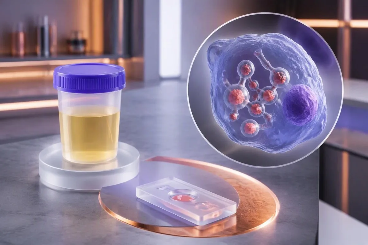
A positive urine or serum antigen result supports histoplasmosis in the right clinical setting, but it cannot reliably identify the fungus by itself. Symptoms, immune status, and independent testing determine what happens next.


- Positive antigen means the assay detected fungal material recognized by its antibodies; it does not independently establish Histoplasma as the cause.
- Cross-reactivity is particularly substantial with Blastomyces; testing a second specimen with a similar assay does not necessarily resolve the species question.
- Urine sensitivity reached 91.8% in disseminated histoplasmosis in one multicenter study, but that percentage does not apply to every pulmonary presentation.
- Antitelaning vaqti differs from antigen timing: Histoplasma antibodies can take approximately 4–8 weeks to become detectable.
- Antitel titrlari of 1:32 or higher can support active histoplasmosis in context; a fourfold rise is stronger evidence than one isolated titer.
- Immune status changes urgency: HIV with a CD4 count below 150 cells/µL, transplantation, and significant immunosuppressive treatment increase concern for dissemination.
- Antigen concentration has no universal ng/mL threshold that independently diagnoses histoplasmosis or determines emergency care.
- Culture timing commonly involves several weeks, often 2–6 weeks; severely ill patients may need treatment before culture confirmation.
- Shoshilinch baholash is appropriate for breathlessness at rest, confusion, fainting, or a new oxygen saturation below 92% at sea level, unless a clinician has established a different baseline.
What does a positive Histoplasma antigen actually establish?
A histoplasma antigen positive result establishes that the assay detected fungal material recognized by its test antibodies—not that Histoplasma is definitively the organism responsible. Histoplasmosis becomes more likely when the result matches symptoms, exposure, and immune status; cross-reacting fungal infections remain possible.
The distinction is practical, not semantic: a positive result can be strong enough to support a working diagnosis and urgent treatment without satisfying every criterion for species-level confirmation. In Hage et al.’s 2011 multicenter evaluation, urine antigen was detected in 91.8% of disseminated histoplasmosis cases, but performance differed across clinical presentations and comparison groups.
Consider a hypothetical 46-year-old receiving a TNF inhibitor who develops fever, weight loss, and diffuse lung abnormalities after disturbing soil: a positive antigen result deserves prompt infectious-disease assessment. The same laboratory flag in an otherwise well person with no plausible exposure deserves verification and a careful history rather than an automatic antifungal prescription.
I’m Thomas Klein, Chief Medical Officer at Kantesti, and my practical rule is to separate 3 questions: was antigen detected, which fungus is responsible, and how sick is the patient? Kantesti is an AI blood test analyzer that can help organize accompanying laboratory findings, not establish fungal identity; our tashkilot va klinik yoʻnalishimiz bu farqni tushuntiradi.
How do urine and serum Histoplasma antigen tests differ?
The Histoplasma urine antigen test measures fungal antigen excreted into urine, while serum testing measures antigen circulating in the liquid portion of a laboratory sample. The 2 specimens provide related evidence, and either can be negative despite infection.
Urine is often a useful first specimen because collection is noninvasive and sensitivity is high in many patients with disseminated disease. Azar and Hage’s 2017 laboratory review describes antigen detection as particularly valuable when fungal burden is substantial; ordering 2 specimen types can improve detection in selected situations, although their results are not statistically independent.
A positive urine result does not mean the kidneys or bladder are necessarily infected. Antigen originating elsewhere can circulate and be excreted, and the Histoplasma assay is separate from a routine siydik dipstick talqini; normal nitrite, leukocyte esterase, or protein findings do not negate a positive fungal antigen test.
Collection instructions are laboratory-specific: many assays use a random urine specimen rather than a 24-hour collection, while serum requires the laboratory’s designated sample preparation. Before interpreting a discrepancy, check whether both samples were collected before treatment, processed by the same laboratory, and analyzed with comparable methods—those 3 details can explain more than the positive-versus-negative labels.
What does the antigen number in ng/mL mean?
A quantitative Histoplasma antigen result estimates antigen concentration, often in ng/ml, within that laboratory’s assay range. There is no universal concentration that proves histoplasmosis, distinguishes Histoplasma from Blastomyces, or determines whether someone needs hospital care.
A report saying detected below the limit of quantification means the signal met the assay’s detection criteria but was too small to measure reliably. For example, an illustrative 0.5 ng/mL result and a 5 ng/mL result are not interchangeable, yet neither becomes a species diagnosis; our sifatli va miqdoriy qo‘llanmamiz explains why detection and measurement are separate claims.
Higher concentrations often accompany greater fungal burden in disseminated histoplasmosis, but overlap between patient groups prevents a clean severity ladder. A value above the upper reporting limit is censored rather than exact: if 2 reports both say above range, the clinician cannot calculate a meaningful percentage change without a laboratory dilution or another validated measurement.
Urine concentration also affects interpretation: a small change between specimens can reflect hydration, collection conditions, or assay variation rather than a true change in disease activity. Understanding urine concentration and dilution is useful, but patients should not invent a creatinine-corrected Histoplasma ratio unless the laboratory has validated that specific approach.
Why can other fungi produce a Histoplasma antigen false positive?
A Histoplasma antigen false positive can occur because other fungi contain antigenic structures recognized by the assay, especially Blastomyces. In that situation, the result may correctly signal a fungal illness while assigning misleading confidence to 1 particular fungal species.
Blastomyces is the central cross-reactivity concern because its geographic distribution and pulmonary symptoms can overlap with histoplasmosis. Hage et al. reported cross-reactivity in 90% of blastomycosis cases in their 2011 study, so a high apparent specificity against nonfungal controls should not be mistaken for equally high specificity against other endemic mycoses.
Talaromyces marneffei and Paracoccidioides can also cross-react in some Histoplasma antigen systems; the exact pattern depends on the assay and the patient’s travel or residence history. A 2-year-old overseas exposure can still matter in an immunosuppressed patient, and asking only where someone lives today can miss a clinically useful clue.
A second positive fungal antigen assay may repeat the same shared-antigen problem rather than independently confirm both infections. Species-directed culture, tissue examination, or validated molecular testing offers a different type of evidence; meanwhile, a positive Histoplasma result is not a diagnosis of household mold toxicity, as our mold testing limitations explain.
How likely is histoplasmosis after a positive result?
The probability of histoplasmosis after a positive antigen result depends on the patient’s pretest ehtimollik, the assay, and competing fungal diagnoses. No single positive predictive value applies to both an asymptomatic screening population and a severely ill, immunocompromised population.
Here is a deliberately hypothetical example: with 95% sensitivity and 95% specificity, testing 1,000 people when only 5% have the target illness produces about 48 true positives and 48 false positives. Roughly half the positive results would therefore represent that illness; these numbers teach probability and are not performance claims for a particular Histoplasma assay.
Raise the hypothetical disease prevalence to 50%, keeping the same assay characteristics, and the positive predictive value becomes approximately 95%. Real Histoplasma interpretation is more complicated because Blastomyces cross-reactivity creates a competing fungal explanation, and the patient’s symptom duration, antifungal exposure, and specimen type can all alter the test’s usefulness.
At Kantesti, we aim to explain why a laboratory flag should trigger a question rather than pretend it answers all 3 diagnostic questions at once. Our AI tahlil texnologiyamiz describes the interpretation workflow; no AI-generated probability should replace an infectious-disease evaluation or be presented as a validated Histoplasma risk score without specific supporting evidence.
How are Histoplasma antigen and antibody results different?
Antigen testing looks for fungal components; antibody testing looks for the patient’s immune response to the fungus. Histoplasma antibodies may take approximately 4–8 weeks to develop, so an early negative antibody result does not cancel a positive antigen result.
Antibodies can remain detectable after a previous infection, whereas antigen more directly reflects the presence of recognized fungal material at the sampling time. Neither test provides an infallible active-versus-past distinction: persistent antigen after treatment, cross-reactivity, and delayed antibody development all complicate the story; our ijobiy antitelalar natijasi qoʻllanmasi separates immune memory from current disease.
For complement-fixation testing, a titer of 1:32 or higher can support active histoplasmosis when symptoms and other findings fit, and a fourfold rise between paired samples is stronger evidence than one isolated low titer. Azar and Hage’s 2017 review also explains the limits of immunodiffusion bands; those older serologic patterns should not be translated directly into modern antibody-assay index values.
A clinician may repeat initially negative antibodies several weeks later, often within a 2–6-week follow-up interval selected for the clinical situation. That repeat is useful only if waiting is safe: transplant recipients or people with advanced HIV may not produce a dependable antibody response, so culture, tissue assessment, and antigen testing can carry greater diagnostic weight.
Which symptoms and exposures make a positive result more convincing?
Fever, cough, chest discomfort, fatigue, and a plausible soil or bird-and-bat-dropping exposure make a positive Histoplasma antigen result more clinically convincing. Acute symptoms commonly begin about 3–17 days after exposure, although that timing does not cover every form of histoplasmosis.
Disturbing contaminated soil matters more than simply seeing a bird or bat: demolition, cave visits, roost cleanup, and excavation can aerosolize fungal particles. A hypothetical family developing fever and cough 10 days after the same dusty renovation provides a stronger shared-exposure clue than one person reporting an ordinary walk outdoors.
Persistent fever, weight loss, gastrointestinal symptoms, enlarged lymph nodes, or abnormalities affecting several organs raise concern for dissemination rather than a lung-limited illness. Breathlessness requires assessment of oxygenation and competing causes—not just another antigen test—and our hansilashni tekshirish bo'yicha qo'llanmamiz explains why 1 laboratory result cannot assess respiratory stability.
Routine inflammatory markers cannot reliably identify the organism: an elevated CRP, a normal white-cell count, or a low procalcitonin does not independently distinguish histoplasmosis from other infections. Recording 2 dates—the likely exposure and the first symptom—often improves interpretation more than repeating nonspecific markers; our WBC va CRP taqqoslashi discusses these discordant patterns.
How does immune status change the urgency of a positive antigen?
A positive Histoplasma antigen result deserves faster assessment when the patient has advanced HIV, transplantation, or substantial immunosuppression because disseminated infection is more likely. In HIV, a CD4 count below 150 cells/µL is a recognized risk marker, not a universal diagnostic cutoff.
TNF inhibitors, prolonged systemic corticosteroids, and some cancer therapies can reduce the cellular immune response needed to contain Histoplasma. The medication name, daily dose, and treatment duration are 3 separate pieces of information; a normal total white-cell count does not prove normal cellular immunity, and antibody testing may be less reassuring in these patients.
HIV viral load and CD4 count answer different questions: viral load measures viral replication, while CD4 count helps characterize immune function. Someone with a previously undetectable viral load can still need assessment if their immune recovery is incomplete; our OIV virusli yuklamasini talqin qilish should not be used as a substitute for reviewing the current CD4 result.
Kantesti is an AI blood test interpretation platform that can help place 2 or more accompanying results in context, but a positive fungal antigen in an immunocompromised person needs clinician-led triage. A broader frequent infection assessment can support the workup; do not abruptly stop transplant medicines or corticosteroids without the prescribing team’s guidance.
Which tests help confirm Histoplasma rather than a cross-reacting fungus?
Culture with organism identification, tissue examination, and appropriately validated molecular tests can help distinguish Histoplasma from cross-reacting fungi. Culture often takes approximately 2–6 weeks, so confirmation and treatment decisions may proceed in parallel when illness is severe.
The best specimen depends on the affected site: respiratory samples may help in pulmonary disease, while specialized cultures or tissue examination may be useful in suspected dissemination. The laboratory should be told that an endemic fungus is suspected because fungal culture requirements and safety procedures differ from routine bacterial processing; our ekma natijasini talqin qilish explains why specimen and method matter.
Histoplasma classically appears as small, approximately 2–4 µm budding yeast, often within macrophages; Blastomyces usually has larger yeast with broad-based budding. Those features require expert interpretation because other organisms and staining artifacts can mimic parts of the picture, and a tissue description alone may need culture or molecular confirmation before species confidence is justified.
PCR availability and clinical validation vary between laboratories, so a negative molecular result is not an all-purpose exclusion test. Before ordering a second procedure, ask which of 2 uncertainties remains: whether fungal infection is present or which species caused it; repeating antigen can help detection, but an independent identification method more directly addresses cross-reactivity.
What do chest imaging and other blood tests add?
Chest imaging identifies the pattern and extent of lung involvement, but it cannot independently prove histoplasmosis. Nodules, enlarged hilar or mediastinal lymph nodes, and diffuse lung abnormalities can overlap with tuberculosis, sarcoidosis, malignancy, and other infections.
Old calcified nodules or lymph nodes can reflect healed granulomatous disease and do not automatically explain a newly positive antigen result. Comparing imaging from 2 time points helps separate stable structural remnants from evolving disease; a new respiratory illness still requires its own assessment rather than being attributed to every old spot on a scan.
Tuberculosis testing illustrates a different diagnostic limitation: a positive IGRA indicates an immune response to tuberculosis antigens but does not by itself distinguish latent infection from active disease. Our IGRA result interpretation explains why a positive IGRA and a positive fungal antigen require 2 parallel clinical questions, not an assumption that either result disproves the other.
An elevated ACE level is also not a reliable shortcut to sarcoidosis, especially when infection remains plausible; our ACE testing limitations covers that trap. In suspected dissemination, low counts across 2 or more blood-cell lines or abnormal liver tests can help identify affected organs, but none of those findings supplies fungal species confirmation.
What should you do today after receiving a positive result?
Contact the ordering clinician promptly after a positive Histoplasma antigen result; seek emergency assessment for breathlessness at rest, confusion, fainting, or rapidly worsening illness. A new oxygen saturation below 92% at sea level is concerning, although altitude, measurement error, and established baseline disease can change interpretation.
A stable, asymptomatic person can usually begin with clinician-directed verification rather than an emergency department visit solely because of the laboratory flag. Conversely, an immunocompromised person with fever or systemic symptoms should seek same-day advice even if the antigen concentration is low; neither 0.5 ng/mL nor 5 ng/mL overrides the patient’s clinical condition.
Prepare 5 items for the discussion: the complete report, symptom-onset date, exposure and travel history, medication list, and immune-status information. Include the specimen type and laboratory reference comments, because a cropped screenshot can omit a below-quantification warning or cross-reactivity statement that materially changes the interpretation.
Kantesti AI can help explain accompanying results, but it should not tell a symptomatic patient to wait for repeat testing instead of urgent assessment. Our klinik validatsiya doirasi addresses interpretation standards; in this setting, 2 genuinely different tests are often more informative than repeatedly measuring the same shared antigen, provided the clinician selects appropriate specimens.
Does a positive antigen mean you need antifungal treatment?
A positive antigen result does not automatically require antifungal treatment; treatment depends on the clinical syndrome, severity, immune status, and likelihood of true disease. Severe pulmonary or disseminated histoplasmosis can require immediate treatment, while selected mild pulmonary cases are managed with observation.
Wheat et al.’s 2007 IDSA guideline provides established treatment principles for moderately severe to severe disseminated histoplasmosis: liposomal amphotericin B at 3 mg/kg daily for about 1–2 weeks, followed by itraconazole for at least 12 months. That is a clinician-managed adult regimen, not an instruction to self-treat; organ involvement, pregnancy, interactions, and newer syndrome-specific guidance can alter the plan.
Itraconazole can substantially increase tacrolimus exposure through drug-metabolism interactions, so transplant teams often need additional level monitoring and dose adjustment. A previous stable tacrolimus value does not guarantee safety after even 1 medication change; our tacrolimus monitoring guide explains why sample timing and a current medication list are essential.
Amphotericin formulations can affect kidney function, potassium, and magnesium, making baseline and repeat laboratory measurements part of treatment rather than optional extras. A magnesium result below the laboratory range—often below approximately 1.7 mg/dL, depending on method—needs clinical interpretation alongside symptoms and other electrolytes; our magniyning kam bo'lish sabablari explains relevant medication-related patterns.
Can Histoplasma antigen stay positive after treatment?
Histoplasma antigen can remain detectable after effective treatment, and persistent low-level positivity does not independently prove treatment failure. A rising trend is more concerning when it accompanies recurrent symptoms or other evidence of active disease; 1 isolated measurement cannot establish relapse.
For disseminated disease, Wheat et al.’s 2007 IDSA guideline describes measuring antigen before therapy, at approximately 2 weeks and 1 month, then about every 3 months during treatment and for 12 months afterward. The appropriate schedule remains individualized; a patient with new fever or worsening breathing needs reassessment sooner than the next planned surveillance sample.
Urine and serum trends are not numerically interchangeable, and changing laboratories can create a false impression of improvement or relapse. Keep the specimen type and method consistent where possible, then compare symptoms, organ tests, and antigen together; our meaningful laboratory trend guide explains why 2 different methods may not produce a valid before-and-after comparison.
Kantesti is an AI lab test interpretation service that can help organize serial reports, but it should not label a small numerical rise as relapse without clinical evidence. Before reacting to a change, I would ask about 3 common confounders—treatment interruption, changed sample conditions, and assay differences—while the clinician considers adherence, drug absorption, interactions, and independent testing.
A practical follow-up checklist and supporting publications
The safest follow-up plan identifies clinical urgency, the need for species confirmation, and how response will be monitored. As of October 14, 2026, no standalone antigen concentration or antibody titer replaces those 3 decisions, and an updated article date does not itself certify medical review.
Write down 3 questions for the appointment: does my presentation suggest active infection, could another fungus explain this assay, and which test would change the treatment plan? Thomas Klein’s editorial emphasis here is deliberately narrow: distinguish detection from diagnosis, and do not turn a reassuring antibody result into permission to ignore systemic illness.
Kantesti’s tibbiy maslahat axboroti describes our clinical advisory structure, while our tibbiy kontent standartlari explain expectations for sourcing and review. This article relies on 3 external histoplasmosis publications listed below; they address diagnostic performance, laboratory limitations, and established treatment principles rather than validating an AI fungal diagnosis.
The 2 supplementary Zenodo publications below concern general urinalysis and iron studies, not Histoplasma assay validation or antifungal efficacy. Our urinalysis reference guide helps distinguish specialized fungal antigen testing from routine urine chemistry; our temirni tekshirish bo'yicha talqim qo'llanmasi provides background for evaluating anemia during prolonged illness, without identifying its infectious cause.
Tez-tez so'raladigan savollar
Histoplasma antigeni natijasi musbat bo‘lsa, demak, menda albatta gistoplazmoz bormi?
Histoplasma antigenining musbat natijasi gistoplazmozni tasdiqlashga yordam beradi, biroq zamburug‘ turini yoki klinik tashxisni mustaqil ravishda tasdiqlamaydi. Blastomyces va ayrim boshqa zamburug‘lar bilan o‘zaro reaktivlik kuzatilishi mumkin, natijani talqin qilishga esa namuna turi, alomatlar va immun holat ta’sir qiladi. 2011-yilda o‘tkazilgan ko‘p markazli tadqiqotda disseminatsiyalangan gistoplazmoz holatlarining 91.8% tasida siydik antigeni aniqlangan, biroq bu ko‘rsatkich o‘pkadagi barcha ko‘rinishlarni ifodalamaydi. Natijani aniqlashtirish uchun klinitsistlar ekma, to‘qimani tekshirish, antitanalar yoki validatsiyalangan molekulyar testlardan foydalanishi mumkin.
0 Blastomyces Histoplasma antigeni natijasi noto‘g‘ri musbat chiqishiga sabab bo‘lishi mumkinmi?
Blastomyces zamburug’lari ayrim tahlillar taniydigan antigenik xususiyatlari o‘xshash bo‘lgani uchun Histoplasma antigeni natijasining musbat chiqishiga sabab bo‘lishi mumkin. Hage va boshqalarning 2011-yildagi tadqiqotida blastomikoz holatlarining 90% qismida o‘zaro reaktivlik qayd etilgan. Shu sababli musbat natija tasodifiy laboratoriya xatosidan ko‘ra haqiqiy zamburug‘li kasallikni aks ettirishi mumkin, ammo kasallikni keltirib chiqargan turni to‘g‘ri aniqlamasligi mumkin. Ekma, to‘qimani tekshirish yoki turi aniq ko‘rsatilgan va validatsiyadan o‘tgan molekulyar tahlil mustaqilroq dalillarni taqdim etishi mumkin.
Histoplasma siydik antigenining normal darajasi qanday?
Histoplasma siydik antigeni testining odatiy salbiy talqini — laboratoriyaning tahlil chegarasiga ko‘ra aniqlanmadi. Barcha laboratoriyalar uchun umumiy bo‘lgan normal ng/mL oralig‘i yoki ijobiy konsentratsiya mavjud emas. Misol tariqasidagi 0.5 ng/mL qiymatini bir usul bilan miqdoriy aniqlash mumkin, ammo u boshqa usulning ishonchli o‘lchash diapazonidan past bo‘lishi mumkin. Tahlillar orasida chegaraviy qiymatlarni ko‘chirish o‘rniga, laboratoriya hisobotidagi ma’lumotlar va klinik holatga tayaning.
Antitanalar manfiy bo‘lganda Histoplasma antigeni ijobiy bo‘lishi mumkinmi?
Histoplasma antigeni, ayniqsa kasallikning ilk bosqichlarida yoki immunitet faoliyati buzilganda, antitanalar manfiy bo‘lsa ham ijobiy chiqishi mumkin. Antitanalar hosil bo‘lishi uchun taxminan 4–8 hafta kerak bo‘lishi mumkin, ayrim immuniteti zaiflashgan bemorlarda esa ishonchli antitana javobi umuman yuzaga kelmaydi. Agar kutish xavfsiz bo‘lsa, shifokor bir necha haftadan keyin serologik tekshiruvni takrorlashi mumkin. Og‘ir alomatlarda antitanalar hosil bo‘lishini kutmasdan, zudlik bilan tibbiy ko‘rikdan o‘tish kerak.
Siydikdagi antigenning ijobiy chiqishi siydik yo‘llari infeksiyasi borligidan dalolat beradimi?
Siydikdagi Histoplasma antigenining musbat chiqishi infeksiya siydik pufagi yoki buyraklarda ekanini anglatmaydi. Boshqa joydagi zamburug‘ antigeni qon aylanishiga tushib, siydik bilan ajralib chiqishi mumkin. Maxsus antigen testi va odatiy siydik tahlili 2 xil savolga javob beradi, shuning uchun nitrit yoki leykotsit esteraza natijalarining me’yorda bo‘lishi zamburug‘ga oid topilmani inkor etmaydi. Qaysi a’zolar zararlanganini alomatlar, ko‘rik, tasviriy tekshiruvlar va qo‘shimcha tahlillar aniqlaydi.
Davolashdan keyin Histoplasma antigeni qancha vaqt davomida musbat bo‘lib qolishi mumkin?
Histoplasma antigen can remain detectable for months after effective treatment, especially after disseminated disease. Persistent low-level positivity alone does not establish relapse, whereas a rise accompanied by recurrent symptoms is more concerning. The 2007 IDSA guideline describes antigen monitoring during treatment and for 12 months afterward in disseminated histoplasmosis. Follow-up timing and treatment duration should be individualized by the treating clinician.
When is a positive Histoplasma antigen result an emergency?
A positive Histoplasma antigen result requires emergency assessment when it accompanies breathlessness at rest, confusion, fainting, or rapidly worsening illness. A new oxygen saturation below 92% at sea level is concerning, although established baseline disease, altitude, and measurement quality affect interpretation. Immunocompromised patients with fever or systemic symptoms should seek same-day clinical advice even with a low antigen concentration. The antigen number itself is not an emergency threshold.
Bugun AI asosidagi qon tahlilini tahlil qilishni oling
Kantesti’ga tezkor va aniq laboratoriya tahlili uchun ishonadigan butun dunyo bo‘ylab 2 milliondan ortiq foydalanuvchiga qo‘shiling. Qon tahlili natijalaringizni yuklang va soniyalar ichida 15,000+ biomarkerlarining to‘liq talqinini oling.
📚 Havola qilingan ilmiy tadqiqot nashrlari
Klein, T., Mitchell, S., & Weber, H. (2026). Siydikdagi urobilinogen: Siydik tahlili bo‘yicha to‘liq qo‘llanma 2026. Kantesti AI tibbiy tadqiqoti.
Klein, T., Mitchell, S., & Weber, H. (2026). Temir tadqiqotlari bo'yicha qo'llanma: TIBC, temirning to'yinganligi va bog'lanish qobiliyati. Kantesti AI tibbiy tadqiqoti.
📖 Tashqi tibbiy manbalar
Hage CA et al. (2011). A multicenter evaluation of tests for diagnosis of histoplasmosis. Clinical Infectious Diseases.
Azar MM, Hage CA. (2017). Laboratory Diagnostics for Histoplasmosis. Journal of Clinical Microbiology.
Wheat LJ et al. (2007). Clinical practice guidelines for the management of patients with histoplasmosis: 2007 update by the Infectious Diseases Society of America. Clinical Infectious Diseases.
⚕️ Tibbiy ogohlantirish
Ushbu maqola faqat ta’lim maqsadlari uchun mo‘ljallangan va tibbiy maslahatni anglatmaydi. Tashxis va davolash bo‘yicha qarorlar uchun har doim malakali sog‘liqni saqlash mutaxassisiga murojaat qiling.
E-E-A-T ishonch signallari
Tajriba
Shifokor boshchiligidagi laboratoriya talqin qilish ish jarayonlarini klinik ko‘rib chiqish.
Tajriba
Laboratoriya tibbiyoti biomarkerlarning klinik kontekstda qanday o‘zini tutishini yoritadi.
Vakolatlilik
Dr. Tomas Klein tomonidan yozilgan, Dr. Sarah Mitchell va Prof. Dr. Hans Weber tomonidan ko‘rib chiqilgan.
Ishonchlilik
Xavotirni kamaytirish uchun aniq keyingi qadamlar yo‘nalishlari bilan dalillarga asoslangan talqin.