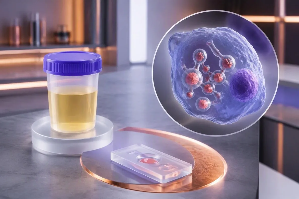
ຜົນກວດແອນຕິເຈນໃນປັດສະວະ ຫຼື ເລືອດທີ່ເປັນບວກ ສະໜັບສະໜູນການວິນິດໄສຮິສໂຕພລາສໂມຊິສໃນບໍລິບົດທາງຄລີນິກທີ່ເໝາະສົມ, ແຕ່ບໍ່ສາມາດລະບຸເຊື້ອລາໄດ້ຢ່າງໜ້າເຊື່ອຖືດ້ວຍຕົນເອງ. ອາການ, ສະຖານະພູມຄຸ້ມກັນ ແລະ ການກວດຢືນຢັນອິດສະຫຼະ ເປັນຕົວກຳນົດວ່າຈະດຳເນີນການຕໍ່ແນວໃດ.


- ແອນຕິເຈນບວກ ໝາຍຄວາມວ່າຊຸດກວດພົບສານຈາກເຊື້ອລາທີ່ແອນຕິບໍດີຂອງມັນຈຳແນກໄດ້; ແຕ່ຜົນນີ້ຢ່າງດຽວບໍ່ສາມາດຢືນຢັນວ່າ Histoplasma ເປັນສາເຫດໄດ້.
- ປະຕິກິລິຍາຂ້າມ ເກີດຂຶ້ນຢ່າງເດັ່ນຊັດເປັນພິເສດກັບ Blastomyces; ການກວດຕົວຢ່າງທີສອງດ້ວຍຊຸດກວດທີ່ຄ້າຍຄືກັນ ບໍ່ຈຳເປັນວ່າຈະຊ່ວຍແຍກຊະນິດເຊື້ອໄດ້.
- ຄວາມໄວໃນການກວດພົບໃນປັດສະວະ ສູງເຖິງ 91.8% ໃນກໍລະນີຮິສໂຕພລາສໂມຊິສແບບແຜ່ກະຈາຍ ໃນການສຶກສາຫຼາຍສູນແຫ່ງໜຶ່ງ, ແຕ່ອັດຕານີ້ບໍ່ໄດ້ນຳໃຊ້ກັບການສະແດງອາການທາງປອດທຸກຮູບແບບ.
- ໄລຍະເວລາຂອງແອນຕິບໍດີ ແຕກຕ່າງຈາກໄລຍະເວລາທີ່ແອນຕິເຈນກວດພົບໄດ້: ແອນຕິບໍດີຕໍ່ Histoplasma ອາດໃຊ້ເວລາປະມານ 4–8 ອາທິດຈຶ່ງກວດພົບໄດ້.
- Antibody titers ລະດັບ 1:32 ຫຼື ສູງກວ່າ ອາດສະໜັບສະໜູນວ່າມີຮິສໂຕພລາສໂມຊິສທີ່ກຳລັງດຳເນີນຢູ່ ເມື່ອພິຈາລະນາໃນບໍລິບົດ; ການເພີ່ມຂຶ້ນສີ່ເທົ່າເປັນຫຼັກຖານທີ່ໜັກແໜ້ນກວ່າຄ່າໄຕເຕີທີ່ສູງພຽງຄັ້ງດຽວ.
- ສະຖານະພູມຄຸ້ມກັນ ປັດໄຈທີ່ເຮັດໃຫ້ຄວາມຮີບດ່ວນປ່ຽນໄປ: ຜູ້ຕິດເຊື້ອ HIV ທີ່ມີຈຳນວນ CD4 ຕ່ຳກວ່າ 150 ເຊວລ໌/µL, ການປູກຖ່າຍອະໄວຍະວະ ແລະການຮັກສາດ້ວຍຢາກົດພູມຄຸ້ມກັນທີ່ສຳຄັນ ເພີ່ມຄວາມກັງວົນວ່າອາດມີການແຜ່ກະຈາຍຂອງເຊື້ອ.
- ຄວາມເຂັ້ມຂຸ້ນຂອງແອນຕິເຈນ ບໍ່ມີຄ່າເກນ ng/mL ທີ່ໃຊ້ໄດ້ທົ່ວໄປ ເຊິ່ງສາມາດວິນິດໄສພະຍາດຮິສໂທພລາສໂມຊິສໄດ້ໂດຍລຳພັງ ຫຼືໃຊ້ກຳນົດວ່າຕ້ອງໄດ້ຮັບການດູແລສຸກເສີນຫຼືບໍ່.
- ໄລຍະເວລາເພາະລ້ຽງເຊື້ອ ໂດຍທົ່ວໄປໃຊ້ເວລາຫຼາຍອາທິດ, ມັກຈະເປັນ 2–6 ອາທິດ; ຜູ້ປ່ວຍທີ່ອາການໜັກອາດຕ້ອງເລີ່ມການຮັກສາກ່ອນຈະຢືນຢັນຜົນເພາະລ້ຽງເຊື້ອ.
- ການປະເມີນດ່ວນ ແມ່ນເໝາະສົມເມື່ອມີອາການຫາຍໃຈລຳບາກຂະນະພັກ, ສັບສົນ, ເປັນລົມ ຫຼືມີຄ່າຄວາມອີ່ມຕົວອອກຊີເຈນໃໝ່ຕ່ຳກວ່າ 92% ໃນລະດັບນ້ຳທະເລ, ເວັ້ນແຕ່ແພດໄດ້ກຳນົດຄ່າພື້ນຖານອື່ນໄວ້.
ຜົນແອນຕິເຈນ Histoplasma ທີ່ເປັນບວກ ຢືນຢັນຫຍັງແທ້?
A ຜົນກວດແອນຕິເຈນ histoplasma ເປັນບວກ ຜົນນີ້ຢືນຢັນວ່າຊຸດກວດພົບສານຈາກເຊື້ອຣາທີ່ແອນຕິບໍດີໃນຊຸດກວດຈຳແນກໄດ້—ບໍ່ໄດ້ຢືນຢັນວ່າ Histoplasma ເປັນເຊື້ອທີ່ກໍ່ໂລກຢ່າງແນ່ນອນ. ພະຍາດຮິສໂທພລາສໂມຊິສມີແນວໂນ້ມເປັນໄປໄດ້ຫຼາຍຂຶ້ນ ເມື່ອຜົນກວດສອດຄ່ອງກັບອາການ, ການສຳຜັດເຊື້ອ ແລະສະຖານະພູມຄຸ້ມກັນ; ການຕິດເຊື້ອຣາຊະນິດອື່ນທີ່ໃຫ້ຜົນຂ້າມກັນຍັງເປັນໄປໄດ້.
ຄວາມແຕກຕ່າງນີ້ເປັນເລື່ອງທາງປະຕິບັດ, ບໍ່ແມ່ນພຽງການໃຊ້ຄຳຕ່າງກັນ: ຜົນບວກອາດມີນ້ຳໜັກພໍທີ່ຈະສະໜັບສະໜູນການວິນິດໄສເບື້ອງຕົ້ນ ແລະການຮັກສາເລັ່ງດ່ວນ ໂດຍບໍ່ຈຳເປັນຕ້ອງຄົບທຸກເກນສຳລັບການຢືນຢັນເຖິງລະດັບຊະນິດເຊື້ອ. ໃນການປະເມີນຫຼາຍສູນກາງຂອງ Hage ແລະຄະນະ ໃນປີ 2011, ກວດພົບແອນຕິເຈນໃນປັດສະວະໄດ້ໃນ 91.8% ຂອງກໍລະນີຮິສໂທພລາສໂມຊິສທີ່ແຜ່ກະຈາຍ, ແຕ່ປະສິດທິພາບແຕກຕ່າງກັນຕາມຮູບແບບທາງຄລີນິກ ແລະກຸ່ມປຽບທຽບ.
ພິຈາລະນາກໍລະນີສົມມຸດຂອງຄົນອາຍຸ 46 ປີ ທີ່ກຳລັງໃຊ້ຢາຍັບຢັ້ງ TNF ແລະເກີດອາການໄຂ້, ນ້ຳໜັກຫຼຸດ ແລະຄວາມຜິດປົກກະຕິແບບກະຈາຍໃນປອດ ຫຼັງຈາກລົບກວນດິນ: ຜົນແອນຕິເຈນເປັນບວກຄວນໄດ້ຮັບການປະເມີນຈາກແພດຜູ້ຊ່ຽວຊານດ້ານພະຍາດຕິດເຊື້ອໂດຍໄວ. ສັນຍານຜົນກວດດຽວກັນໃນຄົນທີ່ສຸຂະພາບດີ ແລະບໍ່ມີປະຫວັດສຳຜັດເຊື້ອທີ່ເປັນໄປໄດ້ ຄວນມີການກວດຢືນຢັນ ແລະຊັກປະຫວັດຢ່າງລະອຽດ ແທນການສັ່ງຢາຕ້ານເຊື້ອຣາໂດຍອັດຕະໂນມັດ.
ຂ້ອຍຊື່ Thomas Klein, ຫົວໜ້າແພດປະຈຳ Kantesti, ແລະຫຼັກປະຕິບັດຂອງຂ້ອຍແມ່ນແຍກ 3 ຄຳຖາມອອກຈາກກັນ: ກວດພົບແອນຕິເຈນຫຼືບໍ່, ເຊື້ອຣາຊະນິດໃດເປັນສາເຫດ ແລະຜູ້ປ່ວຍອາການໜັກປານໃດ? Kantesti ເປັນເຄື່ອງວິເຄາະຜົນກວດເລືອດດ້ວຍ AI ທີ່ຊ່ວຍຈັດລະບຽບຜົນກວດທາງຫ້ອງປະລິກອື່ນໆທີ່ກ່ຽວຂ້ອງ, ແຕ່ບໍ່ສາມາດຢືນຢັນຊະນິດເຊື້ອຣາໄດ້; ຂອງພວກເຮົາ ອົງການຈັດຕັ້ງ ແລະ ເປົ້າໝາຍທາງດ້ານຄລີນິກ อธิบายความแตกต่างนั้น.
ການກວດແອນຕິເຈນ Histoplasma ໃນປັດສະວະ ແລະ ໃນເລືອດ ແຕກຕ່າງກັນແນວໃດ?
ໄດ້ ການກວດແອນຕິເຈນ Histoplasma ໃນປັດສະວະ ວັດແທກແອນຕິເຈນຈາກເຊື້ອຣາທີ່ຖືກຂັບອອກມາໃນປັດສະວະ, ສ່ວນການກວດໃນເຊຣັມວັດແທກແອນຕິເຈນທີ່ໝູນວຽນຢູ່ໃນສ່ວນຂອງແຫຼວຂອງຕົວຢ່າງທີ່ສົ່ງກວດ. ຕົວຢ່າງທັງ 2 ຊະນິດໃຫ້ຫຼັກຖານທີ່ກ່ຽວຂ້ອງກັນ, ແລະຜົນອາດເປັນລົບໄດ້ໃນຕົວຢ່າງຊະນິດໃດຊະນິດໜຶ່ງ ເຖິງແມ່ນຈະມີການຕິດເຊື້ອ.
ປັດສະວະມັກເປັນຕົວຢ່າງທຳອິດທີ່ໃຊ້ໄດ້ດີ ເພາະເກັບຕົວຢ່າງໄດ້ໂດຍບໍ່ຮຸກລ້ຳ ແລະມີຄວາມໄວສູງໃນຜູ້ປ່ວຍຈຳນວນຫຼາຍທີ່ມີພະຍາດແຜ່ກະຈາຍ. ບົດທົບທວນດ້ານຫ້ອງປະລິກຂອງ Azar ແລະ Hage ໃນປີ 2017 ລະບຸວ່າການກວດພົບແອນຕິເຈນມີຄຸນຄ່າເປັນພິເສດເມື່ອປະລິມານເຊື້ອຣາມີຫຼາຍ; ການສັ່ງກວດຕົວຢ່າງ 2 ຊະນິດອາດຊ່ວຍເພີ່ມການກວດພົບໃນບາງສະຖານະການ, ແຕ່ຜົນຂອງທັງສອງບໍ່ເປັນອິດສະຫຼະຕໍ່ກັນທາງສະຖິຕິ.
ຜົນກວດປັດສະວະເປັນບວກບໍ່ໄດ້ໝາຍຄວາມວ່າໄຕ ຫຼືພົກຍ່ຽວຕ້ອງຕິດເຊື້ອ. ແອນຕິເຈນທີ່ມາຈາກບ່ອນອື່ນສາມາດໝູນວຽນໃນເລືອດ ແລະຖືກຂັບອອກມາໄດ້, ແລະຊຸດກວດ Histoplasma ແມ່ນແຍກຕ່າງຫາກຈາກການກວດປັດສະວະປົກກະຕິ การตีความแถบตรวจปัสสาวะ; ຜົນ nitrite, leukocyte esterase ຫຼືໂປຣຕີນທີ່ປົກກະຕິ ບໍ່ໄດ້ລົບລ້າງຜົນບວກຂອງການກວດແອນຕິເຈນຈາກເຊື້ອຣາ.
ຄຳແນະນຳການເກັບຕົວຢ່າງຂຶ້ນກັບແຕ່ລະຫ້ອງປະລິກ: ການກວດຫຼາຍຊະນິດໃຊ້ປັດສະວະສຸ່ມ ແທນການເກັບປັດສະວະ 24 ຊົ່ວໂມງ, ສ່ວນເຊຣັມຕ້ອງກຽມຕົວຢ່າງຕາມວິທີທີ່ຫ້ອງປະລິກກຳນົດ. ກ່ອນຕີຄວາມຜົນທີ່ຂັດແຍ້ງກັນ ໃຫ້ກວດວ່າເກັບຕົວຢ່າງທັງສອງກ່ອນເລີ່ມການຮັກສາຫຼືບໍ່, ປະມວນຜົນໂດຍຫ້ອງປະລິກດຽວກັນຫຼືບໍ່ ແລະວິເຄາະດ້ວຍວິທີທີ່ປຽບທຽບກັນໄດ້ຫຼືບໍ່—ລາຍລະອຽດ 3 ຂໍ້ນີ້ອາດອະທິບາຍໄດ້ຫຼາຍກວ່າປ້າຍຜົນບວກກັບຜົນລົບ.
ຕົວເລກແອນຕິເຈນໃນ ng/mL ໝາຍຄວາມວ່າແນວໃດ?
ຜົນກວດແອນຕິເຈນ Histoplasma ແບບປະລິມານປະເມີນຄວາມເຂັ້ມຂຸ້ນຂອງແອນຕິເຈນ, ໂດຍມັກລະບຸເປັນ ng/ml, ພາຍໃນຊ່ວງການວັດແທກຂອງຊຸດກວດໃນຫ້ອງປະລິກນັ້ນ. ບໍ່ມີຄວາມເຂັ້ມຂຸ້ນມາດຕະຖານທົ່ວໄປທີ່ພິສູດພະຍາດຮິສໂທພລາສໂມຊິສ, ຈຳແນກ Histoplasma ອອກຈາກ Blastomyces ຫຼືກຳນົດວ່າບຸກຄົນນັ້ນຈຳເປັນຕ້ອງນອນໂຮງໝໍຫຼືບໍ່.
ລາຍງານທີ່ລະບຸວ່າກວດພົບຕ່ຳກວ່າຂີດຈຳກັດການວັດປະລິມານ ໝາຍຄວາມວ່າສັນຍານຜ່ານເກນການກວດພົບຂອງຊຸດກວດ ແຕ່ມີຄ່ານ້ອຍເກີນກວ່າຈະວັດແທກໄດ້ຢ່າງເຊື່ອຖື. ຕົວຢ່າງ, ຄ່າສົມມຸດ 0.5 ng/mL ແລະ 5 ng/mL ບໍ່ແມ່ນຄ່າທີ່ໃຊ້ແທນກັນໄດ້, ແຕ່ບໍ່ມີຄ່າໃດກາຍເປັນການວິນິດໄສຊະນິດເຊື້ອ; ຂອງພວກເຮົາ ຄູ່ມືການວິເຄາະທຽບກັບປະລິມານ ອະທິບາຍວ່າການກວດພົບ ແລະການວັດແທກເປັນຂໍ້ອ້າງຄົນລະຢ່າງ.
ຄວາມເຂັ້ມຂຸ້ນທີ່ສູງຂຶ້ນມັກພົບຮ່ວມກັບປະລິມານເຊື້ອຣາທີ່ຫຼາຍຂຶ້ນໃນພະຍາດຮິສໂທພລາສໂມຊິສທີ່ແຜ່ກະຈາຍ, ແຕ່ຄ່າທີ່ຊ້ອນທັບກັນລະຫວ່າງກຸ່ມຜູ້ປ່ວຍບໍ່ອະນຸຍາດໃຫ້ຈັດລະດັບຄວາມຮຸນແຮງຢ່າງຊັດເຈນ. ຄ່າທີ່ສູງກວ່າຂີດຈຳກັດສູງສຸດຂອງການລາຍງານແມ່ນຄ່າທີ່ຖືກຈຳກັດການລາຍງານ ບໍ່ແມ່ນຄ່າແນ່ນອນ: ຖ້າລາຍງານ 2 ສະບັບຕ່າງກໍລະບຸວ່າສູງກວ່າຊ່ວງທີ່ກຳນົດ, ແພດບໍ່ສາມາດຄຳນວນການປ່ຽນແປງເປັນເປີເຊັນທີ່ມີຄວາມໝາຍໄດ້ ຖ້າບໍ່ມີການເຈືອຈາງຕົວຢ່າງໂດຍຫ້ອງປະລິກ ຫຼືການວັດແທກອື່ນທີ່ຜ່ານການຮັບຮອງ.
Urine concentration also affects interpretation: a small change between specimens can reflect hydration, collection conditions, or assay variation rather than a true change in disease activity. Understanding urine concentration and dilution is useful, but patients should not invent a creatinine-corrected Histoplasma ratio unless the laboratory has validated that specific approach.
ເປັນຫຍັງເຊື້ອລາຊະນິດອື່ນຈຶ່ງເຮັດໃຫ້ຜົນແອນຕິເຈນ Histoplasma ເປັນບວກປອມ?
A Histoplasma antigen false positive can occur because other fungi contain antigenic structures recognized by the assay, especially Blastomyces. In that situation, the result may correctly signal a fungal illness while assigning misleading confidence to 1 particular fungal species.
Blastomyces is the central cross-reactivity concern because its geographic distribution and pulmonary symptoms can overlap with histoplasmosis. Hage et al. reported cross-reactivity in 90% of blastomycosis cases in their 2011 study, so a high apparent specificity against nonfungal controls should not be mistaken for equally high specificity against other endemic mycoses.
Talaromyces marneffei and Paracoccidioides can also cross-react in some Histoplasma antigen systems; the exact pattern depends on the assay and the patient’s travel or residence history. A 2-year-old overseas exposure can still matter in an immunosuppressed patient, and asking only where someone lives today can miss a clinically useful clue.
A second positive fungal antigen assay may repeat the same shared-antigen problem rather than independently confirm both infections. Species-directed culture, tissue examination, or validated molecular testing offers a different type of evidence; meanwhile, a positive Histoplasma result is not a diagnosis of household mold toxicity, as our mold testing limitations explain.
ຫຼັງຈາກຜົນກວດເປັນບວກ ມີໂອກາດເປັນຮິສໂຕພລາສໂມຊິສຫຼາຍປານໃດ?
The probability of histoplasmosis after a positive antigen result depends on the patient’s ຄວາມເປັນໄປໄດ້ກ່ອນການກວດ (pretest probability), the assay, and competing fungal diagnoses. No single positive predictive value applies to both an asymptomatic screening population and a severely ill, immunocompromised population.
Here is a deliberately hypothetical example: with 95% sensitivity and 95% specificity, testing 1,000 people when only 5% have the target illness produces about 48 true positives and 48 false positives. Roughly half the positive results would therefore represent that illness; these numbers teach probability and are not performance claims for a particular Histoplasma assay.
Raise the hypothetical disease prevalence to 50%, keeping the same assay characteristics, and the positive predictive value becomes approximately 95%. Real Histoplasma interpretation is more complicated because Blastomyces cross-reactivity creates a competing fungal explanation, and the patient’s symptom duration, antifungal exposure, and specimen type can all alter the test’s usefulness.
At Kantesti, we aim to explain why a laboratory flag should trigger a question rather than pretend it answers all 3 diagnostic questions at once. Our ເທັກໂນໂລຍີການຕີຄວາມໝາຍ AI describes the interpretation workflow; no AI-generated probability should replace an infectious-disease evaluation or be presented as a validated Histoplasma risk score without specific supporting evidence.
ຜົນກວດແອນຕິເຈນ Histoplasma ແລະ ແອນຕິບໍດີ ແຕກຕ່າງກັນແນວໃດ?
Antigen testing looks for fungal components; antibody testing looks for the patient’s immune response to the fungus. Histoplasma antibodies may take approximately 4–8 weeks to develop, so an early negative antibody result does not cancel a positive antigen result.
Antibodies can remain detectable after a previous infection, whereas antigen more directly reflects the presence of recognized fungal material at the sampling time. Neither test provides an infallible active-versus-past distinction: persistent antigen after treatment, cross-reactivity, and delayed antibody development all complicate the story; our ผลบวกของแอนติบอดี separates immune memory from current disease.
For complement-fixation testing, a titer of 1:32 or higher can support active histoplasmosis when symptoms and other findings fit, and a fourfold rise between paired samples is stronger evidence than one isolated low titer. Azar and Hage’s 2017 review also explains the limits of immunodiffusion bands; those older serologic patterns should not be translated directly into modern antibody-assay index values.
A clinician may repeat initially negative antibodies several weeks later, often within a 2–6-week follow-up interval selected for the clinical situation. That repeat is useful only if waiting is safe: transplant recipients or people with advanced HIV may not produce a dependable antibody response, so culture, tissue assessment, and antigen testing can carry greater diagnostic weight.
ອາການ ແລະ ການສຳຜັດກັບເຊື້ອແບບໃດ ທີ່ເຮັດໃຫ້ຜົນກວດບວກໜ້າເຊື່ອຖືຂຶ້ນ?
Fever, cough, chest discomfort, fatigue, and a plausible soil or bird-and-bat-dropping exposure make a positive Histoplasma antigen result more clinically convincing. Acute symptoms commonly begin about 3–17 days after exposure, although that timing does not cover every form of histoplasmosis.
Disturbing contaminated soil matters more than simply seeing a bird or bat: demolition, cave visits, roost cleanup, and excavation can aerosolize fungal particles. A hypothetical family developing fever and cough 10 days after the same dusty renovation provides a stronger shared-exposure clue than one person reporting an ordinary walk outdoors.
Persistent fever, weight loss, gastrointestinal symptoms, enlarged lymph nodes, or abnormalities affecting several organs raise concern for dissemination rather than a lung-limited illness. Breathlessness requires assessment of oxygenation and competing causes—not just another antigen test—and our ຄູ່ມືການທົດສອບອາການຫາຍໃຈຝືດ explains why 1 laboratory result cannot assess respiratory stability.
Routine inflammatory markers cannot reliably identify the organism: an elevated CRP, a normal white-cell count, or a low procalcitonin does not independently distinguish histoplasmosis from other infections. Recording 2 dates—the likely exposure and the first symptom—often improves interpretation more than repeating nonspecific markers; our ການປຽບທຽບ WBC ແລະ CRP ຂອງພວກເຮົາ discusses these discordant patterns.
ສະຖານະພູມຄຸ້ມກັນປ່ຽນລະດັບຄວາມຮີບດ່ວນຂອງຜົນແອນຕິເຈນບວກແນວໃດ?
A positive Histoplasma antigen result deserves faster assessment when the patient has advanced HIV, transplantation, or substantial immunosuppression because disseminated infection is more likely. In HIV, a CD4 count below 150 cells/µL is a recognized risk marker, not a universal diagnostic cutoff.
TNF inhibitors, prolonged systemic corticosteroids, and some cancer therapies can reduce the cellular immune response needed to contain Histoplasma. The medication name, daily dose, and treatment duration are 3 separate pieces of information; a normal total white-cell count does not prove normal cellular immunity, and antibody testing may be less reassuring in these patients.
HIV viral load and CD4 count answer different questions: viral load measures viral replication, while CD4 count helps characterize immune function. Someone with a previously undetectable viral load can still need assessment if their immune recovery is incomplete; our ການ ຕີ ຄວາມ ໝາຍ ປະລິມານ ໄວຣັດ HIV should not be used as a substitute for reviewing the current CD4 result.
Kantesti is an AI blood test interpretation platform that can help place 2 or more accompanying results in context, but a positive fungal antigen in an immunocompromised person needs clinician-led triage. A broader frequent infection assessment can support the workup; do not abruptly stop transplant medicines or corticosteroids without the prescribing team’s guidance.
ການກວດໃດຊ່ວຍຢືນຢັນວ່າແມ່ນ Histoplasma ແທນທີ່ຈະເປັນເຊື້ອລາທີ່ເກີດປະຕິກິລິຍາຂ້າມ?
Culture with organism identification, tissue examination, and appropriately validated molecular tests can help distinguish Histoplasma from cross-reacting fungi. Culture often takes approximately 2–6 weeks, so confirmation and treatment decisions may proceed in parallel when illness is severe.
The best specimen depends on the affected site: respiratory samples may help in pulmonary disease, while specialized cultures or tissue examination may be useful in suspected dissemination. The laboratory should be told that an endemic fungus is suspected because fungal culture requirements and safety procedures differ from routine bacterial processing; our ການແປຜົນການເພາະເຊື້ອ explains why specimen and method matter.
Histoplasma classically appears as small, approximately 2–4 µm budding yeast, often within macrophages; Blastomyces usually has larger yeast with broad-based budding. Those features require expert interpretation because other organisms and staining artifacts can mimic parts of the picture, and a tissue description alone may need culture or molecular confirmation before species confidence is justified.
PCR availability and clinical validation vary between laboratories, so a negative molecular result is not an all-purpose exclusion test. Before ordering a second procedure, ask which of 2 uncertainties remains: whether fungal infection is present or which species caused it; repeating antigen can help detection, but an independent identification method more directly addresses cross-reactivity.
ການຖ່າຍພາບຊວງເອິກ ແລະ ການກວດເລືອດອື່ນໆ ໃຫ້ຂໍ້ມູນເພີ່ມເຕີມຫຍັງ?
Chest imaging identifies the pattern and extent of lung involvement, but it cannot independently prove histoplasmosis. Nodules, enlarged hilar or mediastinal lymph nodes, and diffuse lung abnormalities can overlap with tuberculosis, sarcoidosis, malignancy, and other infections.
Old calcified nodules or lymph nodes can reflect healed granulomatous disease and do not automatically explain a newly positive antigen result. Comparing imaging from 2 time points helps separate stable structural remnants from evolving disease; a new respiratory illness still requires its own assessment rather than being attributed to every old spot on a scan.
Tuberculosis testing illustrates a different diagnostic limitation: a positive IGRA indicates an immune response to tuberculosis antigens but does not by itself distinguish latent infection from active disease. Our IGRA result interpretation explains why a positive IGRA and a positive fungal antigen require 2 parallel clinical questions, not an assumption that either result disproves the other.
An elevated ACE level is also not a reliable shortcut to sarcoidosis, especially when infection remains plausible; our ACE testing limitations covers that trap. In suspected dissemination, low counts across 2 or more blood-cell lines or abnormal liver tests can help identify affected organs, but none of those findings supplies fungal species confirmation.
ຫຼັງຈາກໄດ້ຮັບຜົນກວດບວກ ມື້ນີ້ທ່ານຄວນເຮັດຫຍັງ?
Contact the ordering clinician promptly after a positive Histoplasma antigen result; seek emergency assessment for breathlessness at rest, confusion, fainting, or rapidly worsening illness. A new oxygen saturation below 92% at sea level is concerning, although altitude, measurement error, and established baseline disease can change interpretation.
A stable, asymptomatic person can usually begin with clinician-directed verification rather than an emergency department visit solely because of the laboratory flag. Conversely, an immunocompromised person with fever or systemic symptoms should seek same-day advice even if the antigen concentration is low; neither 0.5 ng/mL nor 5 ng/mL overrides the patient’s clinical condition.
Prepare 5 items for the discussion: the complete report, symptom-onset date, exposure and travel history, medication list, and immune-status information. Include the specimen type and laboratory reference comments, because a cropped screenshot can omit a below-quantification warning or cross-reactivity statement that materially changes the interpretation.
Kantesti AI can help explain accompanying results, but it should not tell a symptomatic patient to wait for repeat testing instead of urgent assessment. Our ກອບການຢືນຢັນທາງຄລີນິກ (clinical validation framework) addresses interpretation standards; in this setting, 2 genuinely different tests are often more informative than repeatedly measuring the same shared antigen, provided the clinician selects appropriate specimens.
ຜົນແອນຕິເຈນບວກໝາຍຄວາມວ່າທ່ານຈຳເປັນຕ້ອງຮັກສາດ້ວຍຢາຕ້ານເຊື້ອລາບໍ?
A positive antigen result does not automatically require antifungal treatment; treatment depends on the clinical syndrome, severity, immune status, and likelihood of true disease. Severe pulmonary or disseminated histoplasmosis can require immediate treatment, while selected mild pulmonary cases are managed with observation.
Wheat et al.’s 2007 IDSA guideline provides established treatment principles for moderately severe to severe disseminated histoplasmosis: liposomal amphotericin B at 3 mg/kg daily for about 1–2 weeks, followed by itraconazole for at least 12 months. That is a clinician-managed adult regimen, not an instruction to self-treat; organ involvement, pregnancy, interactions, and newer syndrome-specific guidance can alter the plan.
Itraconazole can substantially increase tacrolimus exposure through drug-metabolism interactions, so transplant teams often need additional level monitoring and dose adjustment. A previous stable tacrolimus value does not guarantee safety after even 1 medication change; our tacrolimus monitoring guide explains why sample timing and a current medication list are essential.
Amphotericin formulations can affect kidney function, potassium, and magnesium, making baseline and repeat laboratory measurements part of treatment rather than optional extras. A magnesium result below the laboratory range—often below approximately 1.7 mg/dL, depending on method—needs clinical interpretation alongside symptoms and other electrolytes; our ແມກນີຊຽມໃນເລືອດມີລະດັບອ້າງອີງທົ່ວໄປປະມານ 1.7-2.2 mg/dL (0.70-0.95 mmol/L), ແຕ່ມີພຽງແຕ່ປະມານ 1% ຂອງແມກນີຊຽມໃນຮ່າງກາຍເທົ່ານັ້ນທີ່ໄຫຼວຽນຢູ່ໃນເລືອດ. ຜົນໄດ້ຮັບປົກກະຕິບໍ່ໄດ້ຍົກເວັ້ນສະພາບການເກັບຮັກສາທັງໝົດຕ່ຳ, ໃນຂະນະທີ່ຜົນໄດ້ຮັບຕ່ຳແມ່ນມີຄວາມໝາຍທາງດ້ານຄລີນິກ ແລະ ອາດປະກອບສ່ວນເຮັດໃຫ້ເກີດອາການກ້າມເນື້ອ, ຄວາມສ່ຽງຕໍ່ການເຕັ້ນຂອງຫົວໃຈຜິດປົກກະຕິ, ຫລື ຄວາມອ່ອນເພຍ. ອ່ານກ່ຽວກັບ explains relevant medication-related patterns.
ແອນຕິເຈນ Histoplasma ສາມາດຍັງເປັນບວກຫຼັງການຮັກສາໄດ້ບໍ?
Histoplasma antigen can remain detectable after effective treatment, and persistent low-level positivity does not independently prove treatment failure. A rising trend is more concerning when it accompanies recurrent symptoms or other evidence of active disease; 1 isolated measurement cannot establish relapse.
For disseminated disease, Wheat et al.’s 2007 IDSA guideline describes measuring antigen before therapy, at approximately 2 weeks and 1 month, then about every 3 months during treatment and for 12 months afterward. The appropriate schedule remains individualized; a patient with new fever or worsening breathing needs reassessment sooner than the next planned surveillance sample.
Urine and serum trends are not numerically interchangeable, and changing laboratories can create a false impression of improvement or relapse. Keep the specimen type and method consistent where possible, then compare symptoms, organ tests, and antigen together; our meaningful laboratory trend guide explains why 2 different methods may not produce a valid before-and-after comparison.
Kantesti is an AI lab test interpretation service that can help organize serial reports, but it should not label a small numerical rise as relapse without clinical evidence. Before reacting to a change, I would ask about 3 common confounders—treatment interruption, changed sample conditions, and assay differences—while the clinician considers adherence, drug absorption, interactions, and independent testing.
ລາຍການກວດຕິດຕາມແບບປະຕິບັດໄດ້ ແລະ ເອກະສານອ້າງອີງປະກອບ
The safest follow-up plan identifies clinical urgency, the need for species confirmation, and how response will be monitored. As of October 14, 2026, no standalone antigen concentration or antibody titer replaces those 3 decisions, and an updated article date does not itself certify medical review.
Write down 3 questions for the appointment: does my presentation suggest active infection, could another fungus explain this assay, and which test would change the treatment plan? Thomas Klein’s editorial emphasis here is deliberately narrow: distinguish detection from diagnosis, and do not turn a reassuring antibody result into permission to ignore systemic illness.
Kantesti’s ຂໍ້ມູນຄຳແນະນຳທາງການແພດ describes our clinical advisory structure, while our ມາດຕະຖານເນື້ອຫາທາງການແພດ explain expectations for sourcing and review. This article relies on 3 external histoplasmosis publications listed below; they address diagnostic performance, laboratory limitations, and established treatment principles rather than validating an AI fungal diagnosis.
The 2 supplementary Zenodo publications below concern general urinalysis and iron studies, not Histoplasma assay validation or antifungal efficacy. Our urinalysis reference guide helps distinguish specialized fungal antigen testing from routine urine chemistry; our ຄູ່ມືການຕີຄວາມຫມາຍການສຶກສາທາດເຫຼັກ provides background for evaluating anemia during prolonged illness, without identifying its infectious cause.
ຄໍາຖາມທີ່ຖາມເລື້ອຍໆ
ຜົນກວດແອນຕິເຈນ Histoplasma ເປັນບວກ ໝາຍຄວາມວ່າຂ້ອຍເປັນພະຍາດ histoplasmosis ແນ່ນອນບໍ?
ຜົນກວດແອນຕິເຈນ Histoplasma ເປັນບວກ ສະໜັບສະໜູນການວິນິດໄສ histoplasmosis ແຕ່ບໍ່ສາມາດຢືນຢັນຊະນິດຂອງເຊື້ອລາ ຫຼື ການວິນິດໄສທາງຄລີນິກໄດ້ໂດຍລຳພັງ. Blastomyces ແລະເຊື້ອລາບາງຊະນິດອື່ນອາດເກີດປະຕິກິລິຍາຂ້າມໄດ້, ໃນຂະນະທີ່ປະເພດຕົວຢ່າງ, ອາການ ແລະສະຖານະພູມຄຸ້ມກັນກໍມີຜົນຕໍ່ການແປຜົນ. ການສຶກສາຫຼາຍສູນໃນປີ 2011 ກວດພົບແອນຕິເຈນໃນປັດສະວະໃນ 91.8% ຂອງກໍລະນີ histoplasmosis ແບບແຜ່ກະຈາຍ, ແຕ່ຕົວເລກນັ້ນບໍ່ໄດ້ອະທິບາຍການສະແດງອາການທາງປອດທຸກຮູບແບບ. ແພດອາດໃຊ້ການເພາະເຊື້ອ, ການກວດເນື້ອເຍື່ອ, ແອນຕິບໍດີ ຫຼືການກວດທາງໂມເລກຸນທີ່ຜ່ານການຮັບຮອງ ເພື່ອຊ່ວຍຊີ້ແຈງຜົນ.
Blastomyces ສາມາດເຮັດໃຫ້ຜົນກວດແອນຕິເຈນ Histoplasma ເປັນບວກປອມໄດ້ຫຼືບໍ?
ເຊື້ອ Blastomyces ສາມາດເຮັດໃຫ້ຜົນກວດແອນຕິເຈນ Histoplasma ເປັນບວກໄດ້ ເນື່ອງຈາກເຊື້ອຣາທັງສອງມີລັກສະນະແອນຕິເຈນຮ່ວມກັນ ຊຶ່ງການກວດບາງວິທີສາມາດກວດພົບໄດ້. ການສຶກສາປີ 2011 ຂອງ Hage et al. ລາຍງານການເກີດປະຕິກິລິຍາຂ້າມໃນ 90% ຂອງກໍລະນີໂລກ blastomycosis. ດັ່ງນັ້ນ ຜົນກວດເປັນບວກອາດສະທ້ອນເຖິງພະຍາດຈາກເຊື້ອຣາທີ່ມີຢູ່ຈິງ ຫຼາຍກວ່າຄວາມຜິດພາດໃນຫ້ອງປະຕິບັດການແບບສຸ່ມ, ແຕ່ຜົນກວດອາດບໍ່ສາມາດລະບຸຊະນິດທີ່ຖືກຕ້ອງໄດ້. ການເພາະເຊື້ອ, ການກວດຊິ້ນເນື້ອ ຫຼືການກວດລະດັບໂມເລກຸນທີ່ຜ່ານການຮັບຮອງແລະເຈາະຈົງຕໍ່ຊະນິດ ສາມາດໃຫ້ຫຼັກຖານທີ່ເປັນອິດສະຫຼະຫຼາຍຂຶ້ນ.
ລະດັບແອນຕິເຈນ Histoplasma ໃນປັດສະວະປົກກະຕິແມ່ນເທົ່າໃດ?
ການແປຜົນລົບທີ່ພົບເຫັນໂດຍທົ່ວໄປສຳລັບການກວດແອນຕິເຈນ Histoplasma ໃນປັດສະວະ ແມ່ນກວດບໍ່ພົບຕາມຄ່າຕັດສິນຂອງວິທີກວດຂອງຫ້ອງປະຕິບັດການນັ້ນ. ບໍ່ມີຊ່ວງຄ່າ ng/mL ປົກກະຕິ ຫຼື ຄວາມເຂັ້ມຂຸ້ນທີ່ໃຫ້ຜົນບວກແບບສາກົນ ເຊິ່ງໃຊ້ໄດ້ກັບທຸກຫ້ອງປະຕິບັດການ. ຄ່າຕົວຢ່າງ 0.5 ng/mL ອາດວັດປະລິມານໄດ້ດ້ວຍວິທີໜຶ່ງ ແຕ່ອາດຕ່ຳກວ່າຊ່ວງການວັດທີ່ເຊື່ອຖືໄດ້ຂອງອີກວິທີໜຶ່ງ. ໃຫ້ອ້າງອີງຄຳເຫັນກ່ຽວກັບຄ່າອ້າງອີງໃນລາຍງານ ແລະ ບໍລິບົດທາງຄລີນິກ ແທນການນຳຄ່າຕັດສິນຈາກວິທີກວດໜຶ່ງໄປໃຊ້ກັບອີກວິທີໜຶ່ງ.
ການກວດຫາແອນຕິເຈນ Histoplasma ສາມາດໃຫ້ຜົນບວກໄດ້ບໍ ເມື່ອກວດບໍ່ພົບແອນຕິບໍດີ?
ຜົນກວດແອນຕິເຈນ Histoplasma ອາດເປັນບວກໃນຂະນະທີ່ຜົນກວດແອນຕິບໍດີເປັນລົບ, ໂດຍສະເພາະໃນໄລຍະຕົ້ນຂອງການເຈັບ ຫຼື ເມື່ອການເຮັດວຽກຂອງພູມຄຸ້ມກັນບົກພ່ອງ. ການສ້າງແອນຕິບໍດີອາດໃຊ້ເວລາປະມານ 4–8 ອາທິດ, ແລະຜູ້ປ່ວຍທີ່ມີພູມຄຸ້ມກັນບົກພ່ອງບາງຄົນອາດບໍ່ສ້າງການຕອບສະໜອງຂອງແອນຕິບໍດີທີ່ເຊື່ອຖືໄດ້ເລີຍ. ແພດອາດກວດເຊຣັມວິທະຍາຊ້ຳອີກຫຼາຍອາທິດຕໍ່ມາ ຫາກການລໍຖ້າມີຄວາມປອດໄພ. ອາການຮຸນແຮງຕ້ອງໄດ້ຮັບການປະເມີນຢ່າງທັນທ່ວງທີ ແທນການລໍຖ້າໃຫ້ແອນຕິບໍດີປ່ຽນເປັນບວກ.
ຜົນກວດແອນຕິເຈນໃນປັດສະວະເປັນບວກ ເປັນຫຼັກຖານວ່າມີການຕິດເຊື້ອທາງເດີນປັດສະວະບໍ?
ຜົນກວດແອນຕິເຈນ Histoplasma ໃນປັດສະວະເປັນບວກ ບໍ່ໄດ້ໝາຍຄວາມຈຳເປັນວ່າມີການຕິດເຊື້ອຢູ່ໃນພົກຍ່ຽວ ຫຼື ໝາກໄຂ່ຫຼັງ. ແອນຕິເຈນຈາກເຊື້ອລາຢູ່ບ່ອນອື່ນອາດໄຫຼວຽນໃນຮ່າງກາຍ ແລະ ຖືກຂັບອອກມາກັບປັດສະວະ. ການກວດແອນຕິເຈນສະເພາະທາງ ແລະ ການກວດປັດສະວະທົ່ວໄປຕອບຄຳຖາມ 2 ຂໍ້ທີ່ແຕກຕ່າງກັນ, ດັ່ງນັ້ນຜົນໄນໄຕຣດ ຫຼື ລີໂຄໄຊຕ໌ເອສເຕີເຣສທີ່ປົກກະຕິ ຈຶ່ງບໍ່ໄດ້ຫັກລ້າງຜົນທີ່ພົບເຊື້ອລາ. ອາການ, ການກວດຮ່າງກາຍ, ການຖ່າຍພາບວິນິດໄສ ແລະ ການກວດເພີ່ມເຕີມ ຈະຊ່ວຍກຳນົດວ່າອະໄວຍະວະໃດຖືກກະທົບ.
ແອນຕິເຈນ Histoplasma ສາມາດຍັງໃຫ້ຜົນເປັນບວກໄດ້ດົນປານໃດຫຼັງການປິ່ນປົວ?
ແອນຕິເຈນ Histoplasma ອາດຍັງກວດພົບໄດ້ເປັນເວລາຫຼາຍເດືອນຫຼັງຈາກການປິ່ນປົວໄດ້ຜົນ, ໂດຍສະເພາະຫຼັງຈາກມີພະຍາດແຜ່ກະຈາຍ. ການກວດພົບຜົນບວກໃນລະດັບຕ່ຳຢ່າງຕໍ່ເນື່ອງພຽງຢ່າງດຽວບໍ່ໄດ້ຢືນຢັນວ່າພະຍາດກັບຄືນມາ, ແຕ່ການເພີ່ມຂຶ້ນພ້ອມກັບອາການທີ່ກັບມາເກີດຊ້ຳເປັນສິ່ງທີ່ໜ້າກັງວົນຫຼາຍກວ່າ. ແນວທາງຂອງ IDSA ປີ 2007 ລະບຸເຖິງການຕິດຕາມແອນຕິເຈນໃນລະຫວ່າງການປິ່ນປົວ ແລະຕໍ່ອີກ 12 ເດືອນ ສຳລັບພະຍາດ histoplasmosis ແບບແຜ່ກະຈາຍ. ຄວນກຳນົດເວລາຕິດຕາມ ແລະໄລຍະເວລາການປິ່ນປົວໃຫ້ເໝາະສົມສະເພາະລາຍບຸກຄົນໂດຍແພດຜູ້ດູແລ.
ເມື່ອໃດທີ່ຜົນກວດແອນຕິເຈນ Histoplasma ເປັນບວກ ຈຶ່ງຖືເປັນພາວະສຸກເສີນ?
ຜົນກວດແອນຕິເຈນ Histoplasma ເປັນບວກ ຈຳເປັນຕ້ອງໄດ້ຮັບການປະເມີນສຸກເສີນ ເມື່ອມີອາການຫາຍໃຈລຳບາກຂະນະພັກຜ່ອນ, ສັບສົນ, ເປັນລົມ ຫຼື ອາການປ່ວຍຮຸນແຮງຂຶ້ນຢ່າງວ່ອງໄວ. ຄ່າຄວາມອີ່ມຕົວອອກຊີເຈນຄັ້ງໃໝ່ທີ່ຕ່ຳກວ່າ 92% ໃນລະດັບນ້ຳທະເລ ເປັນສິ່ງໜ້າກັງວົນ, ແມ່ນວ່າພະຍາດທີ່ມີຢູ່ເດີມ, ລະດັບຄວາມສູງ ແລະ ຄຸນນະພາບການວັດແທກ ລ້ວນມີຜົນຕໍ່ການແປຜົນ. ຜູ້ປ່ວຍທີ່ມີພູມຄຸ້ມກັນອ່ອນແອແລະມີໄຂ້ ຫຼື ອາການທົ່ວຮ່າງກາຍ ຄວນຂໍຄຳແນະນຳທາງການແພດພາຍໃນມື້ດຽວກັນ ເຖິງແມ່ນຄວາມເຂັ້ມຂຸ້ນຂອງແອນຕິເຈນຈະຕ່ຳກໍຕາມ. ຕົວເລກຄ່າແອນຕິເຈນເອງບໍ່ແມ່ນເກນສຳລັບການສຸກເສີນ.
ຮັບການວິເຄາະຜົນກວດເລືອດດ້ວຍ AI ທັນທີ
ເຂົ້າຮ່ວມຜູ້ໃຊ້ຫຼາຍກວ່າ 2 ລ້ານຄົນທົ່ວໂລກ ທີ່ໄວ້ໃຈ Kantesti ສຳລັບການວິເຄາະການກວດເລືອດທີ່ທັນທີ ແລະຖືກຕ້ອງ. ອັບໂຫຼດຜົນກວດເລືອດຂອງທ່ານ ແລະຮັບການຕີຄວາມໝາຍຢ່າງຄົບຖ້ວນຂອງ biomarker 15,000+ ໃນວິນາທີ.
📚 ບົດຄວາມວິຈັຍທີ່ອ້າງອີງ
Klein, T., Mitchell, S., & Weber, H. (2026). Urobilinogen ໃນການກວດຍ່ຽວ: ຄູ່ມືການກວດຍ່ຽວຄົບຖ້ວນ 2026. ການຄົ້ນຄວ້າທາງການແພດຂອງ AI Kantesti.
Klein, T., Mitchell, S., & Weber, H. (2026). ຄູ່ມືການສຶກສາທາດເຫຼັກ: TIBC, ຄວາມອີ່ມຕົວຂອງທາດເຫຼັກ ແລະ ຄວາມສາມາດໃນການຜູກມັດ. ການຄົ້ນຄວ້າທາງການແພດຂອງ AI Kantesti.
📖 ເອກະສານອ້າງອີງທາງການແພດພາຍນອກ
Hage CA et al. (2011). A multicenter evaluation of tests for diagnosis of histoplasmosis. ວາລະສານ Clinical Infectious Diseases.
Azar MM, Hage CA. (2017). Laboratory Diagnostics for Histoplasmosis. ວາລະສານ Microbiology ທາງຄລີນິກ.
Wheat LJ et al. (2007). Clinical practice guidelines for the management of patients with histoplasmosis: 2007 update by the Infectious Diseases Society of America. ວາລະສານ Clinical Infectious Diseases.
⚕️ ຂໍ້ສັງເກດທາງການແພດ
ບົດຄວາມນີ້ມີຈຸດປະສົງເພື່ອການສຶກສາເທົ່ານັ້ນ ແລະບໍ່ແມ່ນຄຳແນະນຳທາງການແພດ. ຄວນປຶກສາຜູ້ໃຫ້ບໍລິການດ້ານສຸຂະພາບທີ່ມີຄຸນວຸດທິສະເໝີ ສຳລັບການວິນິດໄຊ ແລະ ການຕັດສິນໃຈດ້ານການຮັກສາ.
ສັນຍານຄວາມໄວ້ໃຈ E-E-A-T
ປະສົບການ
ການທົບທວນຄລີນິກຂອງແພດຜູ້ນຳພາ ກ່ຽວກັບຂັ້ນຕອນການຕີຄວາມໝາຍຜົນການກວດໃນຫ້ອງທົດລອງ.
ຄວາມຊ່ຽວຊານ
ວິຊາການແພດທົດລອງ (ການແພດທາງຫ້ອງທົດລອງ) ເນັ້ນໃສ່ວ່າຕົວຊີ້ວັດ (biomarkers) ມີພຶດຕິກຳແນວໃດໃນບັນບົດທາງຄລີນິກ.
ຄວາມເປັນອຳນາດ
ຂຽນໂດຍທ່ານດຣ. Thomas Klein ໂດຍມີການກວດທານໂດຍທ່ານດຣ. Sarah Mitchell ແລະ ສາດສະດາຈານດຣ. Hans Weber.
ຄວາມໜ້າເຊື່ອຖື
ການຕີຄວາມໝາຍອີງຕາມຫຼັກຖານດ້ວຍເສັ້ນທາງຕິດຕາມທີ່ຊັດເຈນ ເພື່ອຫຼຸດການຕົກໃຈ.