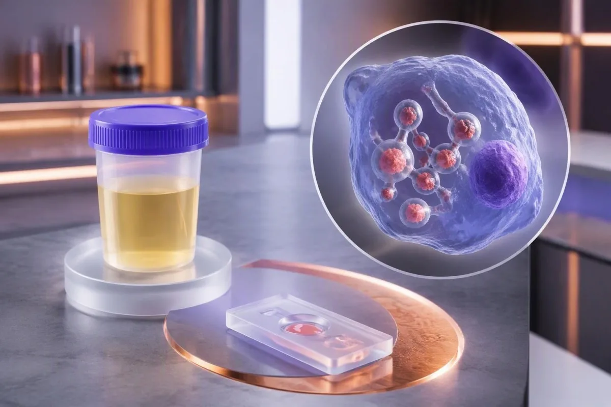
Позитивен резултат на антиген во урина или серум е показател за хистоплазмоза во соодветниот клинички контекст, но самиот по себе не може сигурно да ја идентификува габата. Симптомите, имунолошкиот статус и независните тестирања одредуваат што ќе следи.


- Позитивен антиген значи дека анализата открила габичен материјал што го препознаваат нејзините антитела; тоа само по себе не потврдува дека Histoplasma е причината.
- Вкрстена реактивност е особено изразена со Blastomyces; тестирањето на втор примерок со слична анализа не мора да го разјасни прашањето за видот.
- Чувствителноста во урина достигна 91.8% кај дисеминирана хистоплазмоза во една мултицентрична студија, но тој процент не важи за сите белодробни форми.
- Тајминг на антителата се разликува од времето на појава на антигенот: антителата на Histoplasma може да станат детектибилни дури по приближно 4–8 недели.
- Титри на антитела од 1:32 или повисок може да укажува на активна хистоплазмоза во соодветен контекст; четирикратниот пораст е посилен доказ од една изолирана вредност на титарот.
- Имунолошки статус ја зголемува итноста: ХИВ со број на CD4-клетки под 150 клетки/µL, трансплантација и значителен имуносупресивен третман ја зголемуваат загриженоста за дисеминација.
- Концентрација на антигенот нема универзален праг во ng/mL што самостојно дијагностицира хистоплазмоза или ја одредува итноста на негата.
- Времетраење на култивирањето обично изнесува неколку недели, често 2–6 недели; на тешко болните пациенти може да им биде потребен третман пред да се потврди со култура.
- Потребна е итна проценка е соодветно при недостиг на воздух во мирување, збунетост, несвестица или нова сатурација на кислород под 92% на морско ниво, освен ако лекар не утврдил поинаква вообичаена вредност.
Што всушност потврдува позитивен резултат на антиген за Histoplasma?
A позитивен антиген за Histoplasma резултатот потврдува дека тестот открил габичен материјал препознаен од неговите тестни антитела — но не и дека Histoplasma дефинитивно е причинителот. Хистоплазмозата станува поверојатна кога резултатот се совпаѓа со симптомите, изложеноста и имунолошкиот статус; и понатаму се можни габични инфекции што вкрстено реагираат.
Разликата е практична, а не семантичка: позитивниот резултат може да биде доволно силен за да поддржи работна дијагноза и итен третман, без да ги исполнува сите критериуми за потврда на ниво на вид. Во мултицентричната евалуација на Hage et al. од 2011 година, уринарниот антиген бил откриен кај 91.8% од случаите на дисеминирана хистоплазмоза, но ефикасноста се разликувала во зависност од клиничката слика и споредбените групи.
Да разгледаме хипотетички случај на 46-годишен пациент што прима инхибитор на TNF и развива треска, губење на тежината и дифузни белодробни абнормалности по нарушување на почвата: позитивниот резултат за антиген заслужува навремена проценка од специјалист за инфективни болести. Истата лабораториска ознака кај лице што инаку е здраво и немало веројатна изложеност бара проверка и внимателно земање анамнеза, а не автоматско препишување антимикотик.
Јас сум Thomas Klein, главен медицински директор во Kantesti, и моето практично правило е да ги раздвојам 3-те прашања: дали е откриен антиген, која габа е причинителот и колку е болен пациентот? Kantesti е AI анализатор на крвни тестови што може да помогне во организирањето на придружните лабораториски наоди, но не и да утврди идентитет на габата; нашиот организација и клинички фокус ја објаснуваат таа разлика.
По што се разликуваат тестовите за антиген на Histoplasma во урина и серум?
На тест за антиген на Histoplasma во урина го мери габичниот антиген што се излачува во урината, додека тестирањето на серумот го мери антигенот што циркулира во течниот дел од лабораторискиот примерок. Двата примерока даваат поврзани докази, а резултатот од кој било од нив може да биде негативен и покрај инфекцијата.
Урината често е корисен прв примерок бидејќи собирањето не е инвазивно, а чувствителноста е висока кај многу пациенти со дисеминирана болест. Лабораторискиот преглед на Azar и Hage од 2017 година го опишува откривањето антиген како особено корисно кога габичното оптоварување е значително; нарачувањето 2 вида примероци може да го подобри откривањето во одбрани ситуации, иако нивните резултати не се статистички независни.
Позитивен резултат од урина не значи дека бубрезите или мочниот меур нужно се инфицирани. Антигенот што потекнува од друго место може да циркулира и да се излачува, а анализата за Histoplasma е одделна од рутинската интерпретација на тест лента за урина; нормалните наоди за нитрити, леукоцитна естераза или протеини не го поништуваат позитивниот резултат од тестот за габичен антиген.
Упатствата за собирање примероци се разликуваат меѓу лабораториите: многу анализи користат случаен примерок од урина, а не 24-часовно собирање, додека за серумот е потребна подготовка на примерокот назначена од лабораторијата. Пред да се толкува несовпаѓање, проверете дали двата примерока се собрани пред третманот, обработени во истата лабораторија и анализирани со споредливи методи — овие 3 детали можат да објаснат повеќе од ознаките позитивно и негативно.
Што значи вредноста на антигенот изразена во ng/mL?
Квантитативниот резултат за антиген на Histoplasma ја проценува концентрацијата на антигенот, често во нг/мл, во рамките на опсегот на анализата на таа лабораторија. Не постои универзална концентрација што ја докажува хистоплазмозата, ја разликува Histoplasma од Blastomyces или одредува дали некому му е потребна болничка нега.
Во извештај во кој пишува дека е откриено под границата на квантификација, сигналот ги исполнил критериумите за откривање на анализата, но бил премал за сигурно мерење. На пример, илустративните резултати од 0.5 ng/mL и 5 ng/mL не се заменливи, но ниту еден од нив не е дијагноза на ниво на вид; нашиот водич за квалитативно наспроти квантитативно објаснува зошто откривањето и мерењето се одделни тврдења.
Повисоките концентрации често се поврзани со поголемо габично оптоварување кај дисеминирана хистоплазмоза, но преклопувањето меѓу групите пациенти оневозможува јасна скала на сериозност. Вредност над горната граница на известување е цензурирана, а не точна: ако во 2 извештаи пишува надвор од опсегот, лекарот не може да пресмета значајна процентуална промена без лабораториско разредување или друго валидирано мерење.
Urine concentration also affects interpretation: a small change between specimens can reflect hydration, collection conditions, or assay variation rather than a true change in disease activity. Understanding urine concentration and dilution is useful, but patients should not invent a creatinine-corrected Histoplasma ratio unless the laboratory has validated that specific approach.
Зошто други габи може да дадат лажно позитивен резултат на антиген за Histoplasma?
A Histoplasma antigen false positive can occur because other fungi contain antigenic structures recognized by the assay, especially Blastomyces. In that situation, the result may correctly signal a fungal illness while assigning misleading confidence to 1 particular fungal species.
Blastomyces is the central cross-reactivity concern because its geographic distribution and pulmonary symptoms can overlap with histoplasmosis. Hage et al. reported cross-reactivity in 90% of blastomycosis cases in their 2011 study, so a high apparent specificity against nonfungal controls should not be mistaken for equally high specificity against other endemic mycoses.
Talaromyces marneffei and Paracoccidioides can also cross-react in some Histoplasma antigen systems; the exact pattern depends on the assay and the patient’s travel or residence history. A 2-year-old overseas exposure can still matter in an immunosuppressed patient, and asking only where someone lives today can miss a clinically useful clue.
A second positive fungal antigen assay may repeat the same shared-antigen problem rather than independently confirm both infections. Species-directed culture, tissue examination, or validated molecular testing offers a different type of evidence; meanwhile, a positive Histoplasma result is not a diagnosis of household mold toxicity, as our mold testing limitations explain.
Колкава е веројатноста за хистоплазмоза по позитивен резултат?
The probability of histoplasmosis after a positive antigen result depends on the patient’s претестовна веројатност, the assay, and competing fungal diagnoses. No single positive predictive value applies to both an asymptomatic screening population and a severely ill, immunocompromised population.
Here is a deliberately hypothetical example: with 95% sensitivity and 95% specificity, testing 1,000 people when only 5% have the target illness produces about 48 true positives and 48 false positives. Roughly half the positive results would therefore represent that illness; these numbers teach probability and are not performance claims for a particular Histoplasma assay.
Raise the hypothetical disease prevalence to 50%, keeping the same assay characteristics, and the positive predictive value becomes approximately 95%. Real Histoplasma interpretation is more complicated because Blastomyces cross-reactivity creates a competing fungal explanation, and the patient’s symptom duration, antifungal exposure, and specimen type can all alter the test’s usefulness.
At Kantesti, we aim to explain why a laboratory flag should trigger a question rather than pretend it answers all 3 diagnostic questions at once. Our технологија за толкување со вештачка интелигенција describes the interpretation workflow; no AI-generated probability should replace an infectious-disease evaluation or be presented as a validated Histoplasma risk score without specific supporting evidence.
По што се разликуваат резултатите од тестовите за антиген и антитела на Histoplasma?
Antigen testing looks for fungal components; antibody testing looks for the patient’s immune response to the fungus. Histoplasma antibodies may take approximately 4–8 weeks to develop, so an early negative antibody result does not cancel a positive antigen result.
Antibodies can remain detectable after a previous infection, whereas antigen more directly reflects the presence of recognized fungal material at the sampling time. Neither test provides an infallible active-versus-past distinction: persistent antigen after treatment, cross-reactivity, and delayed antibody development all complicate the story; our водич за позитивен резултат од антитела separates immune memory from current disease.
For complement-fixation testing, a titer of 1:32 or higher can support active histoplasmosis when symptoms and other findings fit, and a fourfold rise between paired samples is stronger evidence than one isolated low titer. Azar and Hage’s 2017 review also explains the limits of immunodiffusion bands; those older serologic patterns should not be translated directly into modern antibody-assay index values.
A clinician may repeat initially negative antibodies several weeks later, often within a 2–6-week follow-up interval selected for the clinical situation. That repeat is useful only if waiting is safe: transplant recipients or people with advanced HIV may not produce a dependable antibody response, so culture, tissue assessment, and antigen testing can carry greater diagnostic weight.
Кои симптоми и изложености го прават позитивниот резултат поуверлив?
Fever, cough, chest discomfort, fatigue, and a plausible soil or bird-and-bat-dropping exposure make a positive Histoplasma antigen result more clinically convincing. Acute symptoms commonly begin about 3–17 days after exposure, although that timing does not cover every form of histoplasmosis.
Disturbing contaminated soil matters more than simply seeing a bird or bat: demolition, cave visits, roost cleanup, and excavation can aerosolize fungal particles. A hypothetical family developing fever and cough 10 days after the same dusty renovation provides a stronger shared-exposure clue than one person reporting an ordinary walk outdoors.
Persistent fever, weight loss, gastrointestinal symptoms, enlarged lymph nodes, or abnormalities affecting several organs raise concern for dissemination rather than a lung-limited illness. Breathlessness requires assessment of oxygenation and competing causes—not just another antigen test—and our водич за тестирање на отежнато дишење explains why 1 laboratory result cannot assess respiratory stability.
Routine inflammatory markers cannot reliably identify the organism: an elevated CRP, a normal white-cell count, or a low procalcitonin does not independently distinguish histoplasmosis from other infections. Recording 2 dates—the likely exposure and the first symptom—often improves interpretation more than repeating nonspecific markers; our споредба на WBC и CRP discusses these discordant patterns.
Како имунолошкиот статус влијае врз итноста при позитивен резултат на антиген?
A positive Histoplasma antigen result deserves faster assessment when the patient has advanced HIV, transplantation, or substantial immunosuppression because disseminated infection is more likely. In HIV, a CD4 count below 150 cells/µL is a recognized risk marker, not a universal diagnostic cutoff.
TNF inhibitors, prolonged systemic corticosteroids, and some cancer therapies can reduce the cellular immune response needed to contain Histoplasma. The medication name, daily dose, and treatment duration are 3 separate pieces of information; a normal total white-cell count does not prove normal cellular immunity, and antibody testing may be less reassuring in these patients.
HIV viral load and CD4 count answer different questions: viral load measures viral replication, while CD4 count helps characterize immune function. Someone with a previously undetectable viral load can still need assessment if their immune recovery is incomplete; our толкувањето на вирусниот товар кај ХИВ should not be used as a substitute for reviewing the current CD4 result.
Kantesti is an AI blood test interpretation platform that can help place 2 or more accompanying results in context, but a positive fungal antigen in an immunocompromised person needs clinician-led triage. A broader frequent infection assessment can support the workup; do not abruptly stop transplant medicines or corticosteroids without the prescribing team’s guidance.
Кои тестови помагаат да се потврди дека станува збор за Histoplasma, а не за габа со вкрстена реактивност?
Culture with organism identification, tissue examination, and appropriately validated molecular tests can help distinguish Histoplasma from cross-reacting fungi. Culture often takes approximately 2–6 weeks, so confirmation and treatment decisions may proceed in parallel when illness is severe.
The best specimen depends on the affected site: respiratory samples may help in pulmonary disease, while specialized cultures or tissue examination may be useful in suspected dissemination. The laboratory should be told that an endemic fungus is suspected because fungal culture requirements and safety procedures differ from routine bacterial processing; our толкување на резултатите од култура explains why specimen and method matter.
Histoplasma classically appears as small, approximately 2–4 µm budding yeast, often within macrophages; Blastomyces usually has larger yeast with broad-based budding. Those features require expert interpretation because other organisms and staining artifacts can mimic parts of the picture, and a tissue description alone may need culture or molecular confirmation before species confidence is justified.
PCR availability and clinical validation vary between laboratories, so a negative molecular result is not an all-purpose exclusion test. Before ordering a second procedure, ask which of 2 uncertainties remains: whether fungal infection is present or which species caused it; repeating antigen can help detection, but an independent identification method more directly addresses cross-reactivity.
Што додаваат снимањето на градниот кош и другите крвни тестови?
Chest imaging identifies the pattern and extent of lung involvement, but it cannot independently prove histoplasmosis. Nodules, enlarged hilar or mediastinal lymph nodes, and diffuse lung abnormalities can overlap with tuberculosis, sarcoidosis, malignancy, and other infections.
Old calcified nodules or lymph nodes can reflect healed granulomatous disease and do not automatically explain a newly positive antigen result. Comparing imaging from 2 time points helps separate stable structural remnants from evolving disease; a new respiratory illness still requires its own assessment rather than being attributed to every old spot on a scan.
Tuberculosis testing illustrates a different diagnostic limitation: a positive IGRA indicates an immune response to tuberculosis antigens but does not by itself distinguish latent infection from active disease. Our IGRA result interpretation explains why a positive IGRA and a positive fungal antigen require 2 parallel clinical questions, not an assumption that either result disproves the other.
An elevated ACE level is also not a reliable shortcut to sarcoidosis, especially when infection remains plausible; our ACE testing limitations covers that trap. In suspected dissemination, low counts across 2 or more blood-cell lines or abnormal liver tests can help identify affected organs, but none of those findings supplies fungal species confirmation.
Што треба да направите денес откако ќе добиете позитивен резултат?
Contact the ordering clinician promptly after a positive Histoplasma antigen result; seek emergency assessment for breathlessness at rest, confusion, fainting, or rapidly worsening illness. A new oxygen saturation below 92% at sea level is concerning, although altitude, measurement error, and established baseline disease can change interpretation.
A stable, asymptomatic person can usually begin with clinician-directed verification rather than an emergency department visit solely because of the laboratory flag. Conversely, an immunocompromised person with fever or systemic symptoms should seek same-day advice even if the antigen concentration is low; neither 0.5 ng/mL nor 5 ng/mL overrides the patient’s clinical condition.
Prepare 5 items for the discussion: the complete report, symptom-onset date, exposure and travel history, medication list, and immune-status information. Include the specimen type and laboratory reference comments, because a cropped screenshot can omit a below-quantification warning or cross-reactivity statement that materially changes the interpretation.
Kantesti AI can help explain accompanying results, but it should not tell a symptomatic patient to wait for repeat testing instead of urgent assessment. Our клиничка рамка за валидација addresses interpretation standards; in this setting, 2 genuinely different tests are often more informative than repeatedly measuring the same shared antigen, provided the clinician selects appropriate specimens.
Дали позитивниот резултат на антиген значи дека ви е потребно антифунгално лекување?
A positive antigen result does not automatically require antifungal treatment; treatment depends on the clinical syndrome, severity, immune status, and likelihood of true disease. Severe pulmonary or disseminated histoplasmosis can require immediate treatment, while selected mild pulmonary cases are managed with observation.
Wheat et al.’s 2007 IDSA guideline provides established treatment principles for moderately severe to severe disseminated histoplasmosis: liposomal amphotericin B at 3 mg/kg daily for about 1–2 weeks, followed by itraconazole for at least 12 months. That is a clinician-managed adult regimen, not an instruction to self-treat; organ involvement, pregnancy, interactions, and newer syndrome-specific guidance can alter the plan.
Itraconazole can substantially increase tacrolimus exposure through drug-metabolism interactions, so transplant teams often need additional level monitoring and dose adjustment. A previous stable tacrolimus value does not guarantee safety after even 1 medication change; our tacrolimus monitoring guide explains why sample timing and a current medication list are essential.
Amphotericin formulations can affect kidney function, potassium, and magnesium, making baseline and repeat laboratory measurements part of treatment rather than optional extras. A magnesium result below the laboratory range—often below approximately 1.7 mg/dL, depending on method—needs clinical interpretation alongside symptoms and other electrolytes; our причините за низок магнезиум explains relevant medication-related patterns.
Може ли антигенот на Histoplasma да остане позитивен по лекувањето?
Histoplasma antigen can remain detectable after effective treatment, and persistent low-level positivity does not independently prove treatment failure. A rising trend is more concerning when it accompanies recurrent symptoms or other evidence of active disease; 1 isolated measurement cannot establish relapse.
For disseminated disease, Wheat et al.’s 2007 IDSA guideline describes measuring antigen before therapy, at approximately 2 weeks and 1 month, then about every 3 months during treatment and for 12 months afterward. The appropriate schedule remains individualized; a patient with new fever or worsening breathing needs reassessment sooner than the next planned surveillance sample.
Urine and serum trends are not numerically interchangeable, and changing laboratories can create a false impression of improvement or relapse. Keep the specimen type and method consistent where possible, then compare symptoms, organ tests, and antigen together; our meaningful laboratory trend guide explains why 2 different methods may not produce a valid before-and-after comparison.
Kantesti is an AI lab test interpretation service that can help organize serial reports, but it should not label a small numerical rise as relapse without clinical evidence. Before reacting to a change, I would ask about 3 common confounders—treatment interruption, changed sample conditions, and assay differences—while the clinician considers adherence, drug absorption, interactions, and independent testing.
Практичен контролен список за следни чекори и публикации како поддршка
The safest follow-up plan identifies clinical urgency, the need for species confirmation, and how response will be monitored. As of October 14, 2026, no standalone antigen concentration or antibody titer replaces those 3 decisions, and an updated article date does not itself certify medical review.
Write down 3 questions for the appointment: does my presentation suggest active infection, could another fungus explain this assay, and which test would change the treatment plan? Thomas Klein’s editorial emphasis here is deliberately narrow: distinguish detection from diagnosis, and do not turn a reassuring antibody result into permission to ignore systemic illness.
Kantesti’s медицински советодавни информации describes our clinical advisory structure, while our стандарди за медицинска содржина explain expectations for sourcing and review. This article relies on 3 external histoplasmosis publications listed below; they address diagnostic performance, laboratory limitations, and established treatment principles rather than validating an AI fungal diagnosis.
The 2 supplementary Zenodo publications below concern general urinalysis and iron studies, not Histoplasma assay validation or antifungal efficacy. Our urinalysis reference guide helps distinguish specialized fungal antigen testing from routine urine chemistry; our упатство за интерпретација на студиите на железо provides background for evaluating anemia during prolonged illness, without identifying its infectious cause.
Често поставувани прашања
Дали позитивен антиген на Histoplasma значи дека дефинитивно имам хистоплазмоза?
Позитивен антиген за Histoplasma ја поддржува дијагнозата на хистоплазмоза, но сам по себе не ги потврдува ниту видот на габата ниту клиничката дијагноза. Blastomyces и некои други габи можат да предизвикаат вкрстена реакција, а видот на примерокот, симптомите и имунолошкиот статус влијаат врз толкувањето. Едно мултицентрично истражување од 2011 година открило антиген во урината кај 91.8% од случаите на дисеминирана хистоплазмоза, но таа бројка не ги опишува сите пулмонални презентации. Клиничарите може да користат култура, испитување на ткиво, антитела или валидирано молекуларно тестирање за да го разјаснат резултатот.
Дали Blastomyces може да предизвика лажно позитивен резултат за антиген на Histoplasma?
Blastomyces може да предизвика позитивен резултат на антигенот за Histoplasma бидејќи габите делат антигенски карактеристики што ги препознаваат некои тестови. Студијата на Hage и соработниците од 2011 година пријавила вкрстена реактивност кај 90% од случаите на бластомикоза. Затоа, позитивниот резултат може да укажува на вистинска габична инфекција, а не на случајна лабораториска грешка, но можеби нема да го идентификува точниот вид. Култура, испитување на ткиво или валидирано молекуларно тестирање насочено кон видот може да обезбеди понезависни докази.
Кое е нормалното ниво на антиген на Histoplasma во урината?
Вообичаеното негативно толкување на тестот за антиген на Histoplasma во урина е „не е детектиран“, според граничната вредност на анализата на конкретната лабораторија. Не постои универзален нормален опсег во ng/mL или концентрација што се смета за позитивна и важи за сите лаборатории. Илустративна вредност од 0.5 ng/mL може да биде мерлива со една метода, но да биде под опсегот на сигурно мерење на друга метода. Користете ги референтните коментари во извештајот и клиничкиот контекст, наместо да пренесувате гранични вредности од една анализа на друга.
Може ли антигенот на хистоплазма да биде позитивен кога антителата се негативни?
Антигенот на Histoplasma може да биде позитивен додека антителата се негативни, особено во раната фаза на болеста или кога имунолошката функција е нарушена. За развој на антитела може да бидат потребни приближно 4–8 недели, а некои имунокомпромитирани пациенти никогаш не создаваат сигурен одговор со антитела. Лекар може да ја повтори серологијата по неколку недели, ако е безбедно да се почека. Тешките симптоми бараат итна процена, наместо да се чека сероконверзија.
Дали позитивниот уринарен антиген е доказ за инфекција на уринарниот тракт?
Позитивен уринарен антиген за Histoplasma не мора да укажува на инфекција во мочниот меур или бубрезите. Габичниот антиген од друго место може да циркулира и да се излачува во урината. Специјализираниот тест за антиген и рутинската анализа на урина даваат одговори на 2 различни прашања, па нормалните резултати за нитрити или леукоцитна естераза не го побиваат наодот за габа. Симптомите, прегледот, снимањето и дополнителните тестирања утврдуваат кои органи се зафатени.
Колку долго може антигенот на Histoplasma да остане позитивен по третманот?
Антигенот на Histoplasma може да остане детектибилен со месеци по ефективното лекување, особено по дисеминирана болест. Самата перзистентна позитивност на ниско ниво не потврдува релапс, додека пораст придружен со повторна појава на симптоми е позагрижувачки. Насоките на IDSA од 2007 година опишуваат следење на антигенот за време на лекувањето и 12 месеци потоа кај дисеминирана хистоплазмоза. Времето на контролите и времетраењето на лекувањето треба да ги индивидуализира лекарот што го лекува пациентот.
Кога позитивниот резултат за антиген на хистоплазма претставува итен случај?
Позитивен резултат од тестот за антиген на Histoplasma бара итна проценка кога е придружен со отежнато дишење во мирување, конфузија, несвестица или брзо влошување на состојбата. Нова сатурација со кислород под 92% на морско ниво е загрижувачка, иако веќе постоечката основна состојба, надморската височина и квалитетот на мерењето влијаат врз толкувањето. Имунокомпромитираните пациенти со треска или системски симптоми треба да побараат медицински совет истиот ден, дури и при ниска концентрација на антигенот. Самата бројчена вредност на антигенот не е праг за итна состојба.
Добијте AI анализа на крв со моќ на вештачка интелигенција денес
Придружете се на над 2 милиони корисници ширум светот кои му веруваат на Kantesti за инстантна, точна анализа на лабораториски тестови. Поставете ги вашите резултати од крвна слика и добијте сеопфатно толкување на 15,000+ биомаркери за секунди.
📚 Реферирани научни публикации
Клајн, Т., Мичел, С., и Вебер, Х. (2026). Тест за уробилиноген во урина: Водич за комплетна анализа на урина 2026. AI Medical Research.
Клајн, Т., Мичел, С., и Вебер, Х. (2026). Водич за студии за железо: TIBC, сатурација на железо и капацитет на врзување. AI Medical Research.
📖 Надворешни медицински референци
Hage CA et al. (2011). A multicenter evaluation of tests for diagnosis of histoplasmosis. Clinical Infectious Diseases.
Azar MM, Hage CA. (2017). Laboratory Diagnostics for Histoplasmosis. Journal of Clinical Microbiology.
Wheat LJ et al. (2007). Clinical practice guidelines for the management of patients with histoplasmosis: 2007 update by the Infectious Diseases Society of America. Clinical Infectious Diseases.
⚕️ Медицинско одрекување од одговорност
Овој напис е само за едукативни цели и не претставува медицински совет. Секогаш консултирајте се со квалификуван здравствен работник за одлуки за дијагноза и третман.
Сигнали за доверба E-E-A-T
Искуство
Клиничка ревизија водена од лекар за работните текови на толкување на лабораториски резултати.
Експертиза
Фокус на лабораториска медицина за тоа како биомаркерите се однесуваат во клинички контекст.
Авторитивност
Напишано од д-р Thomas Klein, со ревизија од д-р Sarah Mitchell и проф. д-р Hans Weber.
Доверливост
Толкување засновано на докази со јасни патеки за следење за да се намали алармирањето.