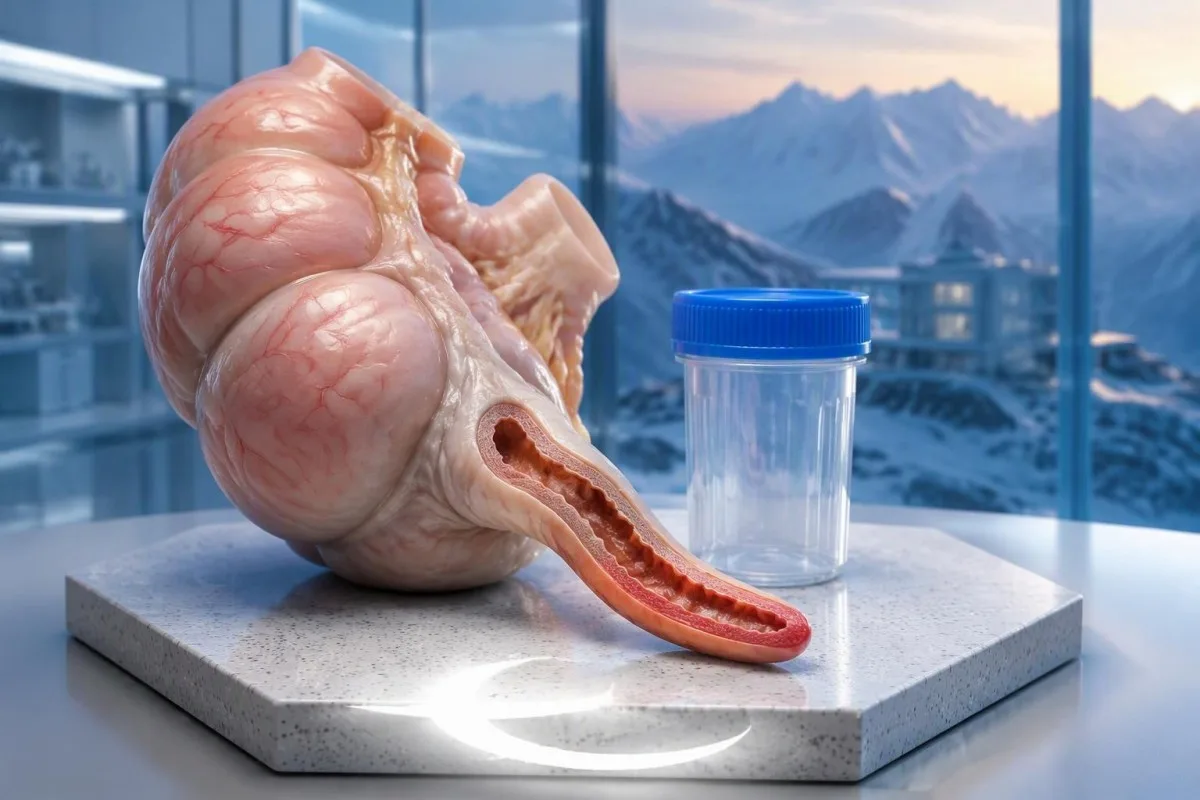
Юк—нормаль ак кан тәнчекләре һәм CRP ышанычлы рәвештә аппендицитны инкарь итә алмый. Лаборатория үзгәрешләренә кадәр симптомнар барлыкка килергә мөмкин, шуңа күрә барлык нәтиҗәләр ышанычлы булса да, корсак авыртуының начарлануына бәя бирергә кирәк.


- Нормаль WBC гадәттә өлкәннәрдә 4.0–11.0 × 10⁹/л тәшкил итә; аппендицит бу интервалда да булырга мөмкин.
- Нормаль CRP еш кына 5 мг/л дан азрак дигәнне аңлата, кайбер лабораторияләр 10 мг/л дан азрак кулланса да; бу чикләрнең берсе дә аппендицитны инкарь итми.
- Башлангыч сынау ялкынсыну реакциясен сагынырга мөмкин, чөнки CRP гадәттә ялкынсыну стимулыннан соң берничә сәгать үткәч, еш кына 6–8 сәгать тирәсендә арта башлый.
- Балалар өчен дәлилләр WBC өчен 10.0 × 10⁹/л сизгерлекне 85% тапты, бу исә якынча 15% авыру балалар бу билгеләнгән чиккә төшүен аңлата.
- Кушылган нәтиҗәләр бер маркерга караганда күбрәк мәгълүмат бирә, ләкин нормаль WBC һәм нормаль CRP тотрыклы урынлы йомшаклыкны яки начарланган авыртуны юкка чыгара алмый.
- Ашыгыч бәяләү начарланган уң-корсак авыртуы, кабатланучы косу, саклану яки хәрәкәт белән көчәйгән авырту өчен кирәк — хәтта 38°C кадәр температура булмаганда да.
- Образлы шиксезлек мөһим: УЗИда күренмәгән аппендикс, нормаль дип ышанычлы күрсәтелгән аппендикс белән бер түгел.
- Куркынычсызлык челтәре махсус яңадан бәяләү планын таләп итә; симптомнар начарланса, башка кан анализын яки AI интерпретациясен көтмәгез.
Нормаль аппендицит кан анализы аны читләтеп уза аламы?
A нормаль аппендицит кан анализы аппендицитны ышанычлы кире кага алмый, аеруча авыруның беренче сәгатьләрендә. Ак кан клеткалары саны һәм CRP организмның җавабын үлчи, аппендиксның үзен түгел; клиницистлар моны авыртуның дәвам итүе, тикшерү нәтиҗәләре һәм кирәк булганда, образлы тикшерүләр белән бергә интерпретацияләргә тиеш.
Кантести - ЯИ анализы WBC һәм CRP нәтиҗәләрен аңлата ала, ләкин ул лаборатор отчеттан аппендицитны кире кага алмый. Безнең оешма һәм клиник юнәлеш нәтиҗәләрне интерпретацияләүгә юнәлтелгән; карынны тикшерү һәм ашыгыч диагностика карары - башка эшләр.
Ике нормаль нәтиҗә, гадәттә, бер нормаль нәтиҗәгә караганда шикне киметә, ләкин бу киметүнең зурлыгы башлангыч рискка бәйле. Йомшак, бетә барган авыртуы булган кеше, икесе дә WBC 7.2 × 10⁹/л булса да, әкренләп начарланган уң як аскы карын төерлелегенә ия булган кешегә тиң түгел.
Лаборатор эталон интервалы - авыруны кире кагу прагьы түгел. Бу фраза нормаль чикләрдә эталон популяциясе белән чагыштыруны тасвирлый; бу һәр орган тикшерелгән дигәнне яки эволюцияләнүче авыру кире кагылган дигәнне аңлатмый.
Минем подход, доктор Томас Клейн буларак, нәтиҗә чыннан да нәрсәгә җавап бирә дип сорау: бу моментта үлчәнә торган системалы җавап бармы? 3 мг/л CRP бу тар сорауга җавап бирә. Ул ни өчен кешедә хәзер локальләшкән авырту барлыгын, хәрәкәтне күтәрә алмавын яки моңа кадәрге бәяләмәдән соң кайтканлыгын аңлатмый.
Аппендицитны ачыклауда нинди кан тестлары клиницистларга ярдәм итә?
Кан анализлары аппендицит диагнозын раслый ала, ләкин бернинди дә гадәти кан маркеры аппендицитны махсус ачыклый алмый. Дифференциалы белән тулы кан анализы һәм CRP еш кулланыла; бөер функциясе, электролитлар һәм башка анализлар дегидратацияне, альтернатив диагнозларны һәм дәвалау яки образлы тикшерүгә әзерлекне бәяләргә ярдәм итә.
.Әр сүзнең лейкоцитлар санына, яки WBC, әйләнештәге ак күзәнәкләрнең гомуми санын үлчи. Дифференциал тулы нейтрофилларны башка күзәнәк төрләреннән аера, бу мөһим, чөнки 9.0 × 10⁹/л гомуми санын югарырак нейтрофил саны һәм азрак лимфоцит саны яшереп торырга мөмкин.
C-реактив аксым, яки CRP, башлыча бавырда ялкынсыну сигналларына җавап итеп җитештерелә. 25 мг/л нәтиҗәсе ялкынсыну реакциясен күрсәтә, ләкин ул бу реакцияне локальләштерми: аппендицит, сидек инфекциясе, пневмония һәм берничә йогышлы булмаган шартлар CRPны арттыра ала.
Гадәти лаборатор интерваллар прибор, популяция һәм отчетлау практикасы буенча төрле була. 10,5 × 10⁹/л WBC бер лаборатория тарафыннан билгеләнергә мөмкин, ә икенчесе тарафыннан билгеләнмәскә мөмкин; төсле кисәтү отчеттан югалганда, клиник мәгънәсе кискен үзгәрми.
Каршылыклы нәтиҗәләр һичшиксез лаборатор хаталар түгел. Түбән CRP белән арткан WBC, төрле реакция вакытларын чагылдырырга мөмкин, шул ук вакытта югары CRP нормаль WBC белән, соңрак яки төрлечә күрсәтелгән реакцияне чагылдырырга мөмкин; безнең кулланмабыз WBC һәм CRP каршылыгы бу аермәне аңлата.
Аппендицит булганда кешедә ничек нормаль WBC булырга мөмкин?
Normal WBC with appendicitis is possible because local inflammation does not always produce immediate or substantial leukocytosis. Symptom duration, baseline cell counts, medicines and immune function influence the result; a normal total count may also coexist with a concerning neutrophil pattern.
A total WBC should be read with the абсолют нейтрофиллар саны, not only the neutrophil percentage. For example, WBC of 8.0 × 10⁹/L with 80% neutrophils gives an absolute neutrophil count of 6.4 × 10⁹/L; the percentage alone can look more alarming than the absolute result.
Fawkner-Corbett et al. (2022) pooled 67 studies involving 34,839 children and found approximately 85% sensitivity for WBC at a cutoff of 10.0 × 10⁹/L. Roughly 15% of children with appendicitis therefore fell below that study cutoff—not necessarily below every laboratory's age-specific upper limit.
Baseline counts change the meaning of a single result. A rise from a person's usual WBC of 3.5 to 8.5 × 10⁹/L may represent a substantial response despite remaining unflagged; neutrophil reference ranges also differ by age and pregnancy status.
Some people have a naturally lower neutrophil baseline, including those with a Дюффи-нул белән бәйле нейтрофиллар саны. That finding is not itself evidence of impaired immunity, but it illustrates why a universal WBC cutoff cannot replace symptoms, examination or comparison with previous results.
Аппендицитның башлангыч стадиясендә CRP дәрәҗәсе нигә нормаль булырга мөмкин?
CRP levels in appendicitis may remain normal early because inflammatory signalling must reach the liver before circulating CRP increases. CRP generally begins rising several hours after an inflammatory stimulus, often around 6–8 hours, but symptom onset and the biological start of inflammation are not identical timestamps.
A CRP of 2 mg/L shortly after pain begins is less reassuring than the same value after a longer, clearly improving illness. There is no reliable clock that lets a clinician say, 'At exactly eight hours, a normal CRP excludes appendicitis.' Biological responses vary, and patients may first notice symptoms at different stages.
Higher CRP can increase concern for more extensive inflammation, particularly when the history and imaging agree. However, CRP of 50 or 100 mg/L does not independently diagnose perforation; duration, another inflammatory condition and the patient's overall physiological state can influence the number.
Standard CRP and high-sensitivity CRP, or hs-CRP, measure the same protein using different analytical sensitivity. Cardiovascular hs-CRP categories such as below 1, 1–3 and above 3 mg/L are not appendicitis decision thresholds; hs-CRP interpretation requires a different clinical question.
A mildly elevated baseline also complicates interpretation. Some medicines, including certain hormonal contraceptives, can influence CRP; our discussion of contraception and CRP results explains why a small elevation should not automatically be attributed to an acute abdominal cause—or used to dismiss that cause.
WBC һәм CRP нормаль булганда бергәләп ышанычлыракмы?
Normal WBC and CRP together are more reassuring than either alone, but they are not a universal rule-out test. Their value depends on symptom duration, examination findings, the population being assessed and the exact cutoffs used; persistent focal pain can still justify imaging or surgical assessment.
In the paediatric meta-analysis by Fawkner-Corbett et al. (2022), combining WBC ≥10.0 × 10⁹/L or CRP ≥10 mg/L produced approximately 97% sensitivity in the studies reporting that combination. This was an 'either marker positive' strategy, not a requirement that both markers be elevated.
A sensitivity of 97% does not mean that a child with two low results has a 3% chance of appendicitis. Sensitivity describes affected patients; the chance of disease after a negative result also depends on how likely appendicitis was before testing and on the specificity of the test strategy.
Thresholds deserve scrutiny. CRP below 10 mg/L counted as negative in that analysis, but a value of 8 mg/L is above the reference limit in many laboratories; calling every study-negative result 'completely normal' would therefore overstate what the evidence actually evaluated.
A clinician may repeat the examination and selected tests during supervised observation, but worsening pain should not wait for a laboratory rise. The timing of blood test changes is useful context, not permission to stay home until WBC or CRP crosses a particular line.
Симтомнар һәм тикшерү диагнозны ничек үзгәртә?
Symptom progression and abdominal examination determine how much weight normal blood tests deserve. Pain migrating toward the right lower abdomen, focal tenderness, guarding and pain with movement increase concern; clinicians may use validated scores to organise these findings, but scores do not replace clinical judgement.
The classic sequence is central abdominal discomfort followed by more localised right-lower pain, but not every patient follows that pattern. An appendix behind the caecum may produce less obvious front-of-abdomen tenderness, while a pelvic appendix can cause urinary or bowel symptoms without the expected textbook presentation.
Consider a hypothetical 24-year-old with WBC of 7.8 × 10⁹/L and CRP of 4 mg/L whose discomfort becomes focal over several hours. Those results may justify caution rather than reassurance if the second examination reveals guarding; the change in the abdomen is new evidence, not a contradiction of the first test.
.Әр сүзнең Alvarado score has a maximum of 10 points, and the Appendicitis Inflammatory Response score has a maximum of 12. Both incorporate clinical features and laboratory information, so a low inflammatory response can lower the score even when an early examination remains concerning.
Di Saverio et al. (2020), in the WSES Jerusalem guidelines, recommend risk-stratified assessment and identify the AIR and Adult Appendicitis Score as useful adult tools. Our guide to unexplained pain testing makes the same practical distinction: a normal inflammatory marker does not explain away a focal symptom.
Нормаль лаборатория нәтиҗәләренә карамастан, кайсы симптомнарны ашыгыч бәяләү таләп ителә?
Worsening or persistent right-lower abdominal pain needs urgent assessment even with normal WBC and CRP. Seek emergency care for severe or spreading pain, a rigid abdomen, repeated vomiting, fainting, confusion or marked weakness; a fever is not required for appendicitis or another abdominal emergency.
Pain aggravated by walking, coughing or ordinary movement can suggest irritation of the abdominal lining. Do not repeatedly press your abdomen or try internet 'jump tests' to prove appendicitis; a negative home manoeuvre cannot exclude it, and a positive one does not identify the cause.
A temperature below 38°C does not exclude appendicitis. Children, older adults and immunosuppressed people may have less typical signs; a child who refuses to walk, has worsening abdominal pain or repeatedly vomits deserves prompt assessment rather than reassurance from an earlier normal panel.
Severe illness requires attention to circulation and breathing as well as abdominal findings. Clinicians may order lactate when shock or sepsis is suspected, but lactate testing limitations mean that neither a normal result nor a mildly elevated result independently settles the appendicitis question.
Call your local emergency number for collapse, confusion, difficulty breathing or severe rapidly worsening pain; in the UK, use 999 яки 112. For less severe but persistent or worsening symptoms, seek urgent same-day medical assessment, and do not delay because you have eaten, taken pain relief or are waiting for an online interpretation.
Сидек, йөклелек һәм химия тестлары нәрсә өсти?
Urine, pregnancy and chemistry tests help identify alternatives and treatment needs, not just appendicitis. Urinalysis can reveal urinary abnormalities, pregnancy testing changes the differential diagnosis and imaging plan, and kidney function or electrolyte tests assess dehydration and other consequences of vomiting.
Urinary white cells or microscopic blood can occur when an inflamed appendix irritates nearby urinary structures. A mildly abnormal urine result therefore does not automatically prove a urinary infection; the сидек анализы буенча тулы кулланма explains why collection quality and the entire urinary pattern matter.
Pregnancy testing is often appropriate for a person who could be pregnant and has acute lower abdominal pain. A negative urine test obtained very early can be misleading; our guide to early pregnancy test limitations explains when serum testing or repeat assessment may be necessary.
An ectopic pregnancy is a separate emergency that must be considered when pregnancy is possible, especially with one-sided pain, vaginal bleeding, dizziness or fainting. WBC of 6.0 × 10⁹/L and CRP of 3 mg/L do not exclude that diagnosis either; laboratory reassurance must not narrow the assessment prematurely.
Кантести - AI кан анализы нәтиҗәләрен аңлату платформасы that can explain reported kidney markers and electrolytes alongside WBC and CRP. Its interpretation cannot determine whether urinary abnormalities arise from the appendix, a stone or infection; that distinction requires the patient's history, examination and sometimes imaging.
УЗИ, КТ яки МРТ кайчан кирәк була?
Imaging is considered when appendicitis remains possible after clinical assessment, including when blood tests are normal. Ultrasound is commonly preferred initially in children and pregnancy, CT is frequently used in non-pregnant adults, and MRI can clarify selected cases without ionising radiation.
Ultrasound findings include appendix size, compressibility, surrounding fat changes and fluid. An outer diameter above approximately 6 mm can support appendicitis, but size alone is not definitive; a mildly enlarged appendix without supporting features is different from a non-compressible appendix with surrounding inflammatory changes.
'Appendix not visualised' is not synonymous with 'normal appendix'. Bowel gas, body habitus, appendix position and operator experience affect visibility; clinicians interpret a non-diagnostic scan with the examination and may choose observation, repeat ultrasound, MRI or CT rather than treating it as a negative answer.
CT can evaluate the appendix and several alternative causes of abdominal pain in one examination. Di Saverio et al. (2020) support selected low-dose CT strategies after inconclusive ultrasound, particularly in adolescents and young adults, while local pathways differ according to expertise, availability and the patient's circumstances.
Imaging choice also depends on where the pain is and what else is plausible. Right-upper abdominal pain may require a different assessment; our article on gallbladder blood test limitations illustrates another condition in which apparently reassuring inflammatory markers cannot substitute for examination and organ-specific imaging.
Нормаль ялкынсыну маркерлары булган очракларда кемгә өстәмә саклык кирәк?
Children, pregnant patients, older adults and immunosuppressed people need particularly careful interpretation of normal results. Symptoms may be atypical, reference intervals may differ and some patients cannot mount a typical inflammatory response; clinical deterioration should carry more weight than an unflagged laboratory panel.
Children need age-specific WBC intervals, not automatic application of an adult 4.0–11.0 × 10⁹/L range. Younger children may struggle to describe migration or localisation of pain, so behaviour, hydration, movement and repeated examination can become more informative than an apparently ordinary laboratory result.
Pregnancy can increase the normal WBC baseline, making mild leukocytosis less specific for appendicitis. Pregnancy also changes the consequences of missed alternative diagnoses and the imaging discussion; ultrasound and MRI are useful options, but necessary diagnostic assessment should not be delayed solely because radiation is a concern.
Older adults may present with less fever or less obvious local tenderness, and delayed recognition can have serious consequences. New persistent abdominal pain in a 78-year-old with WBC of 8.0 × 10⁹/L still needs a broad assessment that considers appendicitis, obstruction, vascular disease and other urgent causes.
Immune-suppressing medicines can alter laboratory responses, while corticosteroids can themselves increase circulating neutrophils. These mixed effects make a single result difficult to interpret; our discussion of children's AI interpretation safety explains why age, symptoms and direct clinical assessment must remain central.
Нормаль кан тестлары белән выпискадан соң нәрсә эшләргә?
Discharge after normal blood tests should include clear return precautions and, when needed, a reassessment plan. Return promptly if pain worsens, localises, spreads, or is accompanied by repeated vomiting, fainting or increasing illness; an earlier reassuring assessment does not guarantee that an evolving condition remains low risk.
Ask what was actually established before discharge: was appendicitis considered unlikely, was a normal appendix demonstrated on imaging, or was ultrasound inconclusive? Those are three different conclusions, and each deserves a different explanation rather than a vague statement that 'the bloods were fine'.
A useful safety plan names the warning signs, where to return and when persistent symptoms should be reviewed. If a clinician recommends reassessment within 6–12 hours, follow that individual plan; it is not a universal instruction to wait that long when pain is getting worse.
Bring the original report, imaging summary, medicine list and a simple symptom timeline. Record when the pain began and how its location changed, not just its severity; our табиб кабул итүе буенча кыскача тикшерү исемлеге helps organise information without replacing the consultation.
At Kantesti AI, we distinguish an explanation of an old result from a decision about new symptoms. A WBC of 7.0 × 10⁹/L from yesterday remains yesterday's measurement; if today's abdomen is more tender or vomiting has developed, another online reading of the same report cannot answer today's clinical question.
Кабат кан тестлары яки тенденцияләр сорауга җавап бирә аламы?
Repeat blood tests can add information, but trends cannot independently confirm or exclude appendicitis. The direction of symptoms and examination findings matters more than a small isolated laboratory change; repeat testing should be part of a clinician-led plan, not a substitute for reassessment or indicated imaging.
A rise in CRP from 3 to 18 mg/L over several hours supports a developing inflammatory response. It still does not identify the appendix as the source; clinicians ask whether the pain, examination and imaging now fit appendicitis or point toward a different abdominal or extra-abdominal diagnosis.
CRP has a circulating half-life of roughly 19 сәгать, so its concentration does not track symptom changes minute by minute. After treatment, CRP may remain elevated despite early clinical improvement; conversely, temporary improvement in discomfort does not prove that an untreated abdominal problem has resolved.
Small WBC differences can reflect hydration, stress, medicines and measurement variation. A change from 8.1 to 8.6 × 10⁹/L should not be portrayed as proof of progression on its own, particularly if different laboratories were used or the patient's fluid status changed between samples.
After hospital treatment, follow the discharge team's instructions rather than chasing a normal CRP independently. Our guide to post-hospital blood test changes explains why recovery curves vary; recurrent fever, worsening abdominal pain or persistent vomiting require assessment even if a previous result was improving.
Ясалма интеллектны аңлату нәрсәгә ярдәм итә ала — һәм нәрсәгә юк?
Кантести - AI нигезендәге кан анализы тикшерү коралы that can explain WBC, neutrophil and CRP results, but it cannot rule out appendicitis or provide an abdominal examination. With worsening abdominal symptoms, seek urgent assessment rather than waiting for a report upload, automated answer or trend comparison.
A useful interpretation checks the reported units before discussing a threshold. WBC of 10 × 10⁹/L equals 10,000 cells/µL, while CRP of 1 mg/dL equals 10 mg/L; confusing either pair can make the same result appear tenfold different or incorrectly classified.
AI should preserve the distinction between a laboratory flag and a diagnostic conclusion. Our AI технологиясе аңлатмасы describes the interpretation workflow, but no laboratory-only workflow can reproduce guarding, percussion tenderness, changing vital signs or the findings of a properly performed imaging study.
Kantesti's report tools can help organise laboratory information for a clinician, including reported reference intervals and previous measurements when available. They should not assign an 'all clear' from WBC of 6.8 × 10⁹/L and CRP of 2 mg/L when the patient describes worsening focal abdominal pain.
If uploading results for a non-urgent explanation, check that the file belongs to the correct person and that dates, units and values are legible. Our нәтиҗә йөкләүнең шәхси саклау чек-листында аңлатыла. covers sensible preparation; during an urgent abdominal episode, obtaining clinical care comes before uploading any document.
Фәнни басмалар: диагностик дәлилләр һәм ЯИ чикләүләре
Appendicitis diagnostic evidence and AI technical benchmarks answer different questions. As of October 4, 2026, this article uses established appendicitis guidelines and diagnostic research; Kantesti's repository publications below should not be treated as proof that an AI system can exclude appendicitis from normal laboratory results.
Andersson's 2004 meta-analysis in the British Journal of Surgery evaluated clinical and laboratory diagnosis and supports combining findings rather than relying on one marker. WSES guidance from 2020 and the 2022 paediatric meta-analysis address different populations and methods; none supports a blanket claim that normal WBC and CRP exclude every case.
A pre-registered, rubric-based automated technical benchmark of the Kantesti blood-test interpretation engine on 100,000 synthetic test cases. (n.d.). Figshare. The DOI-linked record is listed below, with ResearchGate язмасын эзләү һәм Academia басма эзләү links; these are searches, not claims of verified duplicate deposits or peer review.
Clinical validation framework v2.0 (Medical validation page). (n.d.). Zenodo. The DOI-linked record is listed below, with ResearchGate framework search һәм Academia framework search links; a framework describes assessment methods, not an independently established appendicitis rule-out sensitivity.
As Dr. Thomas Klein, my editorial standard is to keep those boundaries explicit: 100,000 synthetic cases are not 100,000 patients prospectively assessed for appendicitis. Our technical validation standards һәм medical advisory structure explain the wider context; neither replaces timely assessment of a person with worsening abdominal pain.
Еш бирелә торган сораулар
Канды анализ аппендицитны ачыклый аламы?
Кандагы тикшерүләр аппендицит диагнозын расларга ярдәм итә ала, ләкин алар аны үзләре генә ышанычлы ачыклый алмыйлар. Күтәрелгән WBC яки CRP ялкынсыну реакциясен күрсәтә, ләкин аның урынын ачыкламыйча. Аппендицит булганда, аеруча авыруның башында, өлкәннәрдә WBC якынча 4,0–11,0 × 10⁹/л булырга мөмкин. Табиблар лаборатория нәтиҗәләрен симптомнарның дәвамлылыгы, карынны тикшерү һәм кирәк булганда имиджлау белән берләштерәләр.
WBC һәм CRP нормаль булганда аппендицит булырга мөмкинме?
Әйе, аппендицит та нормаль WBC һәм нормаль CRP белән булырга мөмкин, аеруча системалы ялкынсыну реакциясе үсеш алганчы. WBC 7.0 × 10⁹/Л һәм CRP 3 мг/л аппендицитны аерым-аерым читләтеп узмас. Ике түбән маркер, гомумән, аз куркынычлы бәяләүдә шикне киметергә мөмкин, ләкин дәвамлы фокуслы сызлану яки авыртуның көчәюе әле дә сурәтләү яки кабат бәяләү өчен нигез булырга мөмкин. Маркерлар күтәрелгәнче көтү урынына симптомнар начарланса, ашыгыч медицина ярдәмен эзләгез.
Перитонит вакытында CRP ничек тиз күтәрелә?
CRP гадәттә ялкынсыну стимулыннан берничә сәгать узгач, еш кына 6–8 сәгать эчендә күтәрелә башлый, әмма аппендицит ышанычлы лаборатория вакыт графигына иярми. Ялкынсынуның биологик башлануы пациентның корсак симптомнарын беренче тапканчы яки аннан соң да булырга мөмкин. Шуңа күрә, авыруның башында 5 мг/л CRPдан түбәнрәк булу аппендицитны инкяр итә алмый. Мәгънәсезләнгән симптомнар, күпме сәгать узганын карамастан, бәяләнергә тиеш.
Appendicitisны нинди WBC саны яки CRP дәрәҗәсе раслый?
Бер генә WBC саны яки CRP дәрәҗәсе дә аппендицитны расламый. WBC күрсәткече якынча 11,0 × 10⁹/л дан югарырак булса, күп кенә олылар өчен нормадан тыш дип санала, һәм CRP күрсәткече 5 яки 10 мг/л дан югарырак булса, лабораториягә карап, арткан дип санала, ләкин мондый үзгәрешләрне башка күп кенә хәлләр дә китереп чыгарырга мөмкин. Хәтта 100 мг/л CRP да аппендицитны яки тишекне бәйсез рәвештә расламый. Раслау гомуми клиник бәяләүгә бәйле, еш кына КТ ярдәме белән.
Нормаль УЗИ аппендицитны кисәтәме?
Узи ярдәмендә ышанычлы диагностика борчуны иң күпкә киметә, әгәр дә аппендикс ачык күренсә һәм башка аз рисклы клиник күрсәткечләргә караганда нормада булса. Аппендикс күренмәде дигән хисап нормаль аппендикска тиң түгел. Диаметры якынча 6 мм дан арткан аппендикс аппендицитны раслый ала, ләкин зурлыкны кысылучанлык һәм тирә-юньдәге табылдыклар белән бергә аңларга кирәк. Баштагы ышанычлы сканерга карамастан, даими яки начарлана барган авырту өстәмә тикшерү таләп итәргә мөмкин.
Гадәттәге аппендицит кан тестларыннан соң кайчан хастаханәгә кайтырга кирәк?
Карын авыртуы көчле булса, уң яктагы аскы корсакка күчсә, таралса яки берничә тапкыр кабатланган косу, хәлсезлек, катыру яки авыруның көчәюе белән бергә булса, тиз арада кире кайтыгыз. 38°C астындагы температура аппендицитны инкар итми, һәм узган көнгә нормаль кан сынаулары бүгенге диагнозны расламый. Хастаханкадан чыгарганда бирелгән үз-үзеңне тикшерү буенча күрсәтмәләрне үтәгез, ләкин хәл начарланса, шул очрашуны көтмәгез. Хәле беткәндә, буталыш булганда, сулыш алу авыр булганда яки авырту бик көчле һәм тиз көчәйсә, җирле ашыгыч ярдәм номерына шалтыратыгыз.
Бүген үк AI белән эшләнгән кан анализы тикшерүе
Дөнья буенча 2 миллионнан артык кулланучы кушылыгыз: алар Kantestiны тиз һәм төгәл лаборатория анализы өчен ышана. Кан анализы нәтиҗәләрегезне йөкләгез һәм 15,000+ биомаркерларын секундлар эчендә тулы аңлатма белән алыгыз.
📚 Сылтама бирелгән тикшеренү басмалары
Klein, T., Mitchell, S., & Weber, H. (2026). Kantesti Кан анализын аңлату двигателе өчен 100 000 синтетик тест очрагы буенча алдан теркәлгән, рубрика нигезендәге автоматлаштырылган техник бенчмарк. Kantesti AI медицина тикшеренүе.
Klein, T., Mitchell, S., & Weber, H. (2026). Клиник валидацияләү структурасы v2.0 (Медицинаны валидацияләү бите). Kantesti AI медицина тикшеренүе.
📖 Тышкы медицина белешмәләре
Fawkner-Corbett D et al. (2022). Diagnostic accuracy of blood tests of inflammation in paediatric appendicitis: a systematic review and meta-analysis. BMJ Open.
⚕️ Медицина кисәтүе
Бу мәкалә бары тик белем бирү максатларында гына һәм медицина киңәше булып тормый. Диагноз һәм дәвалау карарлары өчен һәрвакыт квалификацияле сәламәтлек саклау белгеченә мөрәҗәгать итегез.
E-E-A-T ышаныч сигналлары
Тәҗрибә
Табиб җитәкчелегендә лаборатория нәтиҗәләрен аңлату эш процессларын клиник тикшерү.
Белгечлек
Лаборатория медицинасы: биомаркерларның клиник контекстта үз-үзләрен тотышын аңлау.
Авторититет
Доктор Томас Кляйн тарафыннан язылган, доктор Сара Митчелл һәм профессор доктор Ханс Вебер тарафыннан тикшерелгән.
Ышанычлылык
Ачык күзәтү юллары белән куркуны киметү өчен дәлилләргә нигезләнгән аңлату.