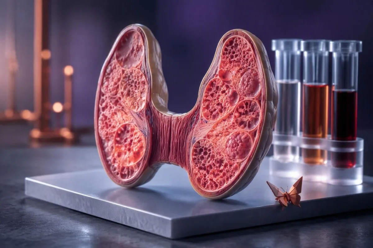
Subacute thyroiditis can make thyroid results look contradictory from one week to the next. The sequence of TSH, free T4, free T3, ESR and CRP usually explains why.


- Early pattern: Free T4 and free T3 rise while TSH falls, because stored hormone leaks from an injured thyroid rather than being newly overproduced.
- TSH lag: TSH can remain below 0.1 mIU/L for 4-12 weeks after free T4 has returned to range.
- การอักเสบ: CRP is commonly above 10 mg/L and ESR often exceeds 50 mm/hour during painful active disease, although neither marker is specific to thyroiditis.
- Low uptake clue: A low radioactive iodine uptake result helps distinguish thyroiditis from Graves disease when blood results alone are unclear.
- Hypothyroid phase: Free T4 may fall below about 0.8 ng/dL before TSH rises, creating a brief period when symptoms and TSH do not yet match.
- Recovery: Most people regain normal thyroid function within 6-12 months, but roughly 5-15% develop lasting hypothyroidism.
- การตรวจซ้ำ: Rechecking TSH and free T4 every 4-6 weeks is usually more informative than chasing symptoms or testing weekly.
What a subacute thyroiditis blood test usually shows
A subacute thyroiditis blood test usually moves through three stages: high free T4/free T3 with low TSH, then low free hormones with a delayed high TSH, then gradual normalisation. This is hormone leakage from injured thyroid follicles, not classic thyroid overproduction.
In the first 1-3 weeks, many adults have free T4 above the laboratory upper limit of roughly 1.8 ng/dL and TSH below 0.1 mIU/L. TSH is a pituitary response, so it reflects what circulating hormone was doing over prior days rather than only what is happening at the moment of collection.
I have seen patients frightened by a TSH of 0.01 mIU/L, assuming they have permanent hyperthyroidism. Dr. Thomas Klein, MD, would read that result alongside neck pain, temperature, pulse, free hormones and inflammatory markers; the combination is much more useful than a single flag.
คันเตสตีเป็น เครื่องวิเคราะห์ผลเลือด AI that places TSH, free T4, free T3, ESR and CRP on a dated timeline rather than treating each result as an isolated verdict. That timeline matters when a result obtained 10 days later appears to tell a completely different story; see our guide to thyroid testing schedules.
The pattern is more diagnostic than one number
A low TSH with high free T4 can occur in Graves disease, thyroiditis, excessive levothyroxine, iodine exposure and assay interference. Painful thyroid enlargement plus raised CRP or ESR makes subacute thyroiditis more likely, but confirmation sometimes needs antibody testing or uptake imaging.
Why free T4 and free T3 rise at the beginning
Free T4 and free T3 rise early because inflamed thyroid follicles release preformed hormone into circulation. The gland is leaking its stored supply, so antithyroid medicines generally do not shorten this phase.
Free T4 often rises more prominently than free T3 in destructive thyroiditis, producing a lower total T3-to-total T4 ratio than is often seen in Graves disease. A total T3 in the upper range or modestly elevated level does not rule thyroiditis out; assay choice and sampling day matter.
The symptoms can feel intense despite a self-limited mechanism: resting pulse above 100 beats/minute, tremor, heat intolerance and poor sleep are common. A clinician may use a beta-blocker such as propranolol for symptoms when appropriate, but it does not repair the thyroid or change the hormone-release curve.
The American Thyroid Association guideline describes thyroiditis-related thyrotoxicosis as a low-uptake state and advises against routine antithyroid drugs because synthesis is not the problem (Ross et al., 2016). If palpitations dominate, our คู่มือการตรวจเลือดสำหรับอาการใจสั่น explains which non-thyroid findings deserve parallel attention.
Why TSH stays low after free T4 improves
TSH commonly remains suppressed for several weeks after free T4 has normalised because pituitary thyrotroph cells recover slowly from hormone exposure. A low TSH alone does not prove ongoing thyroid hormone excess.
A practical reference interval for TSH in many adult laboratories is about 0.4-4.0 mIU/L, yet the report-specific range always wins. After thyrotoxicosis, TSH may stay below 0.4 mIU/L for 4-12 weeks while free T4 is already 0.9-1.4 ng/dL and symptoms are settling.
This creates a common error: increasing surveillance or starting treatment based solely on TSH at week 5. In my experience, repeating ตรวจ TSH และ free T4 พร้อมกัน in 4-6 weeks is safer than interpreting a suppressed TSH as a standalone treatment target.
Biotin supplements can falsely lower some TSH results and falsely raise free T4 or free T3 in certain immunoassays. Hold high-dose biotin for at least 48 hours before testing unless the treating clinician gives a different instruction; our article on ข้อผิดพลาดของการตรวจ free T3 สูง ครอบคลุมกลไก.
Thyroiditis ESR levels and CRP: how high is typical?
Painful subacute thyroiditis often raises ESR above 50 mm/hour และ CRP สูงกว่า 10 mg/L, but neither value measures thyroid damage directly. ESR can remain elevated after pain improves, whereas CRP tends to fall faster with resolving tissue response.
A CRP reference range is commonly below 5 mg/L, although some laboratories use below 8 or 10 mg/L. CRP above 30 mg/L supports active inflammation in the right clinical setting, but bacterial infection, autoimmune disease, injury and obesity can also raise it.
ESR is affected by age, anaemia, pregnancy, immunoglobulins and red-cell shape. An ESR of 70 mm/hour with a CRP of 4 mg/L may reflect slower ESR kinetics or a non-thyroid factor, which is why we do not use ESR as a pain score.
Stasiak and colleagues note that raised ESR and CRP support subacute thyroiditis but diagnosis remains clinical and imaging-supported when uncertain (Stasiak et al., 2019). Compare a persistent result with our explanation of why ESR falls slowly, especially if haemoglobin is also low.
Blood clues that separate thyroiditis from Graves disease
Subacute thyroiditis and Graves disease can both cause low TSH with high free T4, but painful thyroiditis usually has high ESR or CRP and low uptake on nuclear imaging. Graves disease more often has positive TSH-receptor antibodies and increased gland blood flow.
ผลตรวจที่ TRAb or TSI strongly supports Graves disease, though low-level or occasionally transient positivity can complicate real cases. Thyroid peroxidase antibodies may be absent or present in subacute thyroiditis, so TPO antibody positivity does not establish Hashimoto disease or explain the current phase.
Radioactive iodine uptake is typically low in destructive thyroiditis because follicles are not actively trapping iodine to manufacture hormone. Doppler ultrasound often shows reduced or patchy vascularity in affected regions, while Graves disease classically shows diffuse increased flow.
A normal CRP does not fully exclude thyroiditis, particularly after anti-inflammatory treatment or later in the course. When the diagnosis feels uncertain, ask whether the laboratory pattern could represent high free T4 from medicines or testing error rather than assuming one diagnosis fits every result.
The transition phase: when results can look confusing
The transition from thyrotoxicosis to hypothyroidism can produce normal free T4 with persistently low TSH, followed by falling free T4 before TSH rises. This mismatch is expected physiology, not necessarily a laboratory mistake.
A patient may move from free T4 of 2.2 ng/dL to 1.0 ng/dL over 2-4 weeks while TSH remains 0.05 mIU/L. If thyroid reserve has been depleted, free T4 can then drop below 0.8 ng/dL before the pituitary has had time to increase TSH.
Symptoms change direction too. Palpitations may fade, then fatigue, constipation, dry skin and slowed concentration can emerge. These symptoms overlap with recovery from inflammation, so a new symptom alone is less reliable than paired hormone testing.
Do not judge an apparent change using different laboratories if it can be avoided: free-hormone immunoassays have method-dependent ranges. Our guide to การเปลี่ยนแปลงที่มีนัยสำคัญระหว่างการตรวจเลือด explains why assay variation can imitate a biological shift.
Temporary hypothyroid results after thyroiditis
A temporary hypothyroid phase occurs when stored hormone is exhausted and damaged thyroid tissue cannot promptly restore production. Free T4 below the local lower limit with a rising TSH is the clearest laboratory pattern.
TSH may rise above 4.0 mIU/L, and levels above 10 mIU/L with low free T4 usually merit timely clinical review. The decision to prescribe levothyroxine depends on symptoms, TSH level, free T4, pregnancy plans, cardiovascular context and whether the hypothyroidism appears persistent.
Kantesti AI เป็น แพลตฟอร์มการแปลผลตรวจเลือดของ AI that identifies the sequence of low free T4 followed by TSH elevation, a pattern that is easy to miss when results arrive as separate PDFs. It is a prompt for clinician follow-up, not a diagnosis or a substitute for examination.
Some patients are given a time-limited levothyroxine trial for significant symptoms or prolonged hypothyroidism. A later supervised withdrawal, often after 6-12 months, may be needed to determine whether the thyroid recovered; see free T4 ต่ำร่วมกับ TSH ปกติ for the early lag pattern.
Recovery timeline: what changes first and what lingers
Pain and CRP often improve within days to weeks, free T4 usually settles over 1-3 months, and TSH may take 3-6 months to normalise. Complete recovery of thyroid function occurs in most people within 6-12 months.
The classic painful phase lasts about 2-8 weeks, though I have cared for people whose symptoms recur after an apparently quiet fortnight. A rapid CRP fall after anti-inflammatory treatment is reassuring for response, but it does not guarantee that TSH will be normal at the next draw.
Permanent hypothyroidism develops in roughly 5-15% of patients after subacute thyroiditis; published estimates differ because follow-up duration and diagnostic criteria differ. Higher thyroid antibody levels, more severe gland injury and prior autoimmune thyroid disease may increase the chance, but no single early test predicts it perfectly.
Dr. Thomas Klein, MD, advises recording the exact dates of pain onset, treatment changes and each sample. Kantesti AI trend analysis is designed for this kind of serial comparison; it can also support a concise สรุปผลแล็บสำหรับการพบแพทย์แบบกระชับ rather than replacing medical care.
When to repeat thyroid, ESR and CRP tests
Most patients benefit from repeat TSH and free T4 testing every 4-6 weeks during active change, not every few days. ESR and CRP are most useful when pain, fever or diagnostic uncertainty persists.
Repeat testing sooner, often within 1-2 weeks, can be reasonable if free T4 is substantially high, pulse symptoms are difficult to control, pregnancy is possible, or the clinician is changing medication. Stable symptoms with a falling free T4 rarely require weekly thyroid panels.
Use the same laboratory and request the same core set: TSH, free T4 and, when initially high or clinically useful, free T3. Adding total T3, TRAb, TPO antibodies, ESR or CRP repeatedly without a clinical question can generate noise and expense.
Bring all prior reports, including reference intervals, because a value of 1.1 ng/dL can be normal in one free-T4 assay and borderline in another. For practical collection advice, see our baseline blood test preparation เป็นแนวทาง.
How treatment changes the laboratory recovery curve
NSAIDs, corticosteroids and beta-blockers affect symptoms and inflammatory markers differently, so a better CRP does not always mean TSH will normalise quickly. Antithyroid drugs usually do not correct subacute thyroiditis because they block synthesis rather than release.
Nonsteroidal anti-inflammatory treatment may reduce neck pain and CRP within several days. Corticosteroids can produce a striking response, sometimes within 24-72 hours, but recurrence during a too-fast taper is a familiar clinical problem and should be managed by the prescriber.
Beta-blockers lower pulse and tremor but do not lower free T4 directly. This distinction matters when a person feels better at day 5 yet still has free T4 above range; the lab is describing the hormone pool, not the success or failure of symptom control.
Do not start iodine, kelp or so-called thyroid support products in an attempt to speed recovery. Their iodine content can be unpredictable; our review of iodine-rich foods and limits explains why food-level intake is different from concentrated supplements.
When thyroiditis lab results do not fit the expected pattern
Persistent high free T4 with high or normal TSH, or severe symptoms with normal free hormones, is not a typical subacute thyroiditis pattern and deserves reassessment. Assay interference, medication effects, central causes and a second illness can alter the picture.
High free T4 with a non-suppressed TSH raises the possibility of biotin interference, heterophile antibodies, familial binding-protein variants, thyroid hormone resistance or rare pituitary causes. Repeating the sample on another assay platform can be more informative than ordering a large panel immediately.
A low total T3 during acute illness may reflect non-thyroidal illness rather than recovery-stage thyroiditis. Albumin, nutritional status, glucocorticoids and severe systemic illness change hormone transport and conversion; our explanation of T3 ต่ำระหว่างเจ็บป่วย จะช่วยให้เข้าใจบริบทได้มากขึ้น.
Kantesti recognizes internally inconsistent patterns and prompts users to verify units, collection dates and medication lists. A result should never be acted on automatically when it conflicts with clinical findings, particularly if pregnancy, atrial fibrillation or heart disease is in the background.
Results and symptoms that need urgent medical assessment
Chest pain, fainting, severe breathlessness, confusion, a sustained resting pulse above 120 beats/minute, or fever with severe neck swelling need urgent assessment regardless of the thyroid panel. Subacute thyroiditis is usually self-limited, but it should not become a catch-all explanation for dangerous symptoms.
Thyroid storm from destructive thyroiditis is uncommon, but marked thyrotoxicosis can destabilise people with coronary disease, heart failure or atrial fibrillation. An ECG, electrolytes and clinical examination may matter more than repeating TSH, which is predictably low and changes slowly.
High fever, redness over the neck, focal swelling, trouble swallowing or a markedly raised white cell count can suggest an alternative process such as suppurative thyroid infection. That condition is uncommon but requires urgent in-person evaluation rather than home management.
A CRP above 80 mg/L is not an emergency by itself, yet it should trigger a careful search for another source when the history is atypical. Review infection-related blood test clues if a clinician is considering concurrent illness.
Recurrence, pregnancy and pre-existing thyroid disease
Subacute thyroiditis can recur, and pregnancy planning changes the urgency of managing both high and low thyroid hormone states. Anyone using levothyroxine before the illness needs individualised interpretation rather than a standard timeline.
Recurrence is reported in a minority of cases and can occur months or years after a first episode. A second episode should prompt clinicians to reconsider the original diagnosis, medication exposures, antibody profile and whether symptoms could instead reflect autoimmune thyroid disease.
During pregnancy, maternal free T4 and TSH are interpreted using trimester-specific ranges when available. New thyroid pain or thyrotoxic symptoms in pregnancy merits prompt obstetric and endocrine input; do not use non-prescribed iodine or anti-inflammatory medicines without advice.
People with known Hashimoto disease may have a narrower functional reserve after an inflammatory episode. Our article on แอนติบอดีต่อ TPO สูง explains why antibodies indicate risk context, not the speed or severity of today's symptoms.
How to use changing results without overreacting
The safest way to use changing thyroiditis results is to compare dated pairs of TSH and free T4, symptoms, pulse and treatment exposure. A single abnormal result rarely tells you which phase you are in.
As of September 15, 2026, current clinical practice still relies on history, examination, thyroid tests, inflammatory markers and selective imaging rather than a single definitive blood marker. The practical question is whether the curve is moving in the expected direction, not whether every number has already returned to range.
คันเตสตีเป็น แพลตฟอร์มการตีความไบโอมาร์กเกอร์ด้วย AI that compares serial thyroid and inflammatory results while preserving the original laboratory ranges and collection dates. Our medical reviewers set safety boundaries for those comparisons; read about our มาตรฐานการยืนยันทางคลินิก และ คณะกรรมการที่ปรึกษาทางการแพทย์ ของเราเมื่อตัดสินใจว่าจะใช้การตีความที่สนับสนุนโดย AI อย่างไร.
Keep a short record of pulse, temperature, neck pain, new medicines and supplements beside your results. That small habit helps an endocrinologist distinguish recovery, relapse, assay interference and a separate illness far better than a screenshot of TSH alone.
คำถามที่พบบ่อย
ผลตรวจเลือด ที่เป็นลักษณะทั่วไปของภาวะต่อมไทรอยด์อักเสบแบบเฉียบพลันคืออะไร
ไทรอยด์อักเสบชนิดกึ่งเฉียบพลันมักทำให้ค่า free T4 และ free T3 สูงร่วมกับ TSH ต่ำในช่วงแรก ตามมาด้วยค่า free T4 ต่ำและ TSH สูงที่ล่าช้าในช่วงฟื้นตัว ห้องปฏิบัติการหลายแห่งกำหนดค่า TSH ในผู้ใหญ่ไว้ที่ประมาณ 0.4-4.0 mIU/L และ free T4 ไว้ที่ประมาณ 0.8-1.8 ng/dL แต่ควรใช้ช่วงอ้างอิงเฉพาะของรายงาน ในกรณีที่มีอาการปวด ค่า ESR มักจะสูงกว่า 50 mm/hour และ CRP อาจสูงกว่า 10 mg/L รูปแบบการเปลี่ยนแปลงในช่วง 4-12 สัปดาห์จะให้ข้อมูลมากกว่าผลตรวจเพียงครั้งเดียว.
TSH จะต่ำนานแค่ไหนหลังเป็นโรคต่อมไทรอยด์อักเสบแบบเฉียบพลัน
TSH อาจต่ำกว่า 0.4 mIU/L เป็นเวลา 4-12 สัปดาห์หลังจาก free T4 กลับสู่ช่วงอ้างอิง การล่าช้าเกิดขึ้นเนื่องจากต่อมใต้สมองกด TSH หลังจากการได้รับฮอร์โมนไทรอยด์ที่ไหลเวียนมากเกินไป และต้องใช้เวลาในการฟื้นตัว TSH ที่ต่ำร่วมกับ free T4 ปกติในช่วงเวลานี้ไม่ได้หมายความว่าภาวะต่อมไทรอยด์ทำงานเกินจริงอย่างต่อเนื่อง แพทย์มักจะตรวจ TSH และ free T4 ซ้ำร่วมกันหลังจาก 4-6 สัปดาห์.
ค่า ESR และ CRP สูงเสมอในภาวะต่อมไทรอยด์อักเสบแบบเฉียบพลันหรือไม่
ESR และ CRP มักจะสูงในภาวะต่อมไทรอยด์อักเสบเฉียบพลันชนิดไม่ทราบสาเหตุที่มีอาการปวด แต่ก็ไม่เสมอไปที่จะสูงขึ้น และการตรวจทั้งสองอย่างก็ไม่จำเพาะเจาะจงต่อโรคต่อมไทรอยด์ ESR ที่สูงกว่า 50 มม./ชั่วโมง และ CRP ที่สูงกว่า 10 มก./ลิตร บ่งชี้ถึงการตอบสนองของเนื้อเยื่อที่กำลังเกิดขึ้นเมื่อมีอาการปวดคอและฮอร์โมนไทรอยด์ผิดปกติ CRP มักจะลดลงเร็วกว่า ESR เมื่ออาการดีขึ้น CRP ที่ปกติในช่วงท้ายของโรค โดยเฉพาะหลังการรักษาด้วยยาต้านการอักเสบ ไม่สามารถตัดภาวะต่อมไทรอยด์อักเสบออกไปได้ทั้งหมด.
ไทรอยด์อักเสบแบบเฉียบพลันกึ่งแฝงทำให้เกิดภาวะพร่องไทรอยด์ถาวรได้หรือไม่
ไทรอยด์อักเสบชนิดกึ่งเฉียบพลันทำให้เกิดภาวะพร่องไทรอยด์ถาวรในผู้ป่วยประมาณ 5-15% ในขณะที่ส่วนใหญ่จะกลับมาทำงานของต่อมไทรอยด์เป็นปกติภายใน 6-12 เดือน TSH ที่สูงกว่า 10 mIU/L อย่างต่อเนื่อง, free T4 ต่ำ, โรคไทรอยด์ภูมิต้านตนเองก่อนหน้านี้ และอาการ อาจทำให้แพทย์สั่งยา levothyroxine บางคนได้รับการรักษาชั่วคราวและต่อมาได้ทดลองหยุดยาภายใต้การดูแลเพื่อทดสอบการฟื้นตัว ดังนั้น การติดตามผลในระยะยาวจึงมีประโยชน์แม้ว่าอาการปวดคอจะหายไปแล้วก็ตาม.
Why is my free T4 low but my TSH still low after thyroiditis?
Low free T4 with a still-low TSH can occur during the transition from the high-hormone to low-hormone phase of subacute thyroiditis. The thyroid hormone level can fall below about 0.8 ng/dL before the pituitary has had enough time to raise TSH. This pattern should be repeated in 2-4 weeks or sooner if symptoms are substantial, rather than being dismissed as impossible. Medication effects, severe illness and assay interference should also be reviewed.
รับการวิเคราะห์ผลตรวจเลือดด้วย AI วันนี้
เข้าร่วมผู้ใช้งานมากกว่า 2 ล้านคนทั่วโลกที่ไว้วางใจ Kantesti สำหรับการวิเคราะห์ผลตรวจทางห้องแล็บแบบทันทีและแม่นยำ อัปโหลดผลตรวจเลือดของคุณ แล้วรับการอ่านผลตรวจเลือดอย่างครอบคลุมของไบโอมาร์กเกอร์ 15,000+ ภายในไม่กี่วินาที.
📚 งานวิจัยที่อ้างอิง
Klein, T., Mitchell, S., & Weber, H. (2026). คู่มือการตรวจเลือด Complement C3 C4 และค่า ANA Titer.
Klein, T., Mitchell, S., & Weber, H. (2026). คู่มือการตรวจเลือดหาเชื้อไวรัสนิปาห์: การตรวจหาและวินิจฉัยโรคในระยะเริ่มต้น ปี 2026.
📖 อ้างอิงทางการแพทย์ภายนอก
⚕️ ข้อสงวนสิทธิ์ทางการแพทย์
บทความนี้จัดทำเพื่อวัตถุประสงค์ด้านการศึกษาเท่านั้น และไม่ถือเป็นคำแนะนำทางการแพทย์ โปรดปรึกษาผู้ให้บริการด้านสุขภาพที่มีคุณสมบัติเหมาะสมเสมอสำหรับการตัดสินใจด้านการวินิจฉัยและการรักษา.
สัญญาณความน่าเชื่อถือ E-E-A-T
ประสบการณ์
การทบทวนเชิงคลินิกโดยแพทย์ที่นำโดยกระบวนการตีความผลตรวจในห้องแล็บ.
ความเชี่ยวชาญ
โฟกัสด้านเวชศาสตร์ห้องปฏิบัติการเกี่ยวกับพฤติกรรมของไบโอมาร์กเกอร์ในบริบททางคลินิก.
อำนาจ
เขียนโดย ดร. โธมัส ไคลน์ (Dr. Thomas Klein) พร้อมทบทวนโดย ดร. ซาราห์ มิตเชลล์ (Dr. Sarah Mitchell) และ ศ.ดร. ฮันส์ เวเบอร์ (Prof. Dr. Hans Weber).
ความน่าเชื่อถือ
การตีความที่อิงหลักฐาน พร้อมเส้นทางการติดตามที่ชัดเจนเพื่อลดความตื่นตระหนก.