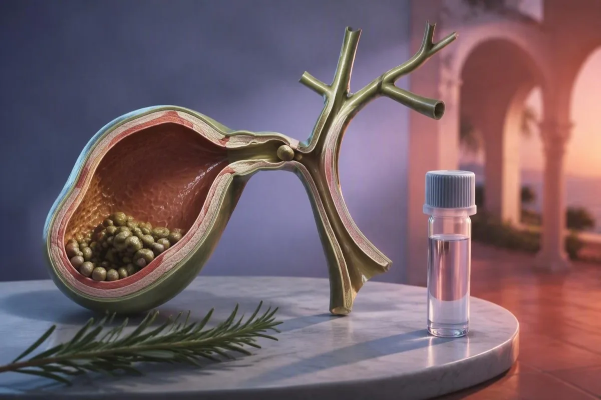
A raised white count, CRP, or liver enzyme can support suspected gallbladder inflammation, but none can diagnose it alone. The more useful question is whether the laboratory pattern fits the pain, examination, and imaging findings.


- vipimo vya damu vya kawaida do not rule out acute cholecystitis, especially during the first 6-12 hours of pain or in older adults.
- Hesabu ya WBC above 11.0 × 10⁹/L supports an inflammatory process but can remain normal in uncomplicated or early gallbladder inflammation.
- CRP above 30 mg/L often adds weight to suspected inflammation, while values above 100 mg/L raise concern for more extensive tissue response.
- ALT na AST may rise briefly when a stone passes through the common bile duct; this does not automatically mean primary liver disease.
- ALP, GGT, and direct bilirubin are more suggestive of bile-flow obstruction than isolated gallbladder inflammation.
- Lipase at least three times the laboratory upper limit suggests acute pancreatitis and changes the urgency and imaging plan.
- Ultrasound is usually the first imaging test when right-upper-abdominal pain suggests gallbladder disease, even if the CBC is normal.
- Tathmini ya haraka is needed for fever, jaundice, confusion, persistent vomiting, low blood pressure, or pain lasting more than 6 hours.
What a cholecystitis blood test can and cannot prove
A cholecystitis blood test can show inflammation, altered bile flow, dehydration, or a competing diagnosis; it cannot confirm an inflamed gallbladder by itself. In practice, a normal CBC and liver panel never overrule persistent right-upper-quadrant pain, fever, guarding, or a positive ultrasound finding.
Tokyo Guidelines diagnose acute cholecystitis from local symptoms, systemic inflammatory evidence, and confirmatory imaging—not laboratory values alone (Yokoe et al., 2018). A WBC above 11.0 × 10⁹/L, CRP above the local reference limit, or fever can satisfy the systemic component, but ultrasound or another imaging study is still needed for a definite diagnosis.
I have seen patients with severe focal pain after a fatty meal whose WBC was 7.4 × 10⁹/L and whose ALT was entirely normal. Their ultrasound nevertheless showed a stone impacted at the gallbladder neck with wall thickening; early obstruction can be anatomically real before the circulating inflammatory signal has caught up.
Kantesti ni Mchambuzi wa mtihani wa damu wa AI that places CBC, liver-panel, bilirubin, and pancreatic results beside one another rather than treating a single flagged result as a diagnosis. Knowing paneli ya ini hujumuisha nini makes it easier to see why normal transaminases do not exclude a gallbladder problem.
How to read the cholecystitis WBC count and differential
A raised neutrophil-predominant WBC count supports acute inflammation but is neither sensitive nor specific for cholecystitis. Many adult laboratories use a total WBC reference interval of roughly 4.0-11.0 × 10⁹/L, although intervals differ by laboratory and age.
A WBC of 12-15 × 10⁹/L with neutrophilia fits uncomplicated acute cholecystitis more comfortably than a normal count, but pneumonia, pyelonephritis, appendicitis, steroid treatment, and even major stress can produce the same pattern. The absolute neutrophil count is more informative than neutrophil percentage when the total WBC is abnormal.
WBC iliyo juu ya 18 × 10⁹/L is one factor that can point to severe disease in the Tokyo severity framework, particularly alongside organ dysfunction or a palpable tender mass. It is a triage clue, not a line between “safe” and “unsafe”; someone can deteriorate with a count of 10.5 × 10⁹/L.
Older patients and people taking prednisolone, chemotherapy, or immune-modifying medicines may mount little leukocytosis. Our guide to normal neutrophil counts explains why a result can look numerically ordinary while the clinical situation remains urgent.
CRP, ESR, and why inflammation markers lag behind pain
CRP often rises 6-12 hours after an inflammatory trigger and peaks about 36-50 hours later, so an early normal CRP can be misleading. CRP is reported as mg/L in most countries; values under 5 mg/L are commonly considered normal in healthy adults.
In suspected cholecystitis, CRP iliyo juu ya 30 mg/L makes active tissue response more plausible, while values above 100 mg/L deserve careful assessment for more extensive inflammation, gangrenous change, perforation, or another infection. No single CRP cutoff reliably separates these conditions, which is why clinicians use a trend and the bedside examination.
ESR changes more slowly than CRP and is rarely useful for deciding whether tonight’s upper-abdominal pain needs imaging. A CRP of 8 mg/L after only 3 hours of symptoms tells me very little; repeating the clinical assessment matters more than repeating the marker at a fixed clock time.
A high-sensitivity CRP test designed for cardiovascular risk should not be used to judge acute abdominal illness. For context on assay purpose and common non-gallbladder causes, see our review of the Uwiano wa CRP hadi albumin.
Cholecystitis liver enzyme results: the patterns that matter
Uncomplicated cystic-duct obstruction may leave ALT, AST, ALP, and bilirubin normal. A mixed or cholestatic liver pattern instead raises the possibility that a stone has entered or compressed the common bile duct.
ALT na AST are intracellular enzymes, not direct measures of bile flow. An ALT of 180 U/L and AST of 140 U/L can occur after transient common-duct obstruction, sometimes before ALP rises; a sharp early spike may fall rapidly within 24-48 hours if the stone passes.
ALP na GGT generally rise more gradually in cholestasis because they reflect enzyme induction at the bile canaliculi and duct epithelium. An isolated ALP of 160 U/L is not enough to diagnose obstruction because bone turnover, pregnancy, recent fracture, and some medicines can also raise ALP.
Dr. Thomas Klein’s practical rule is to compare the direction and timing of all four markers, not to chase one enzyme. Our mwongozo wa vifupisho vya uchunguzi wa ini na maelezo ya high ALT results can help you prepare better questions for the treating clinician.
Bilirubin results that suggest a bile-duct stone
A rising direct bilirubin with elevated ALP and GGT is more concerning for common-bile-duct obstruction than for isolated cholecystitis. Total bilirubin is commonly below 20 µmol/L (1.2 mg/dL), although laboratories set their own limits.
Bilirubin becomes conjugated (direct) in the liver and is normally excreted into bile. When a duct stone blocks that route, conjugated bilirubin can spill back into circulation and darken urine; a total bilirubin of 45 µmol/L with a substantial direct fraction deserves timely imaging and clinical review.
Mild bilirubin elevation alone is surprisingly nonspecific. Fasting, dehydration, and Gilbert syndrome may raise unconjugated bilirubin, often below 50 µmol/L, without a blocked duct; that is a different biochemical story from jaundice plus pale stools, dark urine, and an ALP rise.
A bilirubin result should always be split into total and direct fractions when obstruction is being considered. Read more about ruwaza za bilirubini ya moja kwa moja and the clinical meaning of yellow skin and dark urine.
Why lipase and amylase are checked with gallbladder pain
Lipase is checked because a gallstone can obstruct near the pancreatic duct and cause acute pancreatitis, which needs a different management pathway. Acute pancreatitis is typically supported by lipase at least three times the assay’s upper reference limit plus characteristic pain or imaging.
A lipase of 420 U/L is meaningful only against that laboratory’s upper limit; if the limit is 60 U/L, it is seven times upper limit and strongly supports pancreatitis in the right clinical setting. Mild elevations below threefold occur with kidney impairment, bowel disease, medicines, and sometimes cholecystitis itself.
Amylase is less specific and may normalize sooner than lipase. Normal lipase does not settle every abdominal-pain diagnosis, but it makes established acute pancreatitis less likely when symptoms have been present for more than a day.
I advise patients not to interpret a lipase result as a gallbladder test. The distinction is detailed in our guide to high lipase urgency and our discussion of normal pancreatic enzymes despite pain.
Why normal gallbladder inflammation blood tests do not rule it out
Normal WBC, CRP, liver enzymes, and bilirubin do not exclude acute cholecystitis because the gallbladder can be obstructed locally before systemic markers change. This is particularly true in the first saa 6-12 and in people with a reduced immune response.
The gallbladder’s cystic duct is separate from the common bile duct. A stone lodged only at the cystic duct can create distension and severe pain without raising bilirubin or ALP, while the liver continues to drain bile through the common duct.
People over 75, those with diabetes, and patients receiving corticosteroids may have fever below 38.0°C and a WBC below 11.0 × 10⁹/L despite clinically meaningful disease. The absence of classic findings lowers probability; it does not make the probability zero.
Kantesti AI ni huduma ya kutafsiri vipimo vya maabara ya AI that flags a normal panel as “not diagnostic” when the reported symptoms suggest an urgent structural problem. A laboratory reference interval means the value is statistically common in a comparison population, as explained in our article on what normal lab results can miss.
Other conditions and medicines that can mimic the laboratory pattern
Abnormal liver tests or inflammatory markers do not automatically come from the gallbladder. MASLD, viral hepatitis, alcohol exposure, medicines, urinary infection, pneumonia, and bowel inflammation can produce overlapping results.
A stable ALT of 70 U/L over several years with normal bilirubin and ALP is more often compatible with metabolic dysfunction-associated steatotic liver disease than acute biliary obstruction. In contrast, a new ALT jump from 25 to 250 U/L during colicky pain warrants a search for a transient duct event.
Paracetamol overdose, antibiotics such as amoxicillin-clavulanate, anticonvulsants, and herbal products can cause liver-test abnormalities. Tell the clinician about every regular medicine and supplement, including “natural” products started within the last siku 90.
The useful question is what changed from your own baseline—not whether a result has a red flag. Our MASLD testing guide and discussion of medication-related lab trends show why timing changes the interpretation.
When ultrasound, HIDA scan, CT, or MRCP is needed
Ultrasound is the usual first imaging test for suspected cholecystitis, regardless of whether blood tests are normal or abnormal. It can show gallstones, gallbladder-wall thickening, surrounding fluid, distension, and focal tenderness when the probe is pressed over the gallbladder.
A wall thickness above 3 mm, a distended gallbladder, pericholecystic fluid, and a sonographic Murphy sign support acute cholecystitis, though none is perfect in isolation. A stone plus compatible symptoms is often more decisive than a small liver-enzyme change.
When ultrasound is equivocal but suspicion remains high, clinicians may order a HIDA scan to assess cystic-duct patency. MRCP is especially useful when bilirubin or cholestatic enzymes suggest a common-duct stone; CT is often chosen when complications or an alternative diagnosis are being considered.
The 2020 WSES guideline recommends combining clinical assessment, laboratory findings, and imaging rather than relying on any single test (Pisano et al., 2020). Our overview of ruwaza za vipimo vya damu vya cholestasis explains why MRCP becomes more relevant when ALP, GGT, and bilirubin rise together.
When gallbladder pain and blood-test results need urgent care
Seek urgent same-day assessment for right-upper-abdominal pain lasting more than 6 hours, fever, repeated vomiting, jaundice, faintness, confusion, or a rigid abdomen—even with normal blood results. These features matter more than a single reassuring number.
Joto la 38.0°C au zaidi, pulse persistently above 100 beats/minute, low blood pressure, or new confusion can signal systemic illness. Jaundice plus fever and upper-abdominal pain is particularly concerning for cholangitis, a bile-duct infection that requires urgent hospital assessment.
Avoid eating or drinking if a clinician has advised emergency assessment, because surgery or a procedure may be needed. Do not use repeated high-dose pain medicines to “test” whether symptoms settle; NSAIDs and paracetamol have safe-dose limits and can complicate kidney or liver injury.
Sepsis cannot be diagnosed from WBC alone: a normal count occurs in some serious infections. If you are unsure whether symptoms cross the threshold, our sepsis laboratory warning guide outlines why vital signs and mental state are equally important.
Pregnancy, older age, diabetes, and other atypical presentations
Pregnancy, older age, diabetes, and immunosuppression can alter both symptoms and cholecystitis blood-test patterns. These groups deserve a lower threshold for in-person assessment because fever and leukocytosis may be muted or delayed.
During pregnancy, ALP can rise because the placenta produces the enzyme, so ALP is less useful for diagnosing bile obstruction. A direct bilirubin rise, new itching, jaundice, or right-upper-abdominal pain needs prompt obstetric and medical review; bile acids are a different test used for pregnancy-related cholestasis.
In older adults, reduced pain sensitivity, cognitive impairment, and dehydration can mask the classic symptom sequence. A modest CRP of 25 mg/L paired with functional decline or new confusion may be clinically more significant than the same CRP in a well 30-year-old.
People with diabetes can develop more severe gallbladder disease with fewer warning symptoms, although the evidence does not support assuming every diabetic patient has an atypical presentation. Our article on bile acids in pregnancy separates pregnancy itching from a gallbladder emergency.
How to make your laboratory results useful at the appointment
Bring the complete report, symptom timeline, medication list, and previous liver tests; the trend is often more informative than one result. A clinician can make better decisions when the onset of pain is linked to the time each sample was collected.
Write down whether pain began suddenly, whether it followed food, where it travels, and whether it lasted 30 minutes, 4 hours, or overnight. Note vomiting, fever measurements, stool colour, urine colour, pregnancy possibility, recent travel, and the exact time you last ate.
Ask whether your panel included direct bilirubin, GGT, lipase, and a differential WBC count. A basic metabolic panel alone does not include these tests; it primarily measures electrolytes, glucose, calcium, and kidney markers.
Kantesti can organize a photographed report into a readable trend, but it cannot replace an examination or ultrasound when biliary pain is active. Our dark-urine assessment guide na blood-test timeline checklist may help you record details clinicians commonly need.
Mahali ambapo tafsiri ya AI husaidia—na mahali lazima isimame
AI can identify a concerning combination such as rising bilirubin, ALP, GGT, and neutrophils, but it cannot confirm cholecystitis from a laboratory PDF. Gallbladder inflammation is a diagnosis that requires symptoms, examination, and usually imaging.
Kantesti ni jukwaa la tafsiri ya viashiria vya AI that compares laboratory values with the issuing laboratory’s ranges and with prior results when supplied. A rapid rise in total bilirubin from 12 to 48 µmol/L is clinically more actionable than two isolated values without dates.
Our system should be used as a question generator: “Could this be cholestasis?”, “Was direct bilirubin measured?”, and “Do I need urgent imaging?” It should never be used to delay urgent care for fever, jaundice, persistent pain, or vomiting.
Dr. Thomas Klein reviews these cases with a simple safety principle: a plausible explanation for a blood result is not the same thing as a diagnosis. Readers interested in how our safeguards work can review our mbinu ya uthibitisho wa kimatibabu na mwongozo wetu wa viashiria vya maabara zaidi ya 15,000.
A practical next-step plan after a suspected biliary episode
If symptoms have resolved and there are no warning signs, arrange timely clinical follow-up; if pain persists or red flags appear, seek urgent assessment rather than waiting for repeat blood work. Repeat testing is useful only when it changes a real clinical decision.
For a resolved episode with normal vital signs, clinicians may repeat CBC, CRP, bilirubin, ALT, AST, ALP, and GGT within saa 24-72 if the initial draw was early or abnormal. The interval should be individualized; a repeat panel is not a substitute for ultrasound when symptoms recur.
Until reviewed, avoid very large fatty meals if they consistently trigger pain, stay hydrated, and keep a brief record of episodes. Diet changes may reduce symptom provocation, but they do not dissolve an impacted stone or treat a bacterial complication.
Our medical team encourages readers to use laboratory interpretation as preparation for—not replacement of—care. The Bodi ya Ushauri wa Matibabu provides oversight for Kantesti’s clinical education, while our mwongozo wa teknolojia explains the boundaries of automated pattern analysis.
Maswali Yanayoulizwa Mara Kwa Mara
Unaweza kupata cholecystitis na vipimo vya kawaida vya damu?
Ndiyo. Cholecystitis kali inaweza kutokea ikiwa na WBC, CRP, bilirubini, na ripoti ya kawaida ya vipimo vya ini, hasa ndani ya saa 6-12 za kwanza baada ya maumivu kuanza au wakati jiwe linapoziba tu mrija wa nyongo. WBC ya kawaida chini ya 11.0 × 10⁹/L hupunguza uwezekano wa mwitikio mkali wa kimfumo lakini hauonyeshi kuwa kibofu cha nyongo ni cha kawaida. Maumivu yanayoendelea katika sehemu ya juu kulia ya tumbo, homa, kutapika, au maumivu yanayolenga eneo fulani bado yanahitaji tathmini ya kimatibabu na kwa kawaida ultrasound.
Ni nini hesabu ya WBC ambayo ni kawaida na cholecystitis?
Hesabu ya WBC juu ya takriban 11.0 × 10⁹/L, mara nyingi ikiambatana na neutrophilia, inaweza kuunga mkono cholecystitis papatika wakati dalili zinapofaa. Hesabu za 12-15 × 10⁹/L ni za kawaida katika hali za kawaida za uvimbe, wakati WBC juu ya 18 × 10⁹/L inaweza kuongeza wasiwasi kwa ugonjwa mbaya zaidi au maambukizi mengine makubwa. WBC si maalum, kwa hivyo haiwezi kutofautisha uvimbe wa kibofu cha nyongo kutoka kwa maambukizi mengine mengi au hali za uvimbe.
Je, vimeng'enya vya ini huongezeka na kibofu cha nyongo kilichojaa uvimbe?
Viwango vya vimeng'enya vya ini vinaweza kubaki kawaida katika cholecystitis isiyo na shida kwa sababu kizuizi cha mfereji wa cystic hakizuizi mfumo wa maji taka wa ini. ALT na AST zinaweza kupanda kwa muda mfupi wakati jiwe linapopita kwenye mfereji mkuu wa nyongo, wakati ALP, GGT, na bilirubini moja kwa moja huashiria vizuizi vinavyoendelea vya mtiririko wa nyongo. ALT au AST juu ya 500 U/L huhitaji tathmini ya haraka kwa sababu utambuzi tofauti unajumuisha zaidi ya ugonjwa wa kibofu cha nyongo.
Kiwango gani cha bilirubini kinaonyesha mrija wa nyongo umeziba?
Hakuna kiwango kimoja cha bilirubini kinachoonyesha kuwa njia ya nyongo imezibwa, lakini ongezeko la jumla la bilirubini juu ya kikomo cha juu cha maabara—mara nyingi juu ya 20 µmol/L au 1.2 mg/dL—pamoja na bilirubini moja kwa moja, ALP, na GGT nyingi huongeza wasiwasi. Jumla ya bilirubini karibu 45 µmol/L yenye njano, mkojo mweusi, homa, au maumivu ya juu ya tumbo inahitaji uhakiki wa haraka wa kimatibabu. Kuongezeka kidogo kwa bilirubini isiyojumuishwa pekee kunaweza kutokea na njaa au syndrome ya Gilbert na ina maana tofauti.
Should I go to urgent care if my gallbladder blood tests are normal?
Yes, if pain lasts more than 6 hours, you have fever of 38.0°C or higher, repeated vomiting, jaundice, faintness, confusion, or worsening abdominal tenderness. These warning signs can indicate cholecystitis, cholangitis, pancreatitis, or another acute abdominal condition even when the initial WBC and liver tests are normal. Urgent care clinicians use vital signs, examination, repeat testing, and imaging rather than relying on a single laboratory panel.
Is CRP 100 mg/L high for cholecystitis?
A CRP of 100 mg/L is substantially elevated and indicates a significant inflammatory response, but it does not identify the source by itself. In a person with compatible right-upper-abdominal pain, fever, and ultrasound findings, CRP above 100 mg/L should increase concern for more extensive gallbladder inflammation or a complication. CRP can also reach this range with pneumonia, kidney infection, bowel inflammation, and many other conditions, so urgent clinical context is required.
Pata Uchambuzi wa Vipimo vya Damu kwa AI Leo
Jiunge na zaidi ya watumiaji 2 milioni duniani kote wanaoamini Kantesti kwa uchambuzi wa papo hapo na sahihi wa vipimo vya maabara. Pakia matokeo yako ya vipimo vya damu na upate tafsiri ya kina ya viashiria vya 15,000+ ndani ya sekunde.
📚 Machapisho ya Utafiti Yanayorejelewa
Klein, T., Mitchell, S., & Weber, H. (2026). Ulinganisho wa Kiufundi wa Kiotomatiki uliosajiliwa mapema, unaotegemea Rubric, wa Injini ya Ufafanuzi wa Vipimo vya Damu ya Kantesti kwenye Kesi 100,000 za Majaribio ya Synthetic. Kantesti uchambuzi wa damu kwa AI ya utafiti wa matibabu.
Klein, T., Mitchell, S., & Weber, H. (2026). Mfumo wa Uthibitishaji wa Kitaaluma v2.0 (Ukurasa wa Uthibitishaji wa Tiba). Kantesti uchambuzi wa damu kwa AI ya utafiti wa matibabu.
📖 Marejeo ya Nje ya Tiba
⚕️ Kanusho la Kimatibabu
Makala haya ni kwa madhumuni ya elimu tu na si ushauri wa kimatibabu. Wasiliana na mtoa huduma aliyehitimu kila wakati kwa maamuzi ya utambuzi na matibabu.
E-E-A-T Trust Signals
Uzoefu
Mapitio ya kimatibabu inayoongozwa na daktari ya mifumo ya tafsiri ya maabara.
Utaalamu
Kuzingatia dawa za maabara kuhusu jinsi viashiria (biomarkers) vinavyobadilika katika muktadha wa kliniki.
Mamlaka
Imeandikwa na Dk. Thomas Klein kwa mapitio ya Dk. Sarah Mitchell na Prof. Dk. Hans Weber.
Uaminifu
Tafsiri inayotegemea ushahidi yenye njia zilizo wazi za ufuatiliaji ili kupunguza tahadhari za hofu.