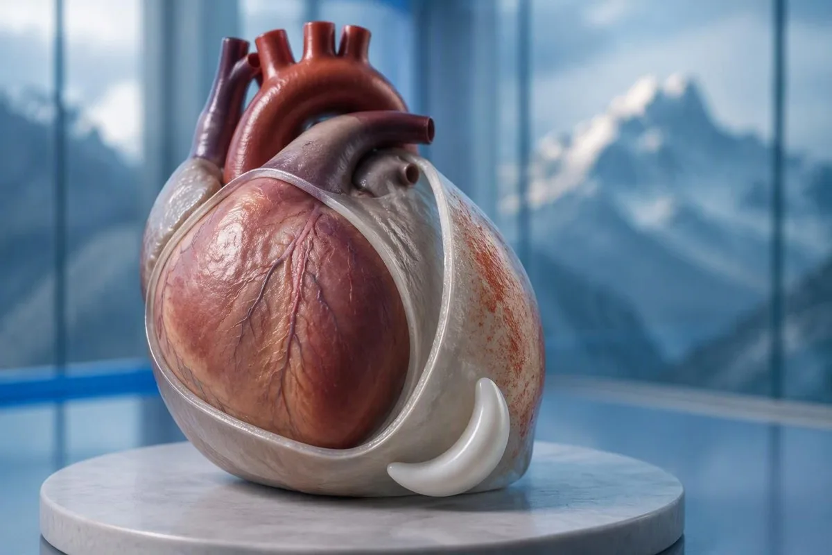
Yes—pericarditis chest pain often worsens lying flat and improves sitting up or leaning forward. But that pattern cannot exclude a heart attack; new unexplained chest pain needs prompt medical assessment.


- Positional pain often worsens lying flat and improves leaning forward, but neither response confirms pericarditis or excludes a heart attack.
- Breathing-related pain can worsen with deep inspiration or coughing; pulmonary embolism and other lung conditions can produce the same pattern.
- ايمرجنسي جون علامتون include severe or persistent chest pain, breathlessness, fainting, cold sweating, or pain spreading to the arm, jaw, or back. Call your local emergency number without waiting for every symptom.
- ECG changes typical of acute pericarditis were reported in up to 60% of cases in the 2015 ESC guideline; a normal ECG does not exclude the condition.
- Echocardiography assesses fluid around the heart and its effect on filling. An effusion greater than 20 mm is a high-risk feature, not a threshold below which all fluid is harmless.
- ٽروپونن above the assay-specific 99th percentile indicates heart muscle injury; the result alone cannot distinguish a heart attack from myocarditis.
- Inflammation tests such as CRP and ESR support assessment and follow-up, but normal results cannot independently rule out pericarditis.
- فالو اپ commonly occurs within about 1 week for appropriately selected outpatients; worsening symptoms require earlier reassessment.
Why can pericarditis chest pain worsen lying down?
Pericarditis chest pain often becomes worse lying down and less intense when sitting up or leaning forward. Changes in posture alter the mechanical relationship between the irritated pericardium and nearby structures, but relief in 1 position is a diagnostic clue—not evidence that a heart attack is impossible.
جي pericardium is a 2-layered sac surrounding the heart, and its outer lining has pain-sensitive connections that the heart muscle itself does not share in the same way. The familiar explanation that the layers simply “rub together” is incomplete: movement, surrounding tissue irritation, and individual sensory responses also influence the pain.
A hypothetical 42-year-old who develops sharp pain after a respiratory illness and cannot comfortably sleep flat has a pattern compatible with pericarditis. I would still ask about the exact onset, exertion, breathlessness, and cardiovascular history before attaching a diagnosis; age and a recent cold do not make coronary disease or pulmonary embolism disappear.
ڪينٽيسٽي هڪ آهي اي آءِ بلڊ ٽيسٽ اينالائيزر that can help explain reported CRP and troponin results after clinical assessment; it cannot decide whether positional chest pain is safe. As Thomas Klein, I frame this distinction plainly: 1 reassuring symptom should never outweigh an unexplained acute chest complaint; information about اسان جي تنظيم ۾ explains our educational role.
Does pericarditis pain get worse with deep breathing?
Pericarditis pain commonly worsens with a deep breath, coughing, or sometimes swallowing. Clinicians call breathing-related pain pleuritic, but that description identifies a pain pattern rather than 1 particular disease; the lungs, chest wall, and pericardium can all generate it.
Deep inspiration moves the diaphragm and changes relationships within the chest, which can aggravate an irritated pericardial lining. Connections involving the phrenic nerves, arising mainly from C3–C5, help explain why some patients feel referred discomfort near the shoulder or trapezius ridge rather than only behind the breastbone.
Pain that catches with every breath needs a broader assessment when it starts suddenly, especially alongside breathlessness or a swollen leg. A pulmonary embolism can produce the same complaint without a classic pressure sensation; even an oxygen saturation of 98% cannot independently exclude it.
Tenderness when a clinician presses on 1 part of the chest makes a chest-wall source more plausible, but it does not provide an absolute coronary rule-out. Our explanation of tests for unexplained pain distinguishes supportive laboratory clues from conditions that require examination and imaging.
Which other pericarditis symptoms matter?
Pericarditis symptoms can include fever, fatigue, breathlessness, and a faster heartbeat as well as chest pain. None is mandatory, and their combination matters more than any 1 symptom; marked weakness, faintness, or worsening breathing changes the urgency of assessment.
هڪ گرمي پد جيڪا مٿي هجي 38°C is a higher-risk feature in the 2015 ESC pericardial guideline, particularly when the cause is uncertain. A patient with 37.4°C and considerable pain may still need urgent assessment, however; temperature thresholds help clinicians organize risk rather than define a safe home-care boundary.
A fast pulse can reflect pain, fever, dehydration, an arrhythmia, or impaired cardiac filling. I would interpret a resting pulse of 110 beats/min differently in someone comfortable and well hydrated than in someone pale and faint; our دل جي ڌڙڪن جي جاچ جي رهنمائي explains why laboratory results are only part of that assessment.
Breathlessness when lying flat is not automatically the same thing as chest pain when lying flat. Someone who needs 3 pillows because they cannot breathe comfortably may have congestion, an effusion, or another cardiopulmonary problem; the distinction is covered in our ساهه کڻڻ ۾ تڪليف جا نتيجا (shortness-of-breath results) ٻڌائين ٿا ته ڪهڙا ليبارٽري نتيجا خارج (exclude) ڪري سگهجن ٿا ۽ ڪهڙا نه., but active breathing difficulty should not wait for online interpretation.
When does positional chest pain need emergency care?
Call emergency services for severe, persistent, or rapidly worsening chest pain, especially with breathlessness, fainting, cold sweating, or pain spreading to the arm, jaw, or back. In the UK call 999 or 112; elsewhere use your local emergency number, and do not drive yourself.
Sudden maximal pain, collapse, new confusion, or coughing up blood also warrants emergency assessment because the differential includes pulmonary embolism and aortic emergencies. Do not wait 5 or 15 minutes to see whether leaning forward fixes the problem; concerning accompanying symptoms make a waiting experiment unsafe.
New unexplained chest pain that is not severe still deserves prompt, usually same-day, medical assessment rather than a routine appointment several weeks away. The 2021 AHA/ACC chest pain guideline emphasizes structured assessment and discourages using supposedly “atypical” symptoms to dismiss acute coronary syndrome (Gulati et al., 2021).
Stop exertion while arranging help, and follow the emergency dispatcher’s instructions about any medication rather than starting treatment for presumed pericarditis. Our ايمرجنسي سينه-درد رت جا امتحان article explains why 1 laboratory result cannot replace an emergency ECG, examination, and appropriately timed testing.
How do clinicians assess suspected pericarditis?
Clinicians assess suspected pericarditis by combining the history, examination, ECG, echocardiography, and selected blood tests. The widely taught 2015 ESC framework requires at least 2 of 4 findings: typical pain, a pericardial rub, characteristic ECG changes, or a new or worsening pericardial effusion (Adler et al., 2015).
Sharp pain, pain worse lying down, and relief leaning forward are not 3 independent diagnostic criteria; they are different features of the same symptom. This distinction prevents a persuasive story from being mistaken for confirmation, especially when the ECG and imaging have not yet been assessed.
A pericardial friction rub may be transient, so a clinician may listen again after an initial examination rather than treating 1 quiet examination as decisive. Vital signs, signs of poor circulation, heart failure, and the history of recent procedures or systemic illness can matter more urgently than whether the rub is audible.
جي 2025 ESC myocarditis and pericarditis guideline gives contemporary imaging and overlapping myocardial involvement a central place in assessment, so the older checklist is not a complete care pathway (Schulz-Menger et al., 2025). Our clinical standards information should be read as documentation about laboratory interpretation—not evidence that software can reproduce this bedside evaluation.
What does an ECG show in pericarditis?
An ECG may show widespread ST-segment elevation and PR-segment depression in acute pericarditis, but these changes are not always present. The 2015 ESC guideline reports characteristic ECG changes in up to 60% of cases, so a normal tracing does not exclude pericarditis (Adler et al., 2015).
A 12-lead ECG records electrical activity from several perspectives; clinicians assess which leads are affected, reciprocal changes, rhythm, and comparison with previous tracings. “The ST segment looks elevated” is not enough to separate pericarditis, early repolarization, myocardial infarction, and other causes without the clinical setting.
A heart attack more often produces changes corresponding to a coronary territory, whereas pericarditis may produce a broader pattern—but neither rule is absolute. ECG abnormalities can evolve over ڪلاڪن, and repeating the tracing during recurrent or changing symptoms may reveal information absent from the first recording.
Electrolyte disturbances can add another layer of confusion, particularly when kidney disease or medication effects are present. A potassium concentration around 6.0 mmol/L کان مٿي can be clinically urgent depending on context, and our potassium and ECG guide explains why ECG interpretation should never be detached from the rest of the assessment.
Why is echocardiography used for pericarditis?
Echocardiography checks for fluid around the heart, ventricular function, and signs that fluid is impairing cardiac filling. A normal echocardiogram does not exclude uncomplicated pericarditis; an effusion greater than 20 mm is a recognized high-risk feature, not the definition of every dangerous effusion.
The 2015 ESC guideline describes effusions as small below 10 ملي ميٽر, moderate at 10–20 mm, and large above 20 mm, using the echo-free space on imaging. These measurements help communicate size, but the distribution of fluid and its hemodynamic effects still require interpretation rather than a number-only decision.
Cardiac tamponade occurs when pericardial pressure compromises filling; how quickly fluid accumulates can matter more than its absolute volume. A collection that develops over ڪلاڪن may be less well tolerated than a larger, slowly developing collection, and the classic low-pressure, raised-neck-vein, quiet-heart-sound triad is not reliably present in every patient.
جي 2025 ESC guideline supports integrating echocardiography with other imaging when the diagnosis or extent of involvement remains uncertain (Schulz-Menger et al., 2025). An elevated NT-proBNP may add information about cardiac strain, but 1 value cannot diagnose tamponade; our NT-proBNP interpretation guide explains several alternative reasons for elevation.
Can troponin distinguish pericarditis from a heart attack?
Troponin identifies heart muscle injury, but an elevated result alone cannot distinguish a heart attack from myocarditis or overlapping myopericarditis. The relevant threshold is the assay-specific 99th percentile مٿئين حوالائي حد (upper reference limit) کان مٿي ڪنهن به قدر کي غيرمعمولي (abnormal) سمجهيو ويندو آهي، پر تشخيص علامتن ۽ وڌڻ-گهٽڻ واري نموني تي دارومدار رکي ٿي، نه ته صرف هڪ الڳ نموني تي؛ انهيءَ ڪري AI کي ڪڏهن به سينه جي سور کي صاف (clear) نه ڪرڻ گهرجي. هڪ, interpreted alongside symptoms, ECG findings, serial measurements, and sometimes imaging.
There is no universal normal troponin number for every laboratory: ٽروپونن I ۽ ٽروپونن T use different assays, and some laboratories apply sex-specific reference limits. If a hypothetical report lists an upper limit of 14 ng/L, a result of 20 ng/L exceeds that particular limit; it does not independently identify the cause, as our troponin I versus T explanation details.
An early normal troponin may precede a detectable rise, which is why emergency departments use validated serial pathways, often with repeat sampling at 1 or 2 hours for suitable high-sensitivity assays. Selected patients can meet validated single-sample rule-out criteria, but that decision depends on assay, symptom timing, ECG, and risk—not a home interpretation of “normal.”
Pericardial symptoms plus raised troponin prompt assessment for myocardial involvement, especially with arrhythmia, breathlessness, or reduced ventricular function. A 30-year-old with a recent viral illness still needs a proper differential; our myocarditis troponin guide explains why neither youth nor the size of the rise settles the diagnosis.
What do CRP, ESR, and blood counts add?
CRP, ESR, and a full blood count can support evidence of inflammation, but they do not confirm pericarditis or exclude a heart attack. Many laboratories use a standard CRP upper reference limit around تقريباً هيٺ آهي, while others use different limits; the printed laboratory range takes precedence.
CRP جو قدر 45 مگ/ايل indicates a substantial inflammatory response in a laboratory with an upper limit of 5 mg/L, but it cannot say whether the source is pericardial, pulmonary, autoimmune, or elsewhere. High-sensitivity CRP measures the same protein at low concentrations; acute illness values should not be interpreted using stable cardiovascular-risk categories.
ڪينٽيسٽي هڪ آهي AI blood test interpretation پليٽ فارم that can explain a CRP result against the supplied reference range and help organize its trend alongside other reported results. If CRP changes from 45 to 12 mg/L, the direction may support improvement, but persistent symptoms still matter; our WBC and CRP comparison discusses why these markers can disagree.
ESR often changes more slowly than CRP and is influenced by age, anemia, pregnancy, and protein concentrations, so comparing 2 different markers as though they were interchangeable can mislead follow-up. The 2015 ESC guideline treats inflammatory tests as supportive evidence rather than standalone diagnostic criteria (Adler et al., 2015); our causes of elevated ESR adds context for an unexpectedly persistent elevation.
Which causes and risk features change the care plan?
Pericarditis may follow a viral illness, occur after cardiac injury, or accompany autoimmune disease, severe kidney disease, tuberculosis, or other conditions. 38°C کان مٿي بخار 38°C, a large effusion, tamponade, or failure to improve with initial treatment are among the features that can justify hospital assessment and a broader search for causes.
جي 2015 ESC guideline lists lack of response to anti-inflammatory treatment within 7 ڏينهن as a major adverse prognostic feature, alongside subacute onset and the imaging findings above (Adler et al., 2015). This is a clinician’s reassessment trigger—not permission to wait a week through worsening pain, fever, or breathlessness.
Autoimmune testing is most useful when the history supplies a reason: recurrent episodes, joint symptoms, rashes, mouth ulcers, or known systemic disease. 1 positive antibody does not prove autoimmune pericarditis; our anti-dsDNA نتيجن جي رهنمائي explains why antibody findings need compatible clinical evidence.
Severe kidney dysfunction can change both the cause of pericarditis and medication safety; an eGFR below 15 mL/min/1.73 m² identifies kidney failure-range function but does not itself diagnose uremic pericarditis. Our urgent low-eGFR symptoms guide is relevant to that background, while chest pain in this setting requires direct medical assessment.
Does improvement with medication confirm pericarditis?
Improvement with ibuprofen, aspirin, or a change in posture does not confirm pericarditis. For appropriately diagnosed uncomplicated cases, clinicians commonly use an anti-inflammatory medicine with colchicine, often for approximately 3 مهينن کان وڌيڪ after a first episode, but the regimen must account for kidney function, interactions, and the underlying cause.
A reduction in pain from 8 out of 10 to 3 out of 10 is useful clinical information, but several chest-pain causes can respond to analgesia. Do not use medication response as a reason to cancel assessment, and do not start high-dose anti-inflammatory treatment for undiagnosed chest pain without professional advice.
Colchicine requires particular care with kidney or liver impairment and certain interacting medicines, including some antibiotics; taking 2 medicines together can alter exposure even when each prescription is familiar. Corticosteroids are not routine first-line treatment for every uncomplicated case, and selected bacterial or systemic causes need a different strategy rather than simply more pain relief.
Kantesti can help organize laboratory follow-up for a clinician through our دوا-حفاظت جي رجحان گائيڊ, but it should not select the treatment or tapering schedule. A plan that includes 1 named prescriber, clear interaction checks, and instructions for worsening symptoms is more useful than a generic dosing chart.
When is exercise safe after pericarditis symptoms improve?
Exercise should be restricted during active pericarditis, and return should follow clinical reassessment rather than pain relief alone. The decision changes substantially if troponin, rhythm testing, or imaging suggests myocardial involvement; 1 symptom-free day is not evidence that strenuous activity is safe.
The older 2015 ESC guideline used a minimum 3 مهينن restriction for athletes with isolated pericarditis, while contemporary guidance places greater emphasis on individualized remission and risk assessment. Patients should not borrow an athlete’s timetable—or a myocarditis timetable—without knowing which diagnosis their clinician has established.
For a hypothetical 28-year-old runner, the difference between isolated pericarditis and myocardial involvement can change follow-up testing and return-to-training decisions. I would ask whether symptoms, inflammatory markers, ventricular function, and any relevant rhythm concerns have resolved; a normal resting pulse of 65 beats/min is only one small part of that picture.
Daily activity, physical work, and competitive sport are 3 different workloads, so a written return plan should specify what the patient can actually do. Our خون جو بائيو مارڪر گائيڊ can explain reported measurements, but no biomarker page, wearable reading, or AI result constitutes exercise clearance.
What if chest pain returns or tests look normal?
Persistent or recurrent chest pain needs reassessment even when an earlier ECG, echocardiogram, or CRP result was normal. Appropriately selected low-risk outpatients are commonly reviewed within about 1 هفتو, but recurrence with faintness, breathlessness, or worsening pain warrants earlier—and sometimes emergency—assessment.
Residual discomfort, recurrent inflammation, chest-wall pain, and a new unrelated problem can feel similar to the patient, so 2 episodes should not automatically receive the same explanation. Cardiac MRI can sometimes identify pericardial or myocardial involvement when first-line tests leave uncertainty; it is selected for a clinical question, not ordered for every ache.
A follow-up note should record symptom timing, temperature, activity tolerance, medication changes, and the actual laboratory reference ranges. CRP values of 8 and 6 mg/L from different laboratories may not represent a meaningful improvement without knowing the methods and context; our post-discharge result changes guide explains why small numerical changes deserve restraint.
ڪينٽيسٽي هڪ آهي AI-powered رت جي ٽيسٽ تجزيي جو اوزار that can help organize reported results for discussion, but it cannot verify the cause of renewed chest pain from a PDF. My advice here, as Thomas Klein, is to agree on 3 things before leaving follow-up: the next review date, permitted activity, and the symptoms that require immediate help.
Clinical boundaries and related research publications
Symptoms and laboratory interpretation cannot safely replace an acute chest-pain assessment. جي مطابق آڪٽوبر 3، 2026, the clinical references below support the distinction between pericardial clues, myocardial injury, and emergency risk; the 2 Zenodo resources are background laboratory guides, not trials validating a pericarditis diagnosis.
Kantesti جا medical advisory information describes the professional expertise behind our educational work, but readers should not equate a medical title with an examination of their own chest pain. A personal care plan still needs 1 accountable treating clinician or team who can examine the patient, review the ECG, and act on imaging or troponin findings.
جي aPTT نارمل رينج: ڊي-ڊائمر، پروٽين سي بلڊ ڪلوٽنگ گائيڊ is available as a Zenodo educational publication with DOI 10.5281/زينوڊو.18262555. Its associated clotting-test interpretation guide is relevant when clinicians consider coagulation questions, but a normal aPTT cannot exclude pulmonary embolism and D-dimer must be used within an appropriate clinical probability pathway.
جي سيرم پروٽين گائيڊ: گلوبولين، البومين ۽ اي/جي تناسب بلڊ ٽيسٽ is a second Zenodo educational publication with DOI 10.5281/زينوڊو.18316300. The associated serum protein interpretation guide supplies background on systemic laboratory findings; neither albumin nor the A/G ratio independently diagnoses pericarditis, and the discovery links below are searches rather than verified copies of these publications.
وچان وچان سوال ڪرڻ
ڇا پريڪارڊائٽس جي سينه جو درد ليٽڻ سان وڌي ٿو؟
Pericarditis جي سيني ۾ سور اڪثر ليٽڻ سان وڌي ويندو آهي ۽ اٿڻ يا اڳتي جهڪڻ سان گهٽجي ويندو آهي. اهو نمونو مشورو ڏيندڙ آهي، پر اهو ڪلينڪل تشخيص جو صرف 1 حصو آهي ۽ دل جي حملي کي رد نٿو ڪري سگهي. ڊاڪٽر تاريخ کي امتحان، ECG، echocardiography، ۽ چونڊيل رت جي ٽيسٽ سان گڏ ڪن ٿا. نئون اڻ ڄاتل سينه جو سور جلدي تشخيص جي ضرورت آهي، سخت درد، ساهه کڻڻ ۾ تڪليف، بي هوشي، يا ٿڌي پسڻ لاءِ هنگامي مدد سان.
ڇا اڳتي جھڪڻ سان سينه جو درد دل جي حملي کي رد ڪري سگهي ٿو؟
اڳتي جھڪڻ سان سيني جي درد ۾ آرام دل جو دورو رد نٿو ڪري. 2021 AHA/ACC چيسٽ پين گائيڊ لائن علامات کي غير ڪلاسيڪل آواز ڏيڻ سبب رد ڪرڻ بجاءِ منظم تشخيص جي حمايت ڪري ٿي. ECG ۽ اعليٰ حساسيت واري ٽروپونين ٽيسٽنگ جي ضرورت ٿي سگھي ٿي، ۽ ابتدائي معمول جو نتيجو خود بخود حتمي ناهي. سخت يا مسلسل درد يا سانس جي تڪليف، بي هوشي، يا ٿڌي پسين جي صورت ۾ ايمرجنسي خدمتون سڏايو.
ڇا عام ECG سان پريڪارڊائٽس ٿي سگهي ٿو؟
ها، پريڪارڊائٽس عام اي سي جي سان ٿي سگهي ٿو. 2015 اي سي سي پريڪارڊيل گائيڊ لائن رپورٽ ڪيو ته ايڪيوٽ ڪيسن جي 60% تائين خاص اي سي جي تبديليون، جنهن جو مطلب آهي ته هڪ وڏو حصو ڪلاسيڪل نموني نه ڏيکاريو. اي سي جي جا نتيجا پڻ تبديل ٿي سگهن ٿا، تنهنڪري ڊاڪٽر علامتن جي تبديلي تي 12-ليڊ ٽريڪنگ ورجائي سگهن ٿا. صرف عام اي سي جي پريڪارڊائٽس يا هر دل جي حملي کي خارج نٿو ڪري سگهي.
ڇا اڪوڪارڊيوگراف هميشه پريڪارڊائٽس ڏيکاريندو؟
اڪوڪارڊيوگرام هميشه سادي پيريڪارڊائٽس نه ڏيکاري سگھي ٿو ڇاڪاڻ ته سوزش ظاهري سيال جي گڏجڻ کان سواء ٿي سگهي ٿي. اڪوڪارڊيوگرافي خاص طور تي پيريڪارڊيل سيال جي ماپ ڪرڻ، وينٽريڪولر فنڪشن جو اندازو لڳائڻ، ۽ ڀرڻ ۾ خرابين جي ڳولا لاءِ مفيد آهي. 2015 ESC ڪلاسيڪيشن ۾ 20 ملي ميٽر کان وڌيڪ effusion کي وڏو سمجهيو ويندو آهي، پر ننڍيون تيزي سان گڏ ٿيندڙ مجموعا اڃا به خطرناڪ ٿي سگهن ٿيون. جڏهن ڪلينڪل سوال حل نه ٿئي ته وڌيڪ تصويرنگي تي غور ڪري سگهجي ٿو.
ڇا عام ٽروپونن جو مطلب آهي ته سيني جو درد سنجيده ناهي؟
دل جي درد جي نارمل ٽروپنن جو نتيجو پاڻمرادو اهو نه ٿو سمجهي ته دل جو درد بي ضرر آهي. وقت، ڪيز خاص 99th پئرسنٽائل حد، ECG جي نتيجن، ۽ مجموعي خطري جو اندازو طئي ڪري ٿو ته نتيجو ڇا خارج ڪري سگهي ٿو. تصديق ٿيل اعلي حساسيت واري ٽروپنن پيٿون اڪثر 1 يا 2 ڪلاڪن تي ورجائي ماپڻ استعمال ڪن ٿا، جيتوڻيڪ چونڊيل مريض سنگل-نموني جي قاعدي کان ٻاهر نڪرڻ جي پيٿون لاء قابليت حاصل ڪري سگهن ٿا. ٽروپنن ان کان علاوه اڪيلائي ۾ پلمونري ايمبولزم، ايورٽڪ ايمرجنسي، يا پريڪارڊائٽس کي خارج نٿو ڪري.
Pericarditis جي علامتون ڪيترو وقت رهي سگهن ٿيون؟
مناسب علاج سان Pericarditis جون علامتون ڏينهن کان هفتن تائين بهتر ٿي سگهن ٿيون، پر وقت جو مدو سبب، ٻيهر ٿيڻ ۽ mycardial ملوث ٿيڻ سان مختلف ٿئي ٿو. Colchicine عام طور تي پهرين سادي واقعي کان تقريبن 3 مهينن تائين لکيو ويندو آهي، جنهن جو مطلب اهو ناهي ته درد انهيءَ عرصي دوران جاري رهڻ گهرجي. 2015 ESC گائيڊ لائن ۾ مخالف سوزش واري علاج جي 7 ڏينهن اندر بهتري نه اچڻ کي ٻيهر تشخيص ڪرڻ جو هڪ تسليم ٿيل سبب سمجهيو ويندو آهي. درد وڌڻ، ساهه کٽڻ، يا بي هوش ٿيڻ جي صورت ۾ شيڊول ٿيل جائزي جو انتظار ڪرڻ بدران اڳتي ئي تشخيص ڪرڻ جي ضرورت آهي.
اڄ ئي AI-طاقتور خون جي جاچ جو تجزيو حاصل ڪريو
دنيا ڀر ۾ 2 ملين کان وڌيڪ استعمال ڪندڙن ۾ شامل ٿيو جيڪي فوري ۽ درست ليب ٽيسٽ تجزيو لاءِ Kantesti تي ڀروسو ڪن ٿا. پنهنجا خون جي جاچ جا نتيجا اپلوڊ ڪريو ۽ سيڪنڊن ۾ 15,000+ بائيو مارڪرز جي جامع تشريح حاصل ڪريو.
📚 حوالا ڏنل تحقيقي اشاعتون
ڪلين، ٽي.، مچل، ايس.، ۽ ويبر، ايڇ. (2026). aPTT نارمل رينج: ڊي-ڊائمر، پروٽين سي بلڊ ڪلوٽنگ گائيڊ. Kantesti AI Medical Research.
ڪلين، ٽي.، مچل، ايس.، ۽ ويبر، ايڇ. (2026). سيرم پروٽين گائيڊ: گلوبولين، البومين ۽ اي/جي تناسب بلڊ ٽيسٽ. Kantesti AI Medical Research.
📖 ٻاهرين طبي حوالا
گولاٽي ايم وغيره. (2021). 2021 AHA/ACC/ASE/CHEST/SAEM/SCCT/SCMR هدايتون: سينه جي درد جي جائزي ۽ تشخيص لاءِ. Circulation.
Schulz-Menger J et al. (2025). 2025 ESC Guidelines for the management of myocarditis and pericarditis. European Heart Journal.
⚕️ طبي دستبرداري
هي آرٽيڪل صرف تعليمي مقصدن لاءِ آهي ۽ طبي مشورو نٿو بڻجي. تشخيص ۽ علاج جي فيصلن لاءِ هميشه ڪنهن قابل صحت فراهم ڪندڙ سان صلاح ڪريو.
E-E-A-T اعتماد جا سگنل
تجربو
ڊاڪٽر جي نگرانيءَ هيٺ ليبارٽري نتيجن جي تشريح واري عمل جو جائزو.
ماهر
ليبارٽري دوائن جو ڌيان ان ڳالهه تي ته بايو مارڪرز ڪلينڪل حوالي سان ڪيئن رويو ڏيکارين ٿا.
اختيار
ڊاڪٽر ٿامس ڪلين لکيو، ۽ ڊاڪٽر ساره مچل ۽ پروف. ڊاڪٽر هانس ويبر طرفان جائزو ورتل.
اعتبار
ثبوتن تي ٻڌل تشريح، جنهن سان خبرداري گهٽائڻ لاءِ واضح پيرويءَ جا رستا موجود هجن.