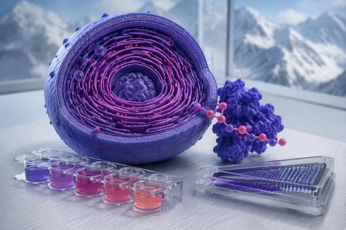
Three tests answer different questions: enzyme function, substrate accumulation, and inherited cause. A reassuring enzyme result can still miss Fabry disease in women.

Key takeaways
- Fabry disease test interpretation combines alpha-galactosidase A activity, lyso-Gb3, and GLA genetic findings; no single screening number answers every diagnostic question.
- Alpha-galactosidase A activity is typically below 1% of mean normal activity in males with classic Fabry disease, although later-onset forms can retain more activity.
- Women with Fabry disease can have normal enzyme activity because different cells express different X chromosomes; normal enzyme activity does not exclude the diagnosis.
- Lyso-Gb3 supports evidence of substrate accumulation, but normal concentrations do not exclude Fabry disease, particularly in women and some later-onset presentations.
- GLA gene testing reports one of 5 standard variant classifications; pathogenic and likely pathogenic findings have a different meaning from a variant of uncertain significance.
- A GLA VUS does not confirm Fabry disease and should not, by itself, determine disease-specific treatment or label relatives as affected.
- Family inheritance is X-linked: a heterozygous mother has a 50% chance of transmitting her GLA variant in each pregnancy.
- Kidney monitoring should include urine albumin-to-creatinine ratio as well as eGFR; albuminuria can appear while eGFR remains above 90 mL/min/1.73 m².
How does a Fabry disease test establish the diagnosis?
A Fabry disease test combines alpha-galactosidase A activity, lyso-Gb3, and GLA genetic testing to assess enzyme function, substrate accumulation, and inherited cause. Normal enzyme activity can miss Fabry disease in women, and a GLA variant of uncertain significance does not confirm diagnosis.
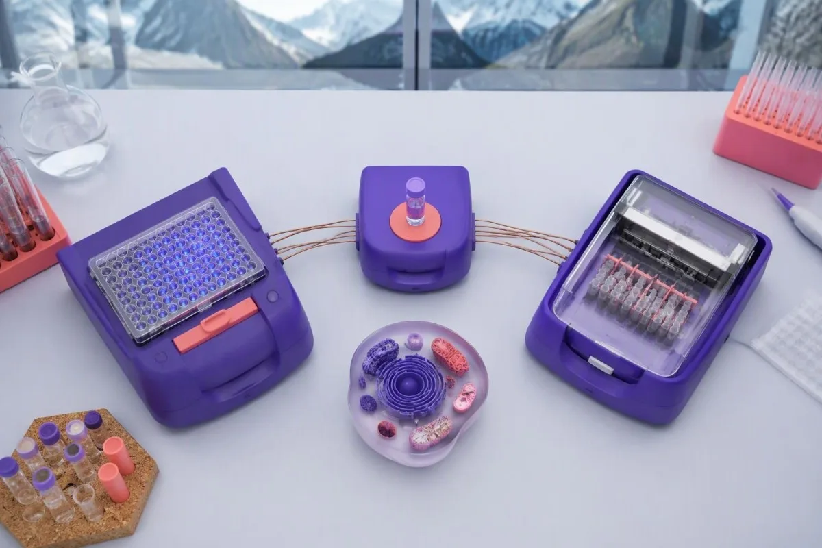
Fabry disease results from disease-causing changes in the GLA gene, which reduce the function of the lysosomal enzyme alpha-galactosidase A and permit accumulation of globotriaosylceramide, also called Gb3. These 3 diagnostic layers are connected but not interchangeable: a genetic finding identifies a potential cause, whereas an enzyme assay measures function in the particular specimen tested.
The starting test depends partly on sex chromosome biology and family history, rather than symptoms alone. In a male with suspected Fabry disease, clinicians commonly assess enzyme activity and confirm the molecular cause; in a woman or anyone with a known familial variant, GLA testing should not be withheld because 1 enzyme result is normal.
Kantesti is an AI blood test analyzer that can help explain the reported values and units across these 3 test types, without replacing specialist diagnosis or genetic variant classification. Our histórico da empresa explains the organization behind the service; clinical genetics or a metabolic disease team should resolve discordant findings before they become a lifelong diagnostic label.
What does low alpha-galactosidase A activity mean?
Baixo alpha-galactosidase A activity indicates reduced enzyme function in the tested specimen, but interpretation requires the specimen type and laboratory method. Males with classic Fabry disease typically have less than 1% of mean normal activity; later-onset disease can retain measurable activity.
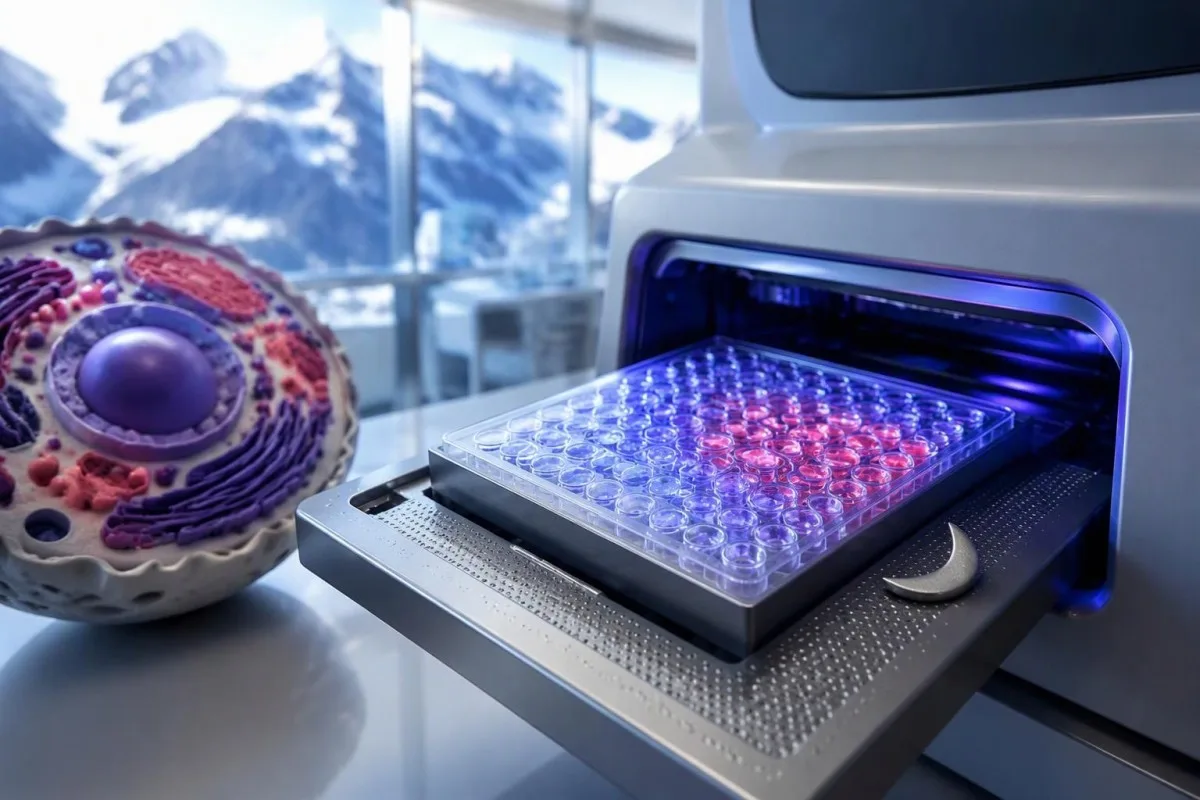
An alpha-galactosidase A test measures catalytic activity, not simply the amount of enzyme protein present. Leukocyte activity may be reported in nmol/h/mg protein, plasma activity in nmol/h/mL, and dried-spot activity in µmol/L/h; these 3 formats cannot be compared numerically without method-specific information, even when every report uses the same enzyme name.
For a clearly hypothetical example, suppose a laboratory's mean normal leukocyte activity is 50 nmol/h/mg protein and a man's result is 0.25 nmol/h/mg protein. His activity is 0.5% of that laboratory's mean normal value, a markedly deficient result that warrants confirmatory investigation—not a reference interval readers should apply to their own report.
A mildly reduced result deserves a different response from near-absent activity: review specimen integrity, repeat in an appropriate confirmatory specimen, and examine GLA findings. Our explanation of enzyme activity versus quantity illustrates that distinction in another condition, although its numerical thresholds do not apply to Fabry testing; 1 low screening result is not a treatment decision.
Why can women have Fabry disease with normal enzyme activity?
Women with a disease-causing GLA variant can have normal alpha-galactosidase A activity because X-chromosome inactivation creates a mixture of cells with different enzyme production. A woman typically has 2 X chromosomes, and an average blood result does not reveal which chromosome is active in every affected tissue.
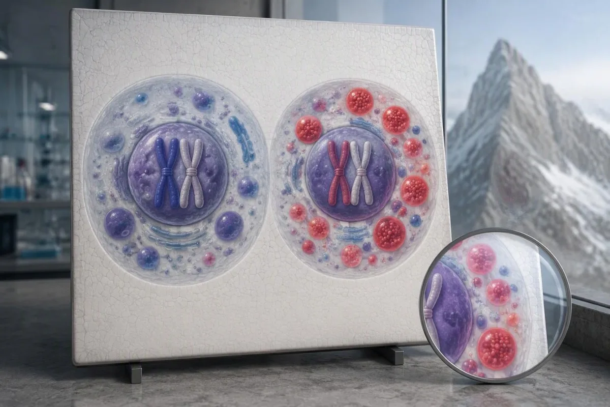
X-chromosome inactivation differs between cells and can differ between tissues. A blood sample may contain enough cells producing functional enzyme to yield a normal laboratory result, while kidney or cardiac cells have a less favorable expression pattern; 1 normal enzyme measurement therefore cannot reliably exclude Fabry disease in a heterozygous woman.
An apparently unaffected woman is not automatically an unaffected carrier: some women develop substantial cardiac, kidney, or neurologic disease, while others remain mildly affected. The limits of X-linked enzyme screening also arise in G6PD deficiency, but G6PD-specific retesting rules and red-cell effects must not be transferred to the GLA test.
Considere um hipotético 38-year-old woman whose father has a confirmed pathogenic GLA variant: her enzyme activity and lyso-Gb3 both fall within their laboratory ranges. She still needs testing for that exact familial variant, because an affected father transmits his X chromosome to all biological daughters; a negative biochemical screen does not undo that inheritance pattern.
What does the lyso-Gb3 test add to enzyme testing?
O lyso-Gb3 test measures a deacylated derivative of Gb3 that often increases in Fabry disease, providing biochemical evidence of substrate accumulation. Laboratories may report results in ng/mL ou nmol/L; an elevated result supports investigation, but a normal result does not exclude Fabry disease.

Lyso-Gb3 concentrations tend to be higher in untreated males with classic Fabry disease than in many women or later-onset cases. Aerts et al., in their 2008 PNAS paper, established elevated globotriaosylsphingosine as a characteristic Fabry finding; the study does not justify turning 1 universal concentration into a diagnostic rule for every patient, assay, or phenotype.
A hypothetical laboratory might use 1,8 ng/mL as its upper reference limit, making 9.0 ng/mL five times that limit. Kantesti AI can help explain that relationship, but we would not compare it directly with a different laboratory's nmol/L report without checking the analyte and method; our guide to alertas laboratoriais fora do intervalo explains why a flag is not itself a diagnosis.
A normal lyso-Gb3 result can occur in genetically confirmed Fabry disease, particularly in women and some later-onset presentations. Repeated testing is most interpretable when the same assay measures the same analyte: a change from 2.0 to 1.6 ng/mL may reflect biological or analytical variation, treatment, or both—not necessarily a meaningful change in organ health.
| Level | Range | What it means |
|---|---|---|
| Within the hypothetical laboratory limit | ≤1.8 ng/mL | Illustrative only: no biochemical elevation by this invented laboratory's rule; Fabry disease is not excluded. |
| Above the hypothetical laboratory limit | >1.8 ng/mL | Illustrative only: review GLA classification, enzyme activity, phenotype, and specimen information. |
| Exemplo resolvido | 9.0 ng/mL | Five times the hypothetical upper limit; this is not a validated severity category or treatment threshold. |
How should you read GLA gene testing results?
GLA gene testing looks for variants that can alter alpha-galactosidase A function and reports their clinical classification. The GLA gene has 7 exons, but the meaning of a detected change depends on evidence of pathogenicity—not simply on whether sequencing found a difference.
Genetic laboratories commonly use 5 categories: pathogenic, likely pathogenic, uncertain significance, likely benign, and benign. A pathogenic or likely pathogenic GLA finding can establish a molecular diagnosis in the appropriate clinical-genetic context, including in a woman with normal enzyme activity; it does not automatically prove that every symptom or organ abnormality is caused by Fabry disease.
The report should identify the exact variant, transcript, classification, and test coverage, because 2 reports saying 'GLA positive' may mean very different things. The distinction between a genetic finding and a disease diagnosis also appears in our guide to interpreting inherited disease variants, although MEFV inheritance and diagnostic criteria differ from Fabry disease.
A negative result is only as broad as the assay: routine sequencing, deletion/duplication analysis, and targeted testing answer different questions. If a relative's precise GLA variant is known, targeted familial testing usually provides a direct answer; if strong biochemical evidence remains after 1 negative sequencing report, a specialist may review coverage, structural changes, or less routinely captured variants.
Why does a GLA VUS not confirm Fabry disease?
A GLA variant of uncertain significance, or VUS, means that available evidence cannot establish whether the variant causes disease. A VUS is 1 of 5 standard classifications, and it must not be treated as a positive Fabry diagnosis or an automatic indication for disease-specific therapy.

The ACMG/AMP sequence-variant framework states that a VUS should not be used in clinical decision-making (Richards et al., 2015). For Fabry disease, 3 useful evidence streams are biochemical findings, a compatible clinical phenotype, and genetic evidence such as segregation or validated functional studies; none should be cherry-picked merely because the variant appears in a disease-associated gene.
Family testing can sometimes help reclassify a VUS, but detecting the same uncertain variant in 2 relatives is not automatically proof of causation. Age-dependent disease, variable expression in women, and common alternative explanations for kidney or cardiac findings complicate segregation analysis; our discussion of genetic findings without overdiagnosis addresses the broader mistake of equating a DNA difference with disease.
For example, a hypothetical 55-year-old man with hypertension, mildly thickened ventricular walls, normal leukocyte enzyme activity, normal lyso-Gb3, and a GLA VUS has an unresolved genetic finding—not confirmed Fabry cardiomyopathy. He still deserves evaluation of his heart findings, while genetics reviews variant evidence and possible reclassification; uncertainty should prompt a plan, not either dismissal or premature treatment.
What if enzyme, lyso-Gb3, and GLA results disagree?
Discordant Fabry results require specialist reconciliation rather than a majority vote between tests. The 3 tests measure different biological layers, so normal biomarkers with a pathogenic GLA variant, or low enzyme activity with uncertain genetics, lead to different diagnostic pathways.
Uma pessoa hipotética 29-year-old man has profoundly deficient leukocyte enzyme activity, clearly elevated lyso-Gb3, and a pathogenic GLA variant: these findings align strongly with Fabry disease. The next question is organ involvement and care planning, not whether a normal creatinine or absence of childhood pain can overturn 3 concordant disease-specific findings.
A different hypothetical 46-year-old woman has a pathogenic GLA variant, normal enzyme activity, and normal lyso-Gb3; that pattern does not erase her molecular diagnosis. Disease burden requires separate assessment, just as the combination of a protein measurement and genotype can be informative in o teste de alfa-1 antitripsina, although that disorder has different biology and thresholds.
Kantesti is an AI lab test interpretation service that can help organize these 3 reported evidence streams without independently reclassifying a GLA variant. Our padrões de interpretação clínica provide context for that boundary; suspected pseudodeficiency, assay interference, or a conflicting laboratory classification requires review by the testing laboratory and a Fabry-experienced specialist.
Can sample handling cause an abnormal Fabry screen?
Sample handling can affect alpha-galactosidase A activity, particularly in dried-spot screening, so an unexpectedly low result may need confirmation in a fresh specimen. At least 2 questions should be checked before interpretation: was the specimen acceptable, and was the method intended for screening or diagnosis?

Dried-spot enzyme results can be influenced by inadequate application, drying, heat, moisture, and specimen composition. A repeat fresh leukocyte assay may be more informative than repeating the same compromised card, especially when more than 1 lysosomal enzyme appears unexpectedly low; the laboratory can explain whether internal quality checks suggest a generalized specimen problem.
Most Fabry-specific enzyme and genetic tests do not inherently require an 8–12 horas, but follow the collection laboratory's instructions when other tests are ordered alongside them. Our first laboratory visit preparation guide helps distinguish general preparation from assay-specific requirements; do not stop prescribed treatment simply to obtain a supposedly cleaner baseline.
Tell the laboratory about Fabry-specific treatment, recent transfusion, or hematopoietic transplantation because these circumstances can complicate enzyme or DNA interpretation. A hypothetical result that changes from 0.4 to 6.0 activity units after recollection needs method and specimen review before being called biological recovery; our guide to sample-quality interference explains why laboratories sometimes recommend a redraw.
Which symptoms and family clues justify Fabry testing?
Fabry testing is most useful when characteristic symptoms, unexplained organ findings, or a confirmed family diagnosis raise suspicion. Childhood heat-triggered burning extremity pain, reduced sweating, corneal changes, or otherwise unexplained kidney or cardiac disease can justify evaluation; 1 nonspecific symptom alone rarely identifies the cause.
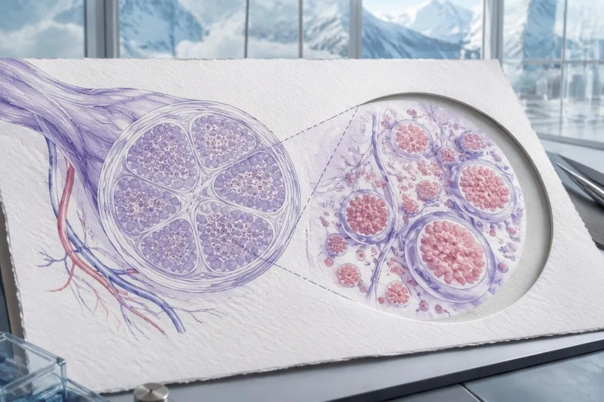
Fabry-related pain often reflects small-fiber involvement, so routine nerve-conduction studies can be normal despite genuine symptoms. In a hypothetical homem de 24 anos with recurrent heat-triggered burning feet and reduced sweating, the pattern merits targeted history and examination; our guide to laboratory clues for unexplained pain also describes alternatives that should not be overlooked.
Cornea verticillata and clusters of angiokeratomas can increase suspicion, but neither should be interpreted outside its clinical context. For example, 2 medications—amiodarone and hydroxychloroquine—can produce corneal changes resembling a Fabry clue, while many skin marks are unrelated; an ophthalmologist or dermatologist can characterize the finding before it becomes genetic evidence.
Later-onset Fabry disease can present without the classic childhood symptom cluster, making unexplained ventricular thickening, proteinuria, or a suggestive pedigree more useful than a childhood checklist. A hypothetical pessoa de 61 anos with isolated cardiac disease needs consideration of common causes and other infiltrative or inherited conditions; blanket Fabry screening for every episode of fatigue or tingling creates more uncertainty than clarity.
Which kidney tests matter alongside Fabry results?
Kidney assessment in Fabry disease should include urine albumin-to-creatinine ratio, creatinine-based eGFR, and clinical context. Uma razão albumina-creatinina de 30–300 mg/g, aproximadamente 3–30 mg/mmol, indicates moderately increased albuminuria; a normal eGFR does not exclude early kidney involvement.
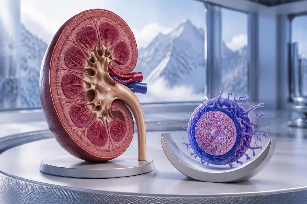
A hypothetical person with confirmed Fabry disease has an eGFR of 98 mL/min/1.73 m² but repeated urine ACR values of 75 mg/g. Filtration is preserved, yet albumin leakage warrants assessment; our urine ACR preparation guide explains how exercise, urinary illness, and collection timing can distort a single measurement.
Creatinine can underestimate kidney impairment in someone with low muscle mass, and cystatin C may improve assessment when creatinine-based estimates appear implausible. At Kantesti, we encourage reading eGFR together with urine findings rather than reassuring someone from 1 creatinine result; our guide to a cistatina C além da creatinina explains the benefits and non-kidney influences of that additional marker.
Kidney impairment must not automatically be attributed to a GLA VUS, even when the patient has proteinuria. If a specialist recommends tissue examination to clarify unexplained kidney disease, lamellated inclusions require expert interpretation because some medications can cause similar phospholipidosis; our guia de BUN e creatinina explains why a ratio alone cannot identify the cause of 1 abnormal kidney profile.
How do heart and neurologic findings affect interpretation?
Cardiac and neurologic findings help assess Fabry disease involvement, but they do not independently prove a GLA variant is pathogenic. Unexplained ventricular thickening, rhythm abnormalities, or stroke can support investigation; 1 elevated cardiac biomarker is neither specific to Fabry disease nor sufficient for genetic diagnosis.
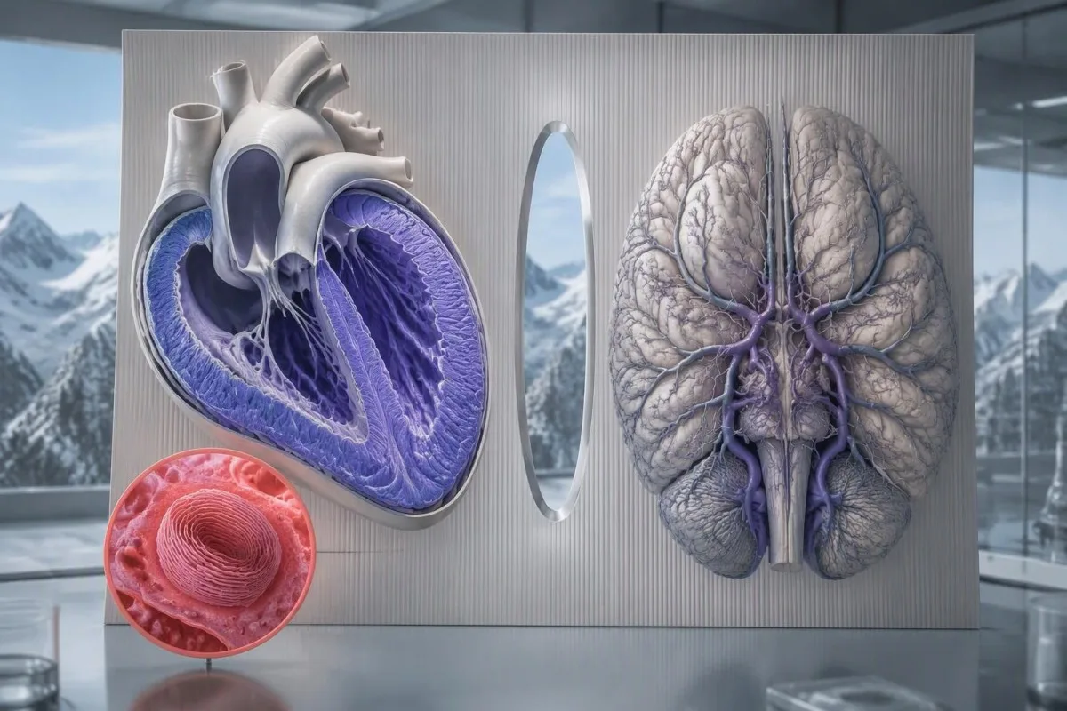
Fabry cardiac evaluation commonly combines electrocardiography, echocardiography, rhythm assessment, and cardiac MRI when appropriate, rather than relying on a single laboratory panel. The 2020 expert consensus on cardiovascular Fabry manifestations describes this multimodal approach (Linhart et al., 2020); 2 useful MRI clues—altered native T1 and characteristic fibrosis distribution—still require interpretation alongside other cardiac diagnoses.
NT-proBNP can rise with cardiac wall stress, but age, atrial fibrillation, and kidney function also influence its concentration. A hypothetical patient with NT-proBNP of 600 pg/mL needs clinical assessment, not a Fabry-specific label; our guide to NT-proBNP causes and cutoffs explains why symptom context and renal function change the meaning of the same number.
Sudden facial weakness, speech difficulty, or new one-sided weakness requires emergency assessment, regardless of pending GLA results or a previous reassuring enzyme test. Family history may shape longer-term prevention, as discussed in family stroke risk assessment, but waiting 1 hora for a genetic explanation is the wrong response to new stroke symptoms; chest pain, collapse, or severe breathlessness also needs urgent care.
Who in the family needs GLA testing?
Cascade testing should focus on the exact familial pathogenic or likely pathogenic GLA variant, supported by genetic counseling. A heterozygous mother has a 50% transmission chance in each pregnancy; an affected father transmits his variant to all biological daughters and none of his biological sons.
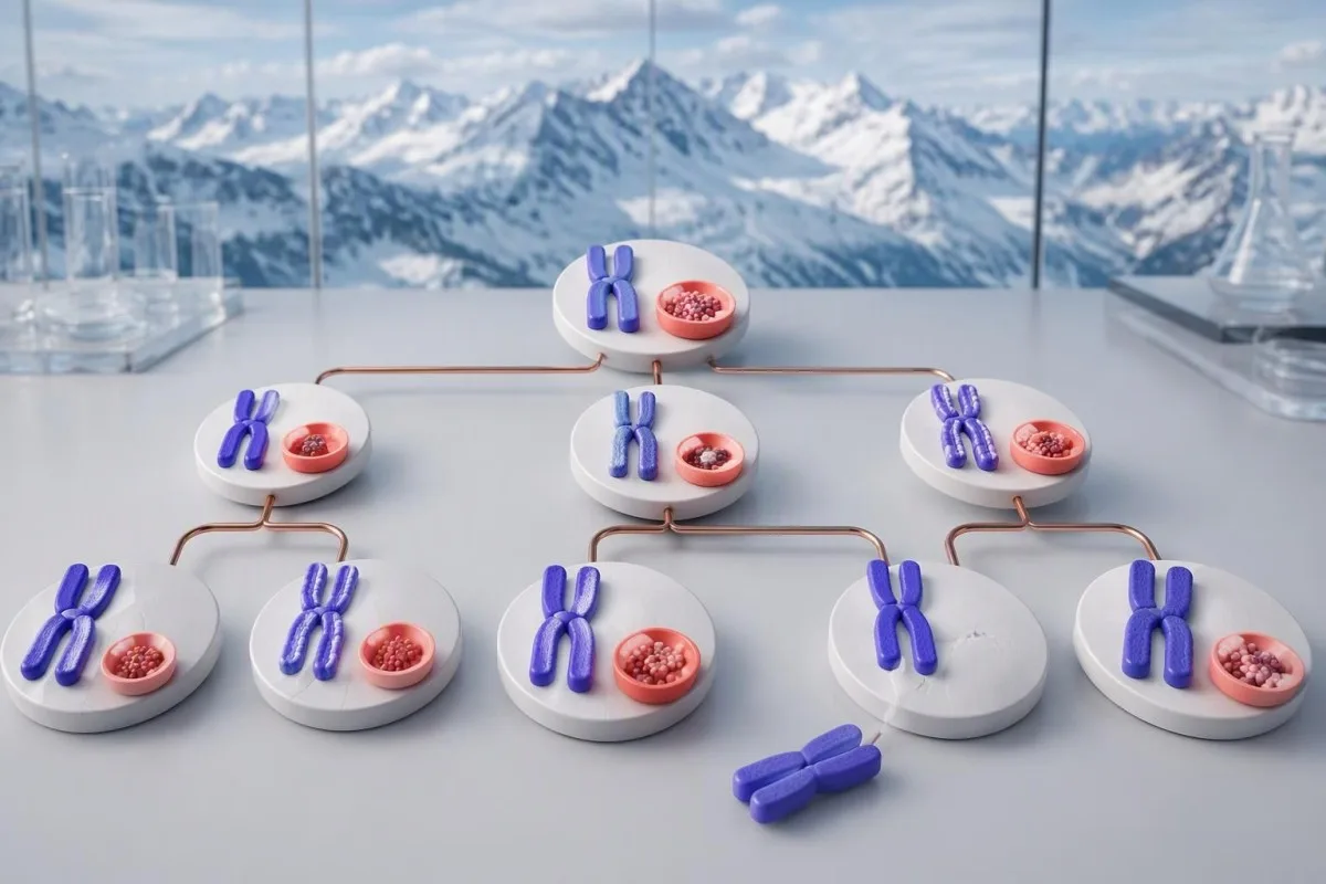
The 50% transmission probability applies independently to each pregnancy, not as a guarantee that half of the children in a particular family will inherit the variant. A daughter who inherits a pathogenic GLA variant may have normal enzyme activity and still need disease-specific assessment; a son's degree of enzyme deficiency and phenotype can differ from a female relative's presentation.
Family testing must distinguish a confirmed disease-causing variant from a VUS found in 1 relative. Testing selected relatives for a VUS may support laboratory reclassification, but it should not be presented as predictive testing for confirmed Fabry disease; our guide to X-linked genetic counseling questions illustrates why the exact condition and assay matter, since FMR1 repeat testing is not a substitute for GLA analysis.
Testing children requires discussion of potential childhood manifestations, benefits of surveillance, consent, and local genetics practice—not an automatic rule to defer until 18 years of age. Reproductive options may include targeted prenatal testing or preimplantation testing when a familial disease-causing variant is established; a genetics team should explain limitations and avoid turning uncertain variant evidence into a reproductive diagnosis.
Do Fabry test results determine treatment or response?
Fabry treatment decisions depend on a confirmed diagnosis, organ involvement, clinical trajectory, and treatment eligibility—not a lyso-Gb3 cutoff alone. Many enzyme replacement regimens are administered every 2 semanas; oral chaperone eligibility requires a disease-causing GLA variant with demonstrated amenability and other appropriate clinical criteria.
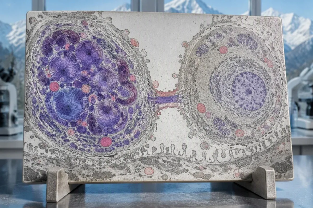
Migalastat amenability and GLA pathogenicity are different questions. A variant can respond in an in-vitro amenability assay without sufficient evidence that it causes Fabry disease, so neither an 'amenable' label nor 1 uncertain genetic result should bypass diagnostic review; specialist prescribing also considers renal function, age eligibility, and the applicable product information.
Lyso-Gb3 often falls with effective Fabry-specific therapy, but biomarker improvement does not prove that established kidney scarring or cardiac fibrosis has reversed. If a hypothetical concentration falls from 40 to 10 ng/mL, that is a 75% reduction; Kantesti AI can help explain the arithmetic while keeping symptoms, eGFR, urine ACR, rhythm findings, and imaging separate from the biomarker trend.
Serial results should be compared using consistent assay methods and documented treatment timing. Uma mudança de 2.1 to 1.9 ng/mL may be much less informative than a sustained multi-visit pattern, and persistent symptoms deserve evaluation even when lyso-Gb3 improves; our guide to mudanças significativas entre testes explains why small numerical shifts should not trigger unsupervised treatment changes.
AI interpretation limits and related research publications
AI can explain a Fabry laboratory report, but it cannot independently confirm Fabry disease, reclassify a GLA VUS, or prescribe disease-specific therapy. Keep the original reports for all 3 diagnostic layers, including specimen details, units, genetic classification, and the laboratory's interpretive comments.

Kantesti is an AI-powered blood test analysis tool that can help organize reported results and questions for a specialist appointment, while a genetic laboratory remains responsible for its variant classification. Before sharing 1 identifiable genetic report, consulte nossa informações sobre privacidade e dados; genetic results can reveal information about relatives as well as the person whose sample was tested.
O 2 DOI-linked publications below concern RDW interpretation and the BUN/creatinine ratio, not clinical validation of a Fabry diagnostic algorithm. Our publicação de interpretação de RDW may help explain accompanying hematology results, but RDW cannot confirm or exclude Fabry disease; a DOI identifies a deposited publication and does not, by itself, establish peer review or diagnostic accuracy.
Nosso padrões de conteúdo médico describe how educational content should separate established facts from uncertainty. When using Kantesti to prepare for care, ask 3 perguntas práticas: does the GLA classification establish a molecular diagnosis, do the biochemical results support it, and what organ monitoring is needed; these answers are more useful than asking whether an AI has declared the report simply positive or negative.
Perguntas frequentes
Qual é o exame mais preciso para a doença de Fabry?
O diagnóstico da doença de Fabry utiliza evidências complementares da atividade da alfa-galactosidase A, do liso-Gb3 e do teste genético de GLA, em vez de um único exame de rastreamento universalmente suficiente. Em homens com a forma clássica da doença, a atividade enzimática está tipicamente abaixo de 1% da atividade média normal, e o teste genético identifica a causa molecular. Nas mulheres, o teste de GLA é particularmente importante, pois a atividade enzimática pode ser normal. Um achado patogênico ou provavelmente patogênico tem um significado diagnóstico diferente do de uma variante de significado incerto.
Uma mulher pode ter doença de Fabry com um teste enzimático normal?
Uma mulher pode ter doença de Fabry apesar da atividade normal da alfa-galactosidase A. As mulheres geralmente têm 2 cromossomos X, e a inativação do cromossomo X cria misturas de células que produzem diferentes quantidades da enzima funcional. A atividade enzimática no sangue não representa de forma confiável a função enzimática em cada célula renal ou cardíaca. Um histórico familiar ou quadro clínico sugestivo justifica a avaliação genética do GLA, mesmo quando o resultado da enzima está dentro dos limites normais.
Qual é um nível normal de liso-Gb3?
A normal lyso-Gb3 result is defined by the testing laboratory's assay-specific reference interval, not a universal Fabry cutoff. For illustration only, a hypothetical laboratory using an upper limit of 1.8 ng/mL would classify 9.0 ng/mL as five times that limit. Those figures must not be substituted for your laboratory's range or compared directly with a different-unit result. Normal lyso-Gb3 does not exclude Fabry disease, particularly in women and some later-onset cases.
Uma variante de significado incerto no gene GLA significa que tenho doença de Fabry?
0 Uma variante de significado incerto no GLA não confirma a doença de Fabry. Uma VUS é uma das 5 classificações padrão de variantes genéticas e significa que as evidências disponíveis não permitem determinar se a variante causa a doença. Especialistas podem avaliar a atividade enzimática, lyso-Gb3, achados clínicos, segregação familiar e evidências funcionais validadas. O tratamento específico da doença e a rotulagem preditiva de familiares como portadores da doença não devem se basear apenas na VUS.
0 Pode um nível normal de creatinina descartar a doença de Fabry nos rins?
0 Creatinina normal não exclui comprometimento renal precoce na doença de Fabry. A relação albumina/creatinina urinária pode se tornar anormal enquanto a eGFR permanece acima de 90 mL/min/1.73 m². Uma ACR de 30–300 mg/g, aproximadamente 3–30 mg/mmol, indica albuminúria moderadamente aumentada e requer interpretação com repetição dos exames e contexto clínico. A proteinúria em alguém com uma VUS no gene GLA ainda requer investigação de outras causas renais.
Os familiares devem ser testados após um diagnóstico de doença de Fabry?
Deve-se oferecer aconselhamento genético e testes direcionados aos familiares quando uma variante patogênica ou provavelmente patogênica familiar no gene GLA for estabelecida. Uma mãe heterozigota tem uma chance de 50% de transmitir a variante em cada gestação. Um pai afetado transmite sua variante a todas as filhas biológicas e a nenhum dos filhos biológicos. Testar os familiares para uma VUS pode ajudar a esclarecer a incerteza, mas não equivale ao teste preditivo para a doença de Fabry confirmada.
Do I need to fast before a Fabry disease test?
Fabry-specific enzyme and GLA genetic tests generally do not inherently require an 8–12-hour fast. Follow the collecting laboratory's instructions because lyso-Gb3 protocols or other tests ordered at the same visit may have additional requirements. Tell the laboratory about Fabry treatment, recent transfusion, or hematopoietic transplantation when relevant. Do not stop prescribed medication or disease-specific treatment solely to prepare for testing without clinical advice.
Get your blood test results explained
Upload your lab report and Kantesti's AI explains each biomarker in plain language within seconds. Used by more than 2 million people worldwide.
Referências
- Klein, T., Mitchell, S., & Weber, H. (2026). RDW Blood Test: Complete Guide to RDW-CV, MCV & MCHC. Kantesti AI Medical Research. DOI
- Klein, T., Mitchell, S., & Weber, H. (2026). Razão BUN/Creatinina Explicada: Guia de Teste de Função Renal. Kantesti AI Medical Research. DOI
- Aerts JM et al. (2008). Elevated globotriaosylsphingosine is a hallmark of Fabry disease. Proceedings of the National Academy of Sciences of the United States of America. DOI PubMed
- Richards S et al. (2015). Standards and guidelines for the interpretation of sequence variants: a joint consensus recommendation of the American College of Medical Genetics and Genomics and the Association for Molecular Pathology. Genetics in Medicine. DOI PubMed
- Linhart A et al. (2020). An expert consensus document on the management of cardiovascular manifestations of Fabry disease. European Journal of Heart Failure.
- 2 milhões+Tests analysed
- 127+Países
- 100+Idiomas
About the medical team


Sinais de confiança E-E-A-T
Experiência
Revisão clínica orientada por médicos dos fluxos de interpretação de exames laboratoriais.
Especialização
Foco em medicina laboratorial sobre como os biomarcadores se comportam no contexto clínico.
Autoridade
Medically reviewed by Dr. Thomas Klein, with contributions from Dr. Sarah Mitchell and Prof. Dr. Hans Weber.
Confiabilidade
Interpretação baseada em evidências, com caminhos de acompanhamento claros para reduzir alarmes.

Medical disclaimer
Este artigo é apenas para fins educacionais e não constitui aconselhamento médico. Consulte sempre um profissional de saúde qualificado para decisões de diagnóstico e tratamento.
Publicado · Contact the editorial team
Published by Kantesti Ltd, registered in England & Wales, company no. 17090423, London, United Kingdom.
