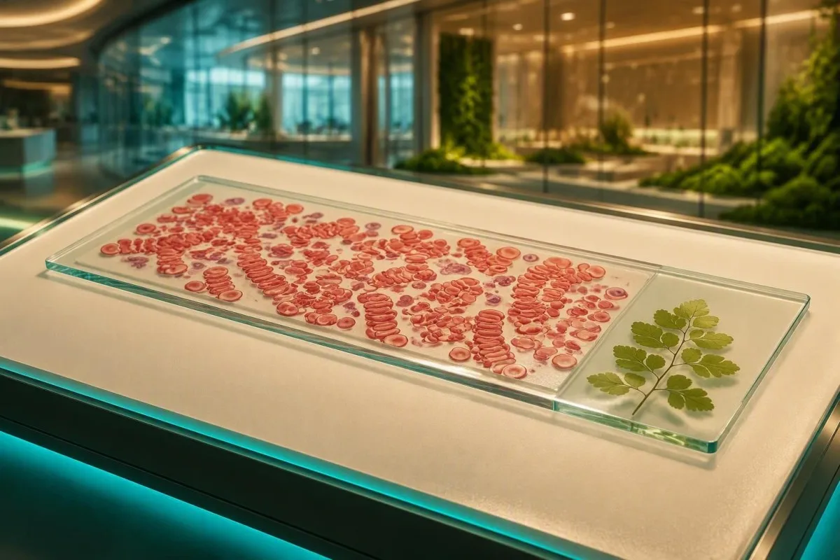
ຈຸລັງເມັດເລືອດແດງສາມາດຊ້ອນກັນຄືກັບຫຼຽນເມື່ອໂປຣຕີນໃນ plasma ປ່ຽນການຖີບອອກຕາມປົກກະຕິ. ຮູບແບບດັ່ງກ່າວມັກຈະເປັນການຕອບສະໜອງ, ແຕ່ຜົນ CBC ແລະ ໂປຣຕີນທີ່ມາຄຽງຄູ່ກັນຈະກໍານົດວ່າມັນຕ້ອງການການຕິດຕາມຢ່າງຮີບດ່ວນຫຼືບໍ່.


- ການສ້າງ rouleaux ໝາຍເຖິງເມັດເລືອດສ້າງເປັນກອງຄ້າຍຫຼຽນ, ໂດຍປົກກະຕິແມ່ນຍ້ອນວ່າ fibrinogen ຫຼື immunoglobulins ຫຼຸດຜ່ອນການຖີບອອກຈາກພື້ນຜິວຕາມປົກກະຕິ.
- ມັນເປັນຮູບແບບ, ບໍ່ແມ່ນການບົ່ງມະຕິ: ການຕິດເຊື້ອເມື່ອບໍ່ດົນມານີ້, ກິດຈະກໍາ autoimmune, ການຖືພາ, ການອັກເສບຊໍາເຮື້ອ, ແລະພະຍາດ plasma-cell ທັງ ໝົດ ສາມາດຜະລິດໄດ້.
- ESR ສາມາດເພີ່ມຂຶ້ນ ເມື່ອມີ rouleaux ເນື່ອງຈາກຈຸລັງທີ່ຊ້ອນກັນຕົກລົງໄວຂຶ້ນ; ESR ຢ່າງດຽວບໍ່ສາມາດລະບຸສາເຫດໄດ້.
- ໂປຣຕີນທັງໝົດ 6.0-8.3 g/dL ແລະ albumin ປະມານ 3.5-5.0 g/dL ແມ່ນຊ່ວງອ້າງອິງຜູ້ໃຫຍ່ທົ່ວໄປ, ເຖິງແມ່ນວ່າຊ່ວງຫ້ອງທົດລອງຈະແຕກຕ່າງກັນ.
- Gamma gap ສູງກວ່າ 4.0 g/dL ອາດຈະສົມເຫດສົມຜົນໃນການຕິດຕາມທີ່ສຸມໃສ່ໂປຣຕີນ, ແຕ່ການຂາດນ້ໍາແລະການອັກເສບກໍ່ສາມາດອະທິບາຍໄດ້ເຊັ່ນກັນ.
- SPEP ດ້ວຍ immunofixation ແລະ serum free light chains ມາ係當紅血球產生滾動現象,同時伴隨貧血、腎臟變化、骨骼疼痛或球蛋白升高時,常用的進一步檢測。.
- 鹽水稀釋 可以幫助實驗室區分滾動現象和紅細胞凝集,後者的鑑別診斷不同。.
- ການປະເມີນດ່ວນ 適用於出現新發的意識模糊、嚴重呼吸困難、視力改變、明顯虛弱或腎功能迅速惡化時。.
Rouleaux ໃນການປິ່ນສະເປັກທີ່ແຜ່ກະຈາຍໝາຍຄວາມວ່າແນວໃດ
血液塗片上的滾動現象形成 表示紅血球排列成短或長的硬幣狀圓柱體,而不是獨立分開。它最常反映的是血漿蛋白增加—特別是纖維蛋白原或免疫球蛋白—而不是紅血球本身的結構缺陷。.
紅血球膜正常帶負電荷,常稱為zeta電位,這能使相鄰的細胞分開。纖維蛋白原和某些免疫球蛋白部分地橋接了這種排斥空間;細胞隨後最容易在製備良好的塗片薄區域排列。.
形態學評論不是測量濃度,也沒有普遍的分級標準。一個實驗室可能報告“偶爾出現滾動現象”,而另一個可能報告“存在”;我會結合血紅蛋白、MCV、總蛋白、白蛋白、肌酐、鈣以及臨床病史來閱讀實際的塗片評論。.
ແຄນເທສຕີ ເປັນ AI ເຄື່ອງວິເຄາະເລືອດ 將塗片評論與相關的數值結果放在一起,而不是將紅血球堆疊視為獨立的診斷。若想重新回顧計數的其他部分,請參閱我們的 ຄູ່ມືຕົວຊີ້ວັດ CBC.
為什麼“形成”這個詞聽起來可能比實際情況更令人擔憂
滾動現象並不意味著身體內部正在形成血栓。它是在實驗室樣本被塗抹在玻璃上後看到的微觀排列,許多患有短期炎症性疾病的人會出現這種現象,而沒有患上血癌或嚴重的循環問題。.
ເປັນຫຍັງຈຶ່ງເກີດການຊ້ອນກັນຂອງເມັດເລືອດແດງ
當循環蛋白減少紅血球之間的靜電分離時,就會發生紅血球堆疊。纖維蛋白原是常見的短期驅動因素,而過多的免疫球蛋白在現象持續存在或與急性疾病不成比例時更為相關。.
在感染、手術、炎症性關節炎或嚴重組織損傷後幾天內,急性期反應會增加纖維蛋白原。這種相同的生物學機制可以提高 ESR 並使塗片看起來很明顯,即使 CBC 指數保持正常;; 觸發因素開始消退 後 ESR 變化緩慢。.
免疫球蛋白較大,當濃度很高或為單株蛋白時,可產生特別明顯的堆疊。Kyle 等人報告說,在一項人群研究中,50 歲及以上的人群中 3.2% 患有意義不明的單株高球蛋白血症 (MGUS),儘管大多數 MGUS 患者從未發展成多發性骨髓瘤。.
在實踐中,僅憑形態學無法區分由纖維蛋白原驅動的炎症與由免疫球蛋白驅動的過程。一個 CRP 為 96 mg/L、總蛋白值正常的 34 歲流感患者,與一個貧血、總蛋白 9.1 g/dL、eGFR 下降的 71 歲患者,需要非常不同的處理方案。.
ສາເຫດທົ່ວໄປທີ່ຕອບສະໜອງ: ການອັກເສບ, ການຕິດເຊື້ອ ແລະ ການຖືພາ
最常見的滾動現象原因是由於暫時性炎症狀態導致纖維蛋白原或多株抗體升高。當其他檢查結果均正常時,呼吸道感染、自身免疫性疾病發作、慢性炎症性疾病和懷孕是常見的解釋。.
蛋白質 C 反應性蛋白迅速升高,而纖維蛋白原和 ESR 可能維持較長時間的升高;這種時間差異在最近出現發燒或疾病發作後很有用。Gabay 和 Kushner 將 CRP 描述為一種快速反應的急性期蛋白,而纖維蛋白原的行為較慢並影響沉降(Gabay 和 Kushner,1999)。.
懷孕會顯著增加纖維蛋白原,在妊娠晚期通常會達到 4-6 g/L 的範圍,因此滾動現象和 ESR 升高不會使用非懷孕期的預期值來解釋。在孕婦患者中,我會在升級塗片評論之前,先查看症狀、血壓、尿蛋白、血紅蛋白趨勢和產科背景。.
អត្ថបទលម្អិតរបស់យើង ຄູ່ມືໂປຣຕີນໃນເລືອດ (serum proteins) 解釋了為什麼白蛋白和球蛋白模式比單獨的總蛋白更重要。這是結果之一,最近感冒可能比單獨的標記更有信息價值。.
ເມື່ອໂປຣຕີນສູງເປັນຄຳອະທິບາຍທີ່ເປັນໄປໄດ້
高免疫球蛋白、高纖維蛋白原或相對於球蛋白的白蛋白降低,可以支持滾動現象的蛋白質相關解釋。高於實驗室上限的總蛋白結果是一個線索,但無法確定責任蛋白。.
Common adult reference intervals are approximately 6.0-8.3 g/dL for total protein ແລະ 3.5-5.0 g/dL for albumin, but the report’s own interval prevails. The calculated globulin value equals total protein minus albumin; an albumin 3.4 g/dL with total protein 8.6 g/dL gives globulins of 5.2 g/dL.
A gamma gap—total protein minus albumin—above 4.0 g/dL is often used as a pragmatic trigger to review chronic infection, autoimmune disease, liver disease, and gammopathy. It is not a diagnosis and is less specific in dehydration, where both albumin and globulins may concentrate; our high total protein guide ຄອບຄຸມຄວາມແຕກຕ່າງນັ້ນ.
ແຄນເທສຕີ ເປັນ ແພລດຟອມການອ່ານຜົນກວດເລືອດຂອງ AI that checks whether an elevated globulin estimate is paired with low albumin, anaemia, renal change, or calcium elevation. That combination carries more clinical weight than rouleaux in a person with otherwise normal values.
ເປັນຫຍັງ rouleaux ຈຶ່ງມາຄຽງຄູ່ກັບ ESR ສູງ
Rouleaux can raise the erythrocyte sedimentation rate because aggregated red cells fall through a vertical tube faster than individual cells. ESR is therefore an indirect measure of altered plasma proteins, not a direct test for one specific disease.
Many laboratories use upper limits near 15 mm/hour for younger adult men ແລະ 20 mm/hour for younger adult women, but age, pregnancy, anaemia, and method alter those figures. An ESR above 100 mm/hour warrants timely clinical evaluation, yet it can occur with infection, autoimmune disease, kidney disease, and monoclonal proteins rather than one single condition.
Anaemia itself can raise ESR because there are fewer red cells interfering with descent. That is why an ESR of 55 mm/hour with haemoglobin 8.7 g/dL cannot be read the same way as 55 mm/hour with haemoglobin 14.2 g/dL; compare haematocrit and haemoglobin ก่อนสรุปข้อใดๆ.
CRP is usually the better marker for short-term change because it can fall substantially within 24-48 hours of effective treatment. ESR may remain high for weeks after symptoms improve, particularly when rouleaux persists.
ການທົດສອບຕິດຕາມເມື່ອໂປຣຕີນ monoclonal ເປັນໄປໄດ້
Clinicians commonly order serum protein electrophoresis when rouleaux accompanies unexplained anaemia, high total protein, kidney dysfunction, high calcium, recurrent infections, or bone pain. SPEP separates major serum protein fractions and can reveal a narrow monoclonal band, often called an M-spike.
An M-spike is not synonymous with multiple myeloma. The International Myeloma Working Group defines symptomatic myeloma using clonal marrow cells or a plasmacytoma plus a myeloma-defining event, such as calcium above 11 mg/dL, creatinine above 2 mg/dL, haemoglobin more than 2 g/dL below normal, or bone lesions (Rajkumar et al., 2014).
If SPEP shows a possible monoclonal component, immunofixation identifies the immunoglobulin type and serum free light chains assess light-chain imbalance. Our SPEP explanation is useful preparation for a clinician conversation, but no online tool can replace haematology review of a confirmed band.
I have seen patients frightened by the word “spike” after a severe infection, only to have repeat testing show a broad polyclonal response. Conversely, a small stable band in an otherwise well person may require scheduled monitoring rather than emergency treatment.
ຕ່ອງແສງຟຣີແລະອິມມູນໂກບູລິນປັບຮູບແບບແນວໃດ
Serum free light-chain testing and quantitative IgG, IgA, and IgM help determine whether protein excess is monoclonal or polyclonal. The reference interval for the kappa-to-lambda free light-chain ratio is commonly about 0.26-1.65, though kidney function and assay method can widen interpretation.
Chronic kidney disease increases both kappa and lambda light chains because clearance falls, so a mildly abnormal ratio requires renal context. A markedly skewed ratio, especially with an M-spike or unexplained anaemia, deserves haematology-led evaluation; see our free light chain ratio guide.
Quantitative immunoglobulins answer a different question from SPEP. Broad elevation of IgG, IgA, or IgM may occur with chronic liver disease, autoimmune conditions, or infection, while suppression of unaffected immunoglobulins can be a more concerning clue in a monoclonal process.
ແຄນເທສຕີ ເປັນ ឧបករណ៍វិភាគតេស្តឈាមដែលដំណើរការដោយ AI used across 127+ countries to connect immunoglobulin, renal, calcium, and CBC results in one report. We deliberately label these patterns as follow-up signals, not diagnoses, because the clinical context changes the meaning.
ເບາະແສຈາກ CBC ທີ່ປ່ຽນລະດັບຄວາມກັງວົນ
Rouleaux is more concerning when it appears with unexplained anaemia, high RDW, low platelets, or a rising creatinine than when the CBC is stable. A normal smear comment does not exclude illness, and rouleaux by itself does not diagnose marrow disease.
For adults, haemoglobin below 13.0 g/dL ສຳລັບຜູ້ຊາຍ ຫຼື 12.0 g/dL ໃນຜູ້ຍິງທີ່ບໍ່ຖືພາ meets a commonly used anaemia definition, but altitude, pregnancy, and laboratory standards matter. New anaemia with rouleaux should prompt a review of MCV, reticulocytes, ferritin, B12, kidney function, and protein studies rather than an assumption that iron deficiency is the answer.
RDW reflects variation in cell size and is often reported near 11.5-14.5% as a reference interval. A high RDW may appear after bleeding, iron treatment, mixed deficiencies, or marrow stress; our ຄູ່ມື MCV ທີ່ບໍ່ຊັດເຈນ shows why MCV needs the full pattern.
Dr. Thomas Klein’s practical rule is simple: ask whether the change is new. A CBC that has been stable for 4 years is usually more reassuring than a single normal value that has fallen sharply since the previous draw.
ສະພາບການ autoimmune, ຕັບ ແລະ ການອັກເສບຊໍາເຮື້ອ
Autoimmune disease, chronic liver disease, and long-standing infections can cause polyclonal immunoglobulin increases that produce rouleaux. These conditions usually create broad protein changes rather than one discrete M-spike, although exceptions occur.
In suspected lupus or immune-complex disease, ANA is a screening test rather than proof of diagnosis; complement C3 and C4 may fall during active immune-complex consumption. The pattern is more persuasive when paired with rash, arthritis, urine protein, cytopenias, or kidney changes than when ANA alone is positive; review our ANA result guide.
Liver disease can lower albumin while increasing immunoglobulins, making the albumin-to-globulin ratio fall below the typical 1.0-2.5 range. A low ratio does not identify the cause, so clinicians often add ALT, AST, ALP, bilirubin, hepatitis testing, and protein electrophoresis according to symptoms.
For clinically focused reading, our C3, C4 and ANA research guide explains what complement results can and cannot settle. The evidence is honestly mixed on broad screening in people without suggestive symptoms.
ເປັນຫຍັງການສຶກສາທາດເຫຼັກຈຶ່ງເຮັດໃຫ້ເຂົ້າໃຈຜິດໃນລະຫວ່າງ rouleaux
Inflammation that produces rouleaux can also raise ferritin and lower serum iron or transferrin, masking iron deficiency. Ferritin is both an iron-storage marker and an acute-phase reactant, so it must be interpreted with CRP, transferrin saturation, and the CBC.
ferritin ຕ່ຳກວ່າ 15 ng/mL is highly specific for depleted iron stores in many adults, while values below 30 ng/mL often support iron deficiency in clinical practice. During active inflammation, ferritin can be normal or high despite iron-restricted erythropoiesis, especially if CRP is elevated.
Transferrin saturation ទាបជាង 20% can support iron restriction, but it does not separate absolute iron deficiency from inflammatory iron sequestration by itself. Soluble transferrin receptor may help in selected cases because it is less affected by inflammation; our ferritin ແລະ CRP ຊີ້ນຳ walks through the common mismatch.
Do not start high-dose iron solely because a smear reports stacking. In my experience, patients do best when treatment follows a documented deficiency and a reason for it—heavy menstrual loss, gastrointestinal loss, diet, malabsorption, or increased demand.
Rouleaux ທຽບກັບ agglutination ແລະ artifact ສະໄລ້
Rouleaux is usually reversible with saline on a properly prepared slide, while red-cell agglutination from cold-reactive antibodies generally persists. Distinguishing these appearances matters because agglutination can interfere with automated CBC results and suggests a different work-up.
True rouleaux forms orderly linear stacks; agglutination produces more irregular grape-like clumps. Cold agglutinins can create spuriously high MCHC, low red-cell count, and high MCV on an analyser because the machine counts clusters incorrectly—an elevated MCHC pattern deserves laboratory confirmation.
Smear quality matters. A thick area of a poorly spread or slowly dried slide may exaggerate apparent stacking, which is why experienced morphologists assess the monolayer rather than the slide edge alone.
Rouleaux is not the same as schistocytes, spherocytes, or sickled cells. Fragmented cells with low platelets and haemolysis markers are a very different, sometimes urgent finding; our ຄູ່ມື schistocyte explains that safety distinction.
ແຜນການຕິດຕາມທີ່ປະຕິບັດໄດ້ຫຼັງຈາກລາຍງານ rouleaux
A sensible follow-up plan starts with confirming the clinical setting, then checking whether inflammation, anaemia, or protein abnormalities are present. For a well person with a recent viral illness and otherwise normal results, repeat testing after recovery is often more informative than an immediate extensive work-up.
First review the CBC, CMP or renal panel, total protein, albumin, calcium, and CRP or ESR. If symptoms have resolved and values are normal, clinicians may repeat CBC and proteins in 4-8 ອາທິດ; there is no single mandatory interval, so earlier retesting is reasonable when abnormalities are progressing.
Second, order SPEP, immunofixation, free light chains, quantitative immunoglobulins, or urine protein testing when the initial pattern suggests excess immunoglobulin. A urine albumin-creatinine ratio above 30 mg/g is abnormal and makes renal context more relevant, though it does not specifically explain rouleaux.
Kantesti AI can organise prior results so meaningful changes are easier to spot across time; our វិធីសាស្ត្រធ្វើឲ្យមានសុពលភាពផ្នែកព្យាបាល (clinical validation) explains the safeguards and limits of that interpretation. Dr. Thomas Klein recommends bringing the original smear wording, not just a screenshot of highlighted values, to the appointment.
ເມື່ອ rouleaux ຕ້ອງການການປະເມີນຢ່າງຮີບດ່ວນແທນທີ່ຈະເປັນການປະເມີນປົກກະຕິ
Rouleaux itself is rarely an emergency, but certain accompanying symptoms or laboratory combinations require urgent assessment. New confusion, severe breathlessness, chest pain, fainting, major visual change, rapidly declining urine output, or profound weakness should not wait for routine repeat testing.
Seek same-day medical advice for a new haemoglobin below 8 g/dL, ແຄວຊຽມສູງກວ່າ 12 mg/dL, creatinine rising quickly, or symptoms suggestive of hyperviscosity such as headache with blurred vision and mucosal bleeding. These values do not prove a plasma-cell disorder, but they warrant prompt clinician-led assessment.
Persistent back or rib pain, recurrent bacterial infections, unintentional weight loss, night sweats, neuropathy, or foamy urine also change the threshold for follow-up. The concern comes from the combination of symptoms and objective findings, not from coin stacks on a slide alone.
A reliable interpretation must preserve uncertainty. Kantesti’s ຄູ່ມືເທັກໂນໂລຍີ AI describes how our system surfaces escalation patterns while directing urgent symptoms to in-person care rather than offering false reassurance.
ควรนำอะไรไปในการนัดหมายติดตามผล
Bring the complete laboratory report, previous CBC and protein results, a medication list, and a brief timeline of infections or inflammatory symptoms. These details often determine whether rouleaux is a transient reactive finding or a reason for targeted protein testing.
Write down whether you had fever, dental work, surgery, vaccination, joint swelling, rash, weight change, recurrent infections, or reduced urine output during the preceding 2-8 ອາທິດ. Also list supplements, especially iron, B12, biotin, and immune products, because they can complicate interpretation of related tests.
Ask three focused questions: Is my CBC changing? Are total protein and albumin abnormal? Do I need repeat testing, SPEP, or referral? Most patients find those questions more productive than asking whether the smear finding is “good” or “bad.”
As of September 15, 2026, our ຄະນະທີ່ປຶກສາທາງການແພດ continues to emphasise that AI-guided pattern recognition should support—not replace—clinical examination and laboratory review. Keep a copy of results, dates, and symptoms; trends are often the missing diagnostic clue.
ຄໍາຖາມທີ່ຖາມເລື້ອຍໆ
Is rouleaux formation on a blood smear cancer?
Rouleaux formation ບໍ່ແມ່ນມະເຮັງ ແລະມັກກ່ຽວຂ້ອງກັບການອັກເສບ, ລະດັບ fibrinogen ສູງ, ຫຼືການເພີ່ມຂຶ້ນຂອງພູມຕ້ານທານຢ່າງກວ້າງຂວາງ. ມັນສາມາດເກີດຂຶ້ນກັບຄວາມຜິດປົກກະຕິຂອງໂປຣຕີນໂມໂນໂຄຣໂນນເຊັ່ນ MGUS ຫລື multiple myeloma, ນີ້ແມ່ນເຫດຜົນທີ່ທ່ານໝໍພິຈາລະນາ SPEP ເມື່ອມີອາການ rouleaux ພ້ອມກັບອາການໂລກເລືອດຈາງ, ໂປຣຕີນທັງໝົດສູງກວ່າປະມານ 8.3 g/dL, ໝາກໄຂ່ຫລັງເຮັດວຽກຜິດປົກກະຕິ, ລະດັບແຄລຊຽມສູງ, ຫລືອາການເຈັບກະດູກ. ລັກສະນະຂອງຮອຍເປື້ອນພຽງຢ່າງດຽວບໍ່ສາມາດວິນິດໄສຄວາມຜິດປົກກະຕິຂອງຈຸລັງພລາສມາໄດ້. ຜົນການກວດໂປຣຕີນປົກກະຕິ ແລະການຕິດເຊື້ອບໍ່ດົນມານີ້ມັກເຮັດໃຫ້ຄຳອະທິບາຍແບບໂຕ້ຕອບມີຄວາມເປັນໄປໄດ້ຫລາຍຂຶ້ນ.
ການຂາດນ້ຳສາມາດເຮັດໃຫ້ເກີດການສ້າງເປັນແຖວຂອງເມັດເລືອດແດງໄດ້ບໍ?
Dehydration can concentrate serum proteins and make rouleaux more noticeable, particularly if total protein and albumin rise together. It is not one of the strongest explanations for marked persistent rouleaux, because fibrinogen and immunoglobulin changes more directly alter red-cell spacing. Repeating a CBC and protein panel after normal hydration can clarify a borderline pattern. A total protein above 8.3 g/dL that remains elevated after hydration deserves clinician review.
rouleaux ໝາຍຄວາມວ່າ ESR ຂອງຂ້ອຍຈະສູງບໍ?
rouleaux ມັກຈະເພີ່ມ ESR ເນື່ອງຈາກຈຸລັງເມັດເລືອດແດງທີ່ຈັດລຽນຕັ້ງລົງໄວຂຶ້ນກ່ວາຈຸລັງແຍກອອກໃນທໍ່ການຕົກຕະກອນ. ESR ຍັງສາມາດເພີ່ມຂຶ້ນຈາກໂລກເລືອດຈາງ, ການຖືພາ, ອາຍຸ, ພະຍາດไต, ການຕິດເຊື້ອ, ແລະກິດຈະກໍາຂອງພູມຕ້ານທານ, ດັ່ງນັ້ນ rouleaux ບໍ່ໄດ້ຮັບປະກັນຜົນໄດ້ຮັບສູງ. ESR ສູງກວ່າ 100 mm/ຊົ່ວໂມງໂດຍທົ່ວໄປແລ້ວຮຽກຮ້ອງໃຫ້ມີການປະເມີນຜົນທັນເວລາສໍາລັບສາເຫດທີ່ຢູ່ພາຍໃຕ້. CRP ມັກຈະປ່ຽນແປງໄວກວ່າ ESR ເມື່ອການອັກເສບແບບສ້ວຍແຫຼມດີຂຶ້ນ.
ການກວດໃດທີ່ຄວນຕິດຕາມການກໍ່ຕົວຂອງ rouleaux?
ການທົດສອບຕິດຕາມຜົນທົ່ວໄປລວມມີ CBC ພ້ອມດ້ວຍດັດຊະນີ້, protein ທັງໝົດ, albumin, ການຄິດໄລ່ globulin, CRP ຫຼື ESR, creatinine, calcium, ແລະ ການທົດສອບຕັບ. ເມື່ອມີຄວາມຜິດປົກກະຕິຂອງ protein ທີ່ໜ້າເປັນຫ່ວງຫຼືອາການທີ່ໜ້າເປັນຫ່ວງ, ທ່ານໝໍອາດເພີ່ມ SPEP, immunofixation, quantitative immunoglobulins, ແລະ serum free light chains; ໄລອັດຕາສ່ວນຂອງ serum free light chain ປົກກະຕິແມ່ນປະມານ 0.26-1.65. ຊຸດທີ່ຖືກຕ້ອງແມ່ນຂຶ້ນກັບວ່າມີໂລກເລືອດຈາງ, ໝາກໄຂ່ຫຼັງເຮັດວຽກຜິດປົກກະຕິ, calcium ສູງ, ການຕິດເຊື້ອ, ຫຼືອາການຂອງພູມຄຸ້ມກັນຜິດປົກກະຕິຫຼືບໍ່. ການທົດສອບຄືນໃໝ່ອາດເປັນປະໂຫຍດຖ້າຫາກຕົວຢ່າງເດີມມີຂໍ້ຈຳກັດທາງດ້ານເຕັກນິກ.
rouleaux ແຕກຕ່າງຈາກ cold agglutination ແນວໃດ?
Rouleaux forms orderly coin-like chains and usually disperses when saline is added to the slide preparation. Cold agglutination creates irregular clumps that may persist with saline and can produce misleading CBC values, including a falsely high MCHC or MCV. Warming the sample and repeating the analyser run may be needed when cold-reactive antibodies are suspected. The distinction is made by laboratory morphology and testing, not by looking at a patient’s symptoms alone.
ຂ້ອຍຄວນກວດເລືອດຄືນຫຼັງຈາກການກໍ່ຕົວຂອງ rouleaux ບໍ?
ການກວດຊ້ຳຄືນແມ່ນເປັນເລື່ອງປົກກະຕິທີ່ສົມເຫດສົມຜົນໃນອີກປະມານ 4-8 ອາທິດ ເມື່ອມີການປາກົດຂອງ rouleaux ໃນລະຫວ່າງການຕິດເຊື້ອທີ່ຊັດເຈນ, ກໍາລັງດີຂຶ້ນ ແລະ CBC, ໝາກໄຂ່ຫຼັງ, ແຄລຊຽມ, ອາລບູມິນ, ແລະຜົນໂປຣຕີນອື່ນໆແມ່ນໜ້າພໍໃຈ. ການກວດກ່ອນໜ້ານີ້ແມ່ນເໝາະສົມ ຖ້າຫາກມີເລືອດຈາງຫຼຸດລົງ, ໂປຣຕີນທັງໝົດສູງ, ຄຣີອາຕິນິນມີການປ່ຽນແປງ, ຫຼືມີອາການຕ່າງໆເຊັ່ນ: ອາການເຈັບກະດູກ, ຫາຍໃຈບໍ່ສະດວກ, ຫຼືມີອາການສັບສົນ. Rouleaux ທີ່ຍັງຄົງຢູ່ຄວນໄດ້ຮັບການປະເມີນຕາມສະພາບແວດລ້ອມແທນທີ່ຈະຖືກລະເລີຍ. ແພດຂອງທ່ານສາມາດເລືອກໄລຍະເວລາທີ່ສັ້ນກວ່າໂດຍອີງຕາມຄວາມຮຸນແຮງເດີມ ແລະປະຫວັດທາງການແພດຂອງທ່ານ.
ຮັບການວິເຄາະຜົນກວດເລືອດດ້ວຍ AI ທັນທີ
ເຂົ້າຮ່ວມຜູ້ໃຊ້ຫຼາຍກວ່າ 2 ລ້ານຄົນທົ່ວໂລກ ທີ່ໄວ້ໃຈ Kantesti ສຳລັບການວິເຄາະການກວດເລືອດທີ່ທັນທີ ແລະຖືກຕ້ອງ. ອັບໂຫຼດຜົນກວດເລືອດຂອງທ່ານ ແລະຮັບການຕີຄວາມໝາຍຢ່າງຄົບຖ້ວນຂອງ biomarker 15,000+ ໃນວິນາທີ.
📚 ບົດຄວາມວິຈັຍທີ່ອ້າງອີງ
Klein, T., Mitchell, S., & Weber, H. (2026). ຄູ່ມືໂປຣຕີນໃນເລືອດ: ການກວດເລືອດກ່ຽວກັບໂກລບູລິນ, ອາລະບູມິນ ແລະ ອັດຕາສ່ວນ A/G. ການຄົ້ນຄວ້າທາງການແພດຂອງ AI Kantesti.
Klein, T., Mitchell, S., & Weber, H. (2026). ຄູ່ມືການກວດເລືອດ C3 C4 Complement & ຄ່າທິດສະດີ ANA. ການຄົ້ນຄວ້າທາງການແພດຂອງ AI Kantesti.
📖 ເອກະສານອ້າງອີງທາງການແພດພາຍນອກ
⚕️ ຂໍ້ສັງເກດທາງການແພດ
ບົດຄວາມນີ້ມີຈຸດປະສົງເພື່ອການສຶກສາເທົ່ານັ້ນ ແລະບໍ່ແມ່ນຄຳແນະນຳທາງການແພດ. ຄວນປຶກສາຜູ້ໃຫ້ບໍລິການດ້ານສຸຂະພາບທີ່ມີຄຸນວຸດທິສະເໝີ ສຳລັບການວິນິດໄຊ ແລະ ການຕັດສິນໃຈດ້ານການຮັກສາ.
ສັນຍານຄວາມໄວ້ໃຈ E-E-A-T
ປະສົບການ
ການທົບທວນຄລີນິກຂອງແພດຜູ້ນຳພາ ກ່ຽວກັບຂັ້ນຕອນການຕີຄວາມໝາຍຜົນການກວດໃນຫ້ອງທົດລອງ.
ຄວາມຊ່ຽວຊານ
ວິຊາການແພດທົດລອງ (ການແພດທາງຫ້ອງທົດລອງ) ເນັ້ນໃສ່ວ່າຕົວຊີ້ວັດ (biomarkers) ມີພຶດຕິກຳແນວໃດໃນບັນບົດທາງຄລີນິກ.
ຄວາມເປັນອຳນາດ
ຂຽນໂດຍທ່ານດຣ. Thomas Klein ໂດຍມີການກວດທານໂດຍທ່ານດຣ. Sarah Mitchell ແລະ ສາດສະດາຈານດຣ. Hans Weber.
ຄວາມໜ້າເຊື່ອຖື
ການຕີຄວາມໝາຍອີງຕາມຫຼັກຖານດ້ວຍເສັ້ນທາງຕິດຕາມທີ່ຊັດເຈນ ເພື່ອຫຼຸດການຕົກໃຈ.