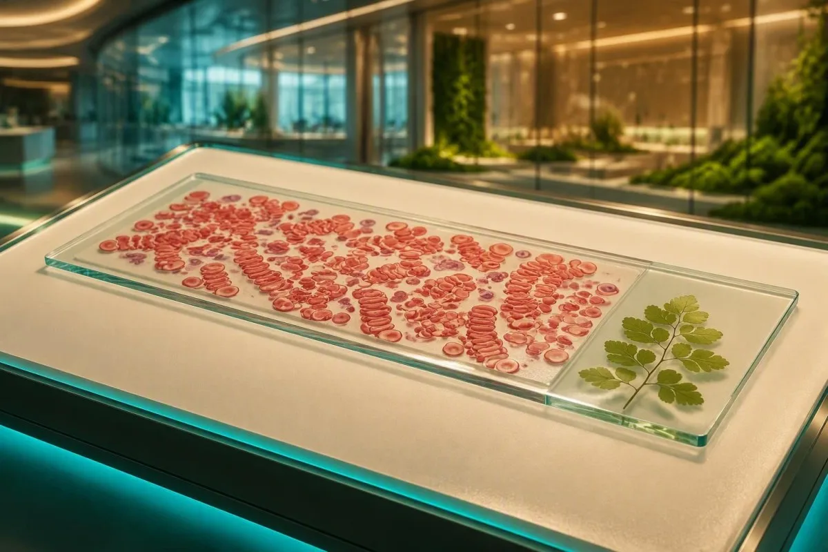
ප්ලාස්මා ප්රෝටීන වල සාමාන්ය විකර්ෂණය වෙනස් වන විට රතු රුධිර සෛල කාසි මෙන් ගොඩ ගැසිය හැකිය. මෙම රටාව බොහෝ විට ප්රතික්රියාශීලී වේ, නමුත් ඒ සමඟ එන CBC සහ ප්රෝටීන් ප්රතිඵල එයට කඩිනම් අනුගාමී වීමක් අවශ්යද යන්න තීරණය කරයි.


- රූලොක්ස් සෑදීම යනු රතු සෛල කාසි වැනි ගොඩවල් සෑදීමයි, සාමාන්යයෙන් ෆයිබ්රිනොජන් හෝ ප්රතිශරීර හෝමෝන ඒවායේ සාමාන්ය මතුපිට විකර්ෂණය අඩු කරන නිසාය.
- එය රෝග විනිශ්චයක් නොව රටාවකි: මෑතකාලීන ආසාදනය, ස්වයං ප්රතිශක්තිකරණ ක්රියාකාරිත්වය, ගැබ් ගැනීම, නිදන්ගත දැවිල්ල සහ ප්ලාස්මා-සෛල ආබාධ සියල්ල එය ඇති කළ හැකිය.
- ESR ඉහළ යා හැක රූලොක්ස් පවතින විට ගොඩ ගැසූ සෛල වේගයෙන් පදිංචි වන බැවින්; ESR පමණක් හේතුව හඳුනාගත නොහැක.
- සම්පූර්ණ ප්රෝටීන් 6.0-8.3 g/dL සහ ඇල්බුමින් දළ වශයෙන් 3.5-5.0 g/dL යනු සාමාන්ය වැඩිහිටි යොමු විරාමයන් වන අතර, රසායනාගාරයේ පරාසයන් වෙනස් වේ.
- 4.0 g/dL ට වඩා වැඩි ගෑමා පරතරයක් ප්රෝටීන්-අවධානය යොමු කරන අනුගාමී වීමක් සාධාරණීකරණය කළ හැකිය, නමුත් විජලනය හා දැවිල්ල ද එය පැහැදිලි කළ හැකිය.
- immunofixation සමඟ SPEP සහ serum free light chains රුධිරයේ රතු රුධිරාණු එකිනෙක මත තැන්පත් වීම (rouleaux) රුධිරයේ සීනි අඩුවීම, වකුගඩු වෙනස්කම්, අස්ථි වේදනාව හෝ ඉහළ ග්ලෝබුලින් සමඟ ඇතිවන පොදු පරීක්ෂණ වේ.
- Saline dispersion රුධිරයේ රතු රුධිරාණු එකිනෙක මත තැන්පත් වීම (rouleaux) රතු රුධිර සෛල සමූහගත වීමෙන් වෙන්කර හඳුනා ගැනීමට රසායනාගාරයකට උපකාරී විය හැකි අතර, එයට වෙනස් රෝග විනිශ්චයක් ඇත.
- හදිසි ඇගයීමක් නව ව්යාකූලත්වය, දැඩි හුස්ම හිරවීම, පෙනීමේ වෙනස්කම්, කැපී පෙනෙන දුර්වලතා හෝ වේගයෙන් නරක අතට හැරෙන වකුගඩු ක්රියාකාරිත්වය සඳහා සුදුසු වේ.
පර්යන්ත ස්පුබ් එකක රූලොක්ස් ඇත්තෙන්ම අදහස් කරන්නේ කුමක්ද?
රුධිර ආලේපනයක රතු රුධිරාණු එකිනෙක මත තැන්පත් වීම (Rouleaux formation on a blood smear) රතු රුධිර සෛල වෙන වෙනම පිහිටීමට වඩා, කෙටි හෝ දිගු කාසි මෙන් තීරු ලෙස පෙළ ගැසී ඇති බව අදහස් වේ. එය බොහෝ විට රතු සෛලවලම ඇති ව්යුහාත්මක දෝෂයකට වඩා, ප්ලාස්මා ප්රෝටීන් - විශේෂයෙන් ෆයිබ්රිනොජන් හෝ ඉමියුනොග්ලොබුලින් - වැඩි වීම පිළිබිඹු කරයි.
රතු රුධිර සෛල පටලය සාමාන්යයෙන් සෘණ විද්යුත් ආරෝපණයක් දරයි, එයට සෙල්ටා විභවය (zeta potential) ලෙසද කියනු ලැබේ, එය අසල්වැසි සෛල වෙන්ව තබා ගනී. ෆයිබ්රිනොජන් සහ සමහර ඉමියුනොග්ලොබුලින් එම විකර්ෂක අවකාශය අර්ධ වශයෙන් සම්බන්ධ කරයි; ඉන්පසු සෛල හොඳින් සකස් කරන ලද ස්ලයිඩයක සිහින් ප්රදේශයේ පහසුවෙන්ම පෙළ ගැසෙයි.
රූප විද්යා අදහස් යනු මනින ලද සාන්ද්රණයක් නොවන අතර විශ්වීය ශ්රේණිගත කිරීමේ පරිමාණයක් නොමැත. එක් රසායනාගාරයක් “අද්ධික rouleaux” වාර්තා කළ හැකි අතර, අනෙක “පවතී” වාර්තා කළ හැකිය; මම රක්තහීමොග්ලොබින්, MCV, මුළු ප්රෝටීන්, ඇල්බියුමින්, ක්රියේටිනින්, කැල්සියම් සහ සායනික කතාව අසල ඇති සැබෑ ස්ලයිඩ් අදහස් කියවමි.
කන්ටෙස්ටි යනු AI රුධිර පරීක්ෂණ විශ්ලේෂකය රතු රුධිර සෛල ස්ථරීකරණය හුදකලා රෝග විනිශ්චයක් ලෙස සැලකීමට වඩා, ස්මියර් අදහසක් අදාළ සංඛ්යාත්මක ප්රතිඵල අසල තබයි. ගණන් කිරීමේ ඉතිරි කොටස පිළිබඳ ප්රයෝජනවත් පුනරීක්ෂණයක් සඳහා, අපගේ බලන්න CBC මාර්කර් මාර්ගෝපදේශය.
“Formation” යන වචනය එයට වඩා භයානක ලෙස ඇසෙන්නේ ඇයිද යන්න
Rouleaux යනු ශරීරය තුළ රුධිර කැටියක් සෑදෙන බව අදහස් නොවේ. එය රසායනාගාර නියැදියක් වීදුරුව මත විහිදුණු විට දැකිය හැකි අන්වීක්ෂණ සැකැස්මකි, රුධිර පිළිකාවක් හෝ භයානක සංසරණ ගැටලුවක් නොමැතිව කෙටි කාලීන දැවිල්ලක් ඇති බොහෝ අය එය පෙන්වයි.
රතු රුධිර සෛල ගොඩ ගැසෙන්නේ ඇයි?
රතු රුධිර සෛල ස්ථරීකරණය සිදුවන්නේ සංසරණ ප්රෝටීන රතු සෛල අතර ඇති විද්යුත්ස්ථිතික දුර අඩු කරන විටය. ෆයිබ්රිනොජන් යනු පොදු කෙටි කාලීන ධාවකය වන අතර, සොයා ගැනීමක් ස්ථීර හෝ උග්ර රෝගයකට අනුපාතික නොවන විට අතිරික්ත ඉමියුනොග්ලොබුලින් වඩාත් අදාළ වේ.
ආසාදනය, ශල්යකර්මය, දැවිල්ල සහිත ආතරයිටිස් හෝ ප්රධාන පටක හානියක දින කිහිපයක් ඇතුළත ෆයිබ්රිනොජන් වැඩි කළ හැක. මෙම ජීව විද්යාව ESR වැඩි කිරීමට සහ CBC දර්ශක සාමාන්යව පවතින විට පවා ආලේපනය කැපී පෙනෙන ලෙස පෙනී යාමට හේතු විය හැක; ESR වෙනස්කම් සෙමින් සිදු වේ ප්රේරකය විසඳීමට පටන් ගැනීමෙන් පසුව.
ඉමියුනොග්ලොබුලින් විශාල වන අතර, ඉහළ සාන්ද්රණයකදී හෝ මොනොක්ලෝනල් ප්රෝටීනයක් ලෙස විශේෂයෙන් කැපී පෙනෙන ස්ථරීකරණයක් ඇති කළ හැකිය. Kyle et al. වාර්තා කළේ, වයස අවුරුදු 50 ත් ඊට වැඩි පුද්ගලයින්ගෙන් 3.2% ට මොනොක්ලෝනල් ගැමොපති, නොතේරුම් වැදගත්කමක් ඇති බව, ජනගහන අධ්යයනයකදී (Kyle et al., 2006) දක්වා ඇති බවයි, MGUS ඇති බොහෝ පුද්ගලයන්ට මයිලෝමා වර්ධනය නොවේ.
පුහුණුව, ෆයිබ්රිනොජන්-ධාවනය වන දැවිල්ල ඉමියුනොග්ලොබුලින්-ධාවනය වන ක්රියාවලියකින් වෙන්කර හඳුනාගත නොහැක. ඉන්ෆ්ලුවෙන්සා, CRP 96 mg/L, සහ සාමාන්ය ප්රෝටීන් අගයන් සහිත අවුරුදු 34 ක් වයසැති අයෙකුට, රක්තහීනතාවය, මුළු ප්රෝටීන් 9.1 g/dL, සහ පහත වැටෙන eGFR සහිත අවුරුදු 71 ක් වයසැති අයෙකුට වඩා ඉතා වෙනස් සැලැස්මක් අවශ්ය වේ.
පොදු ප්රතික්රියාශීලී හේතු: දැවිල්ල, ආසාදනය සහ ගැබ් ගැනීම
රතු රුධිරාණු එකිනෙක මත තැන්පත් වීම (rouleaux formation) වල වඩාත් පොදු හේතු වන්නේ ෆයිබ්රිනොජන් හෝ බහු අවයවික ප්රතිදේහ ඉහළ නංවන තාවකාලික දැවිල්ල තත්වයන්ය. ඉතිරි පැනලය තහවුරු කරන විට ශ්වසන ආසාදන, ස්වයං ප්රතිශක්තිකරණ ඉදිමීම්, නිදන්ගත දැවිල්ල සහිත රෝග සහ ගැබ් ගැනීම නිතරම පැහැදිලි වේ.
C-reactive protein ඉක්මනින් ඉහළ යන අතර, ෆයිබ්රිනොජන් සහ ESR දිගටම ඉහළ මට්ටම්වල පැවතිය හැකිය; මෑතකදී උණ හෝ ඉදිමීමක් ඇති වූ පසු එම කාල වෙනස ප්රයෝජනවත් වේ. Gabay සහ Kushner CRP විස්තර කළේ වේගයෙන් ප්රතිචාර දක්වන තීව්ර-අවධි ප්රෝටීනයක් ලෙස වන අතර, ෆයිබ්රිනොජන් මන්දගාමීව හැසිරෙන අතර අවසාදනයට බලපායි (Gabay and Kushner, 1999).
ගැබ් ගැනීම ෆයිබ්රිනොජන් සැලකිය යුතු ලෙස වැඩි කළ හැක, බොහෝ විට ප්රමාද වූ ගැබ් ගැනීම් වන විට 4-6 g/L පරාසයට, එබැවින් rouleaux සහ ඉහළ ESR ගැබ් නොගත් අපේක්ෂාවන් භාවිතා නොකර අර්ථකථනය නොකෙරේ. ගැබිනි රෝගියෙකු තුළ, ස්මියර් අදහසක් උග්ර කිරීමට පෙර, රෝග ලක්ෂණ, රුධිර පීඩනය, මුත්රා ප්රෝටීන්, හීමොග්ලොබින් ප්රවණතාව සහ ප්රසව සාධකය මම සොයා බලමි.
අපගේ විස්තරාත්මක සෙරුම් ප්රෝටීන මාර්ගෝපදේශය ඇල්බියුමින් සහ ග්ලෝබුලින් රටා මුළු ප්රෝටීනයටම වඩා වැදගත් වන්නේ ඇයිද යන්න පැහැදිලි කරයි. මෙය මෑතකදී සෙම්ප්රතිශ්යාව වැළඳීම හුදකලා වූ කොඩියකට වඩා තොරතුරු සපයන ප්රතිඵල වලින් එකකි.
ඉහළ ප්රෝටීන වල සම්භාවිතා පැහැදිලි කිරීම
ඉහළ ඉමියුනොග්ලොබුලින්, ඉහළ ෆයිබ්රිනොජන්, හෝ ග්ලෝබුලින් වලට සාපේක්ෂව අඩු ඇල්බුමින් රුධිරයේ රතු රුධිරාණු එකිනෙක මත තැන්පත් වීම (rouleaux) සඳහා ප්රෝටීන් සම්බන්ධ පැහැදිලි කිරීමකට සහාය විය හැකිය. රසායනාගාරයේ ඉහළ සීමාවට වඩා මුළු ප්රෝටීන් ප්රතිඵලය දර්ශකයක් වේ, නමුත් එය වගකිව යුතු ප්රෝටීනය හඳුනා ගත නොහැක.
Common adult reference intervals are approximately 6.0-8.3 g/dL for total protein සහ 3.5-5.0 g/dL for albumin, but the report’s own interval prevails. The calculated globulin value equals total protein minus albumin; an albumin 3.4 g/dL with total protein 8.6 g/dL gives globulins of 5.2 g/dL.
A gamma gap—total protein minus albumin—above 4.0 g/dL is often used as a pragmatic trigger to review chronic infection, autoimmune disease, liver disease, and gammopathy. It is not a diagnosis and is less specific in dehydration, where both albumin and globulins may concentrate; our high total protein guide එම වෙනස පැහැදිලි කරයි.
කන්ටෙස්ටි යනු AI රුධිර පරීක්ෂණ ප්රතිඵල වේදිකාව that checks whether an elevated globulin estimate is paired with low albumin, anaemia, renal change, or calcium elevation. That combination carries more clinical weight than rouleaux in a person with otherwise normal values.
රූලොක්ස් බොහෝ විට ඉහළ ESR සමඟ යන්නේ ඇයි?
Rouleaux can raise the erythrocyte sedimentation rate because aggregated red cells fall through a vertical tube faster than individual cells. ESR is therefore an indirect measure of altered plasma proteins, not a direct test for one specific disease.
Many laboratories use upper limits near 15 mm/hour for younger adult men සහ 20 mm/hour for younger adult women, but age, pregnancy, anaemia, and method alter those figures. An ESR above 100 mm/hour warrants timely clinical evaluation, yet it can occur with infection, autoimmune disease, kidney disease, and monoclonal proteins rather than one single condition.
Anaemia itself can raise ESR because there are fewer red cells interfering with descent. That is why an ESR of 55 mm/hour with haemoglobin 8.7 g/dL cannot be read the same way as 55 mm/hour with haemoglobin 14.2 g/dL; compare haematocrit and haemoglobin සමඟ ප්රතිඵලය සංසන්දනය කරන්න.
CRP is usually the better marker for short-term change because it can fall substantially within 24-48 hours of effective treatment. ESR may remain high for weeks after symptoms improve, particularly when rouleaux persists.
මොනොක්ලෝනල් ප්රෝටීනයක් විය හැකි විට අනුගාමී පරීක්ෂණ
Clinicians commonly order serum protein electrophoresis when rouleaux accompanies unexplained anaemia, high total protein, kidney dysfunction, high calcium, recurrent infections, or bone pain. SPEP separates major serum protein fractions and can reveal a narrow monoclonal band, often called an M-spike.
An M-spike is not synonymous with multiple myeloma. The International Myeloma Working Group defines symptomatic myeloma using clonal marrow cells or a plasmacytoma plus a myeloma-defining event, such as calcium above 11 mg/dL, creatinine above 2 mg/dL, haemoglobin more than 2 g/dL below normal, or bone lesions (Rajkumar et al., 2014).
If SPEP shows a possible monoclonal component, immunofixation identifies the immunoglobulin type and serum free light chains assess light-chain imbalance. Our SPEP explanation is useful preparation for a clinician conversation, but no online tool can replace haematology review of a confirmed band.
I have seen patients frightened by the word “spike” after a severe infection, only to have repeat testing show a broad polyclonal response. Conversely, a small stable band in an otherwise well person may require scheduled monitoring rather than emergency treatment.
නිදහස් ආලෝක දාම සහ ප්රතිශරීර හෝමෝන පින්තූරය පරිපූර්ණ කරන්නේ කෙසේද?
Serum free light-chain testing and quantitative IgG, IgA, and IgM help determine whether protein excess is monoclonal or polyclonal. The reference interval for the kappa-to-lambda free light-chain ratio is commonly about 0.26-1.65, though kidney function and assay method can widen interpretation.
Chronic kidney disease increases both kappa and lambda light chains because clearance falls, so a mildly abnormal ratio requires renal context. A markedly skewed ratio, especially with an M-spike or unexplained anaemia, deserves haematology-led evaluation; see our free light chain ratio guide.
Quantitative immunoglobulins answer a different question from SPEP. Broad elevation of IgG, IgA, or IgM may occur with chronic liver disease, autoimmune conditions, or infection, while suppression of unaffected immunoglobulins can be a more concerning clue in a monoclonal process.
කන්ටෙස්ටි යනු AI මගින් බලගැන්වූ රුධිර පරීක්ෂණ විශ්ලේෂණ මෙවලම used across 127+ countries to connect immunoglobulin, renal, calcium, and CBC results in one report. We deliberately label these patterns as follow-up signals, not diagnoses, because the clinical context changes the meaning.
අවධානයේ මට්ටම වෙනස් කරන CBC ඉඟි
Rouleaux is more concerning when it appears with unexplained anaemia, high RDW, low platelets, or a rising creatinine than when the CBC is stable. A normal smear comment does not exclude illness, and rouleaux by itself does not diagnose marrow disease.
For adults, haemoglobin below පුරුෂයන් තුළ 13.0 g/dL හෝ ගර්භණී නොවන කාන්තාවන් තුළ 12.0 g/dL ට අඩු meets a commonly used anaemia definition, but altitude, pregnancy, and laboratory standards matter. New anaemia with rouleaux should prompt a review of MCV, reticulocytes, ferritin, B12, kidney function, and protein studies rather than an assumption that iron deficiency is the answer.
RDW reflects variation in cell size and is often reported near 11.5-14.5% as a reference interval. A high RDW may appear after bleeding, iron treatment, mixed deficiencies, or marrow stress; our සීමා මායිමේ MCV මාර්ගෝපදේශය shows why MCV needs the full pattern.
Dr. Thomas Klein’s practical rule is simple: ask whether the change is new. A CBC that has been stable for 4 years is usually more reassuring than a single normal value that has fallen sharply since the previous draw.
ස්වයං ප්රතිශක්තිකරණය, අක්මාව සහ නිදන්ගත දැවිල්ල පිළිබඳ සන්දර්භයන්
Autoimmune disease, chronic liver disease, and long-standing infections can cause polyclonal immunoglobulin increases that produce rouleaux. These conditions usually create broad protein changes rather than one discrete M-spike, although exceptions occur.
In suspected lupus or immune-complex disease, ANA is a screening test rather than proof of diagnosis; complement C3 and C4 may fall during active immune-complex consumption. The pattern is more persuasive when paired with rash, arthritis, urine protein, cytopenias, or kidney changes than when ANA alone is positive; review our ANA result guide.
Liver disease can lower albumin while increasing immunoglobulins, making the albumin-to-globulin ratio fall below the typical 1.0-2.5 range. A low ratio does not identify the cause, so clinicians often add ALT, AST, ALP, bilirubin, hepatitis testing, and protein electrophoresis according to symptoms.
For clinically focused reading, our C3, C4 and ANA research guide explains what complement results can and cannot settle. The evidence is honestly mixed on broad screening in people without suggestive symptoms.
රූලොක්ස් වලදී යකඩ අධ්යයනයන් ඇයි වැරදි විය හැක්කේ?
Inflammation that produces rouleaux can also raise ferritin and lower serum iron or transferrin, masking iron deficiency. Ferritin is both an iron-storage marker and an acute-phase reactant, so it must be interpreted with CRP, transferrin saturation, and the CBC.
ferritin අගය අඩු වන්නේ නම් 15 ng/mL is highly specific for depleted iron stores in many adults, while values below 30 ng/mL often support iron deficiency in clinical practice. During active inflammation, ferritin can be normal or high despite iron-restricted erythropoiesis, especially if CRP is elevated.
Transferrin saturation පහළින් තිබේ නම් 20% can support iron restriction, but it does not separate absolute iron deficiency from inflammatory iron sequestration by itself. Soluble transferrin receptor may help in selected cases because it is less affected by inflammation; our ferritin සහ CRP මඟ පෙන්වයි walks through the common mismatch.
Do not start high-dose iron solely because a smear reports stacking. In my experience, patients do best when treatment follows a documented deficiency and a reason for it—heavy menstrual loss, gastrointestinal loss, diet, malabsorption, or increased demand.
රූලොක්ස් එදිරිව ඇග්ලූටිනේෂන් සහ ස්ලයිඩ් ආර්ටිෆැක්ට්
Rouleaux is usually reversible with saline on a properly prepared slide, while red-cell agglutination from cold-reactive antibodies generally persists. Distinguishing these appearances matters because agglutination can interfere with automated CBC results and suggests a different work-up.
True rouleaux forms orderly linear stacks; agglutination produces more irregular grape-like clumps. Cold agglutinins can create spuriously high MCHC, low red-cell count, and high MCV on an analyser because the machine counts clusters incorrectly—an elevated MCHC pattern deserves laboratory confirmation.
Smear quality matters. A thick area of a poorly spread or slowly dried slide may exaggerate apparent stacking, which is why experienced morphologists assess the monolayer rather than the slide edge alone.
Rouleaux is not the same as schistocytes, spherocytes, or sickled cells. Fragmented cells with low platelets and haemolysis markers are a very different, sometimes urgent finding; our Vodič za schistocite explains that safety distinction.
රූලොක්ස් වාර්තාවක් ලැබීමෙන් පසු ප්රායෝගික අනුගාමී සැලැස්මක්
A sensible follow-up plan starts with confirming the clinical setting, then checking whether inflammation, anaemia, or protein abnormalities are present. For a well person with a recent viral illness and otherwise normal results, repeat testing after recovery is often more informative than an immediate extensive work-up.
First review the CBC, CMP or renal panel, total protein, albumin, calcium, and CRP or ESR. If symptoms have resolved and values are normal, clinicians may repeat CBC and proteins in සති 4-8; there is no single mandatory interval, so earlier retesting is reasonable when abnormalities are progressing.
Second, order SPEP, immunofixation, free light chains, quantitative immunoglobulins, or urine protein testing when the initial pattern suggests excess immunoglobulin. A urine albumin-creatinine ratio above ට අඩු is abnormal and makes renal context more relevant, though it does not specifically explain rouleaux.
Kantesti AI can organise prior results so meaningful changes are easier to spot across time; our අපගේ සායනික වලංගුකරණ ප්රවේශය explains the safeguards and limits of that interpretation. Dr. Thomas Klein recommends bringing the original smear wording, not just a screenshot of highlighted values, to the appointment.
රූලොක්ස් සඳහා සාමාන්ය තක්සේරුවකට වඩා හදිසි තක්සේරුවක් අවශ්ය වන්නේ කවදාද?
Rouleaux itself is rarely an emergency, but certain accompanying symptoms or laboratory combinations require urgent assessment. New confusion, severe breathlessness, chest pain, fainting, major visual change, rapidly declining urine output, or profound weakness should not wait for routine repeat testing.
Seek same-day medical advice for a new haemoglobin below 8 g/dL, කැල්සියම් අගය ඉහළ 12 mg/dL, creatinine rising quickly, or symptoms suggestive of hyperviscosity such as headache with blurred vision and mucosal bleeding. These values do not prove a plasma-cell disorder, but they warrant prompt clinician-led assessment.
Persistent back or rib pain, recurrent bacterial infections, unintentional weight loss, night sweats, neuropathy, or foamy urine also change the threshold for follow-up. The concern comes from the combination of symptoms and objective findings, not from coin stacks on a slide alone.
A reliable interpretation must preserve uncertainty. Kantesti’s AI තාක්ෂණික මාර්ගෝපදේශය describes how our system surfaces escalation patterns while directing urgent symptoms to in-person care rather than offering false reassurance.
ඔබේ අනුගමන හමුවීමට ගෙන ආ යුත්තේ කුමක්ද?
Bring the complete laboratory report, previous CBC and protein results, a medication list, and a brief timeline of infections or inflammatory symptoms. These details often determine whether rouleaux is a transient reactive finding or a reason for targeted protein testing.
Write down whether you had fever, dental work, surgery, vaccination, joint swelling, rash, weight change, recurrent infections, or reduced urine output during the preceding සති 2-8ක් තුළ. Also list supplements, especially iron, B12, biotin, and immune products, because they can complicate interpretation of related tests.
Ask three focused questions: Is my CBC changing? Are total protein and albumin abnormal? Do I need repeat testing, SPEP, or referral? Most patients find those questions more productive than asking whether the smear finding is “good” or “bad.”
As of September 15, 2026, our වෛද්ය උපදේශක මණ්ඩලය continues to emphasise that AI-guided pattern recognition should support—not replace—clinical examination and laboratory review. Keep a copy of results, dates, and symptoms; trends are often the missing diagnostic clue.
නිතර අසන ප්රශ්න
රුධිර ආලේපනයක රූලොෆෝමේෂන් පිළිකාවක්ද?
රුලේක්ස් සෑදීම පිළිකාවක් නොවන අතර, එය බොහෝ විට දැවිල්ල, ෆයිබ්රිනොජන් වැඩිවීම, හෝ ප්රතිදේහවල පුළුල් ಏರಿಕೆක් සමඟ සම්බන්ධ වේ. එය MGUS හෝ multiple myeloma වැනි මොනොක්ලෝනල් ප්රෝටීන් ආබාධ සමඟ සිදුවිය හැකිය, එබැවින් රුලේක්ස් රක්තහීනතාවය, 8.3 g/dL ට වඩා මුළු ප්රෝටීන්, වකුගඩු ආබාධ, ඉහළ කැල්සියම්, හෝ අස්ථි වේදනාව සමඟ ඇති වූ විට වෛද්යවරු SPEP සලකති. ස්මියර් පෙනුමෙන් පමණක් ප්ලාස්මා-සෛල ආබාධයක් රෝග විනිශ්චය කළ නොහැක. සාමාන්ය ප්රෝටීන් පැනලය සහ මෑතකදී ඇති වූ ආසාදනයක් බොහෝ විට ප්රතික්රියාකාරී පැහැදිලි කිරීමක් වඩාත් පිළිගත හැකි කරයි.
විජලනය රූලොක්ස් සෑදීමට හේතු විය හැකිද?
විජලනය සෙරුම් ප්රෝටීන් සංකේන්ද්රණය කළ හැකි අතර රූලොක්ස් වඩාත් කැපී පෙනෙන ලෙස කළ හැකිය, විශේෂයෙන් මුළු ප්රෝටීන් සහ ඇල්බුමින් එකට ඉහළ ගියහොත්. සලකුණු කරන ලද අඛණ්ඩ රූලොක්ස් සඳහා වන ප්රබලම පැහැදිලි කිරීම් වලින් එකක් නොවේ, මන්ද ෆයිබ්රිනොජන් සහ ඉමියුනොග්ලොබුලින් වෙනස්කම් රතු රුධිර සෛල පරතරය සෘජුවම වෙනස් කරන බැවිනි. සාමාන්ය සජලනයෙන් පසු CBC සහ ප්රෝටීන් පැනලයක් නැවත නැවත කිරීම මායිම් රටාවක් පැහැදිලි කළ හැකිය. 8.3 g/dL ට වඩා වැඩි මුළු ප්රෝටීනයක් සජලනයෙන් පසු ඉහළ මට්ටම්වල පැවතීම වෛද්යවරයෙකුගේ සමාලෝචනයට සුදුසුය.
Rouleaux යන්නෙන් මගේ ESR ඉහළ යනුද?
රුධිර කැටිති සෑදීම නිසා, එකිනෙක ගෑවී ඇති රතු රුධිරාණු, තැන්පත් කිරීමේ නළය තුළ තනි තනි රුධිරාණු වලට වඩා වේගයෙන් පදිංචි වන බැවින්, ESR ඉහළ යාමට හේතුවේ. රක්තහීනතාවය, ගැබ් ගැනීම, වයස, වකුගඩු රෝග, ආසාදනය සහ ස්වයං ප්රතිශක්තිකරණ ක්රියාකාරිත්වය නිසා ද ESR ඉහළ යා හැකිය. එබැවින්, රුධිර කැටිති සෑදීම ඉහළ ප්රතිඵලයක් සහතික නොකරයි. සාමාන්යයෙන් පැයකට මිලිමීටර් 100 ට වැඩි ESR, මූලික හේතුවක් සඳහා කාලෝචිත ඇගයීමක් ඉල්ලා සිටී. උග්ර දැවිල්ල වැඩි දියුණු වන විට CRP, ESR ට වඩා වේගයෙන් වෙනස් වේ.
රුදැල්ලක් ඇතිවීමෙන් පසු කළ යුතු පරීක්ෂණ මොනවාද?
සාමාන්ය අනුගාමී පරීක්ෂණ අතර දර්ශක සමඟ CBC, මුළු ප්රෝටීන්, ඇල්බුමින්, ග්ලෝබුලින් ගණනය කිරීම, CRP හෝ ESR, ක්රියේටිනින්, කැල්සියම් සහ අක්මා පරීක්ෂණ ඇතුළත් වේ. ප්රෝටීන් අසාමාන්යතා හෝ සැලකිල්ලට ලක්වන රෝග ලක්ෂණ පවතී නම්, වෛද්යවරු SPEP, immunofixation, quantitative immunoglobulins, සහ serum free light chains එකතු කළ හැකිය; සාමාන්ය free light-chain අනුපාතය ආසන්න වශයෙන් 0.26-1.65 වේ. රක්තහීනතාවය, වකුගඩු ආබාධ, ඉහළ කැල්සියම්, ආසාදනය, හෝ ස්වයංක්රීය රෝග ලක්ෂණ තිබේද යන්න මත නිවැරදි කට්ටලය රඳා පවතී. මුල් සාම්පල තාක්ෂණික වශයෙන් සීමිත නම් නැවත ආලේප කිරීම ප්රයෝජනවත් විය හැකිය.
රුලොව සීතල එකතුවෙන් වෙනස් වන්නේ කෙසේද?
රූලො ෆෝම්ස් (Rouleaux forms) පිළිවෙලින් කාසියක් වැනි දම්වැල් සාදයි. මෙය සාමාන්යයෙන් ස්ලයිඩයේ සේලයින් (saline) එකතු කළ විට විසිරී යයි. කෝල්ඩ් ඇග්ලූටිනේෂන් (Cold agglutination) මඟින් අක්රමවත් පොකුරු ඇතිවේ. මේවා සේලයින් (saline) සමඟ ද පැවතිය හැකි අතර, වැරදි CBC අගයන්, උදාහරණයක් ලෙස, උසස් MCHC හෝ MCV, ඇති කිරීමට හේතු විය හැක. සීතලට ප්රතික්රියා කරන ප්රතිදේහ (antibodies) සැක කරන විට, සාම්පල රත් කර විශ්ලේෂණය නැවත ධාවනය කිරීම අවශ්ය විය හැකිය. රෝගියාගේ රෝග ලක්ෂණ පමණක් බැලීමෙන් නොව, රසායනාගාරයේ රූප විද්යාව (morphology) සහ පරීක්ෂණ මඟින් මෙය වෙන් කර හඳුනා ගනී.
රුධිර කැටිති ඇතිවීමෙන් පසු මම නැවත රුධිර පරීක්ෂණයක් කළ යුතුද?
රුධිරයේ රතු රුධිරාණු එකට ගොඩ ගැසීම (rouleaux) පැහැදිලි, සුවවන ආසාදනයක් අතරතුරේදී පෙනී සිටින විට සහ CBC, වකුගඩු, කැල්සියම්, ඇල්බුමින් සහ ප්රෝටීන් ප්රතිඵල සාමාන්ය නම්, සති 4-8 ක් පමණ සාමාන්යයෙන් නැවත පරීක්ෂා කිරීම සාධාරණ ය. හිමොග්ලොබින් පහත වැටෙන්නේ නම්, මුළු ප්රෝටීන් ඉහළ ගියහොත්, ක්රියේටිනින් වෙනස් වුවහොත්, හෝ අස්ථි වේදනාව, හුස්ම හිරවීම, හෝ ව්යාකූලත්වය වැනි රෝග ලක්ෂණ ඇති වුවහොත් කලින් පරීක්ෂා කිරීම සුදුසු ය. පවතින රුධිරයේ රතු රුධිරාණු එකට ගොඩ ගැසීම (rouleaux) නොසලකා හැරීමට වඩා සන්දර්භය තුළ තක්සේරු කළ යුතුය. මුල් තීව්රතාවය සහ ඔබේ වෛද්ය ඉතිහාසය මත පදනම්ව ඔබේ වෛද්යවරයාට කෙටි කාල පරතරයක් තෝරා ගත හැකිය.
අදම AI බලයෙන් රුධිර පරීක්ෂණ විශ්ලේෂණය ලබාගන්න
තත්පර කිහිපයකින් ක්ෂණික හා නිවැරදි රසායනාගාර පරීක්ෂණ විශ්ලේෂණය සඳහා Kantesti විශ්වාස කරන ලොව පුරා මිලියන 2කට වැඩි පරිශීලකයන්ට එක්වන්න. ඔබගේ රුධිර පරීක්ෂණ ප්රතිඵල උඩුගත කර, තත්පර කිහිපයකින් 15,000+ ජෛව සලකුණු පිළිබඳ සවිස්තර අර්ථකථනය ලබාගන්න.
📚 යොමු කර ඇති පර්යේෂණ ප්රකාශන
ක්ලයින්, ටී., මිචෙල්, එස්., සහ වෙබර්, එච්. (2026). සෙරුම් ප්රෝටීන් මාර්ගෝපදේශය: ග්ලෝබියුලින්, ඇල්බියුමින් සහ A/G අනුපාත රුධිර පරීක්ෂාව. Kantesti AI වෛද්ය පර්යේෂණ.
ක්ලයින්, ටී., මිචෙල්, එස්., සහ වෙබර්, එච්. (2026). C3 C4 අනුපූරක රුධිර පරීක්ෂණය සහ ANA ටයිටර් මාර්ගෝපදේශය. Kantesti AI වෛද්ය පර්යේෂණ.
📖 බාහිර වෛද්ය යොමු
⚕️ වෛද්ය වියාචනය
මෙම ලිපිය අධ්යාපනික අරමුණු සඳහා පමණක් වන අතර වෛද්ය උපදෙස් ලෙස නොසැලකේ. රෝග නිદાન සහ ප්රතිකාර තීරණ සඳහා සෑම විටම සුදුසුකම් ලත් සෞඛ්ය සේවා සපයන්නෙකුගෙන් උපදෙස් ලබාගන්න.
E-E-A-T විශ්වාස සංඥා
අත්දැකීම්
වෛද්යවරයා විසින් මෙහෙයවන ලද රසායනාගාර අර්ථකථන ක්රියාවලි පිළිබඳ සමාලෝචනය.
ප්රවීණතාව
සායනික සන්දර්භය තුළ ජෛව සලකුණු (biomarkers) හැසිරෙන ආකාරය පිළිබඳ රසායනාගාර වෛද්ය විද්යා අවධානය.
අධිකාරීත්වය
ආචාර්ය තෝමස් ක්ලයින් විසින් ලියන ලද අතර ආචාර්ය සාරා මිචෙල් සහ මහාචාර්ය ආචාර්ය හෑන්ස් වෙබර් විසින් සමාලෝචනය කරන ලදී.
විශ්වසනීයත්වය
අනතුරු ඇඟවීම් අඩු කිරීමට පැහැදිලි පසුකැඳවීම් මාර්ග සහිත සාක්ෂි-පාදක අර්ථකථනය.