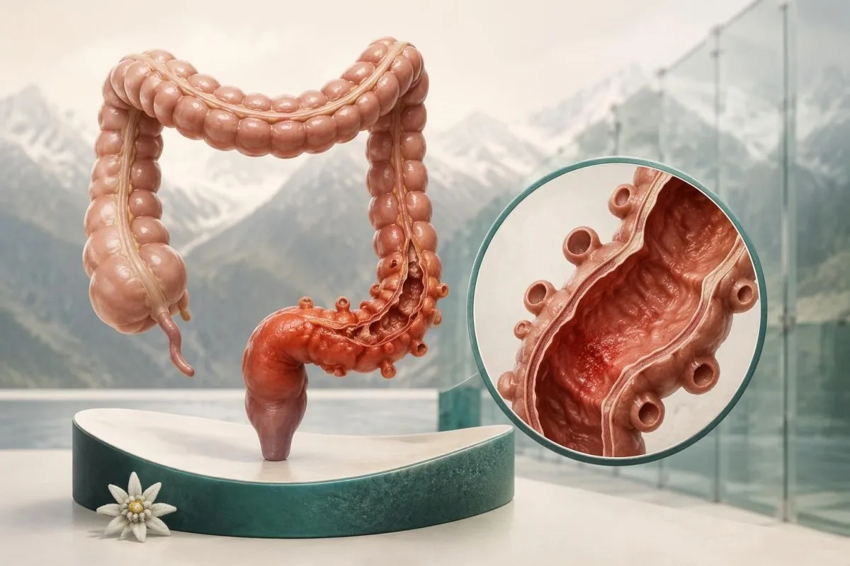
ಜ್ವರ ಅಥವಾ ಮಲವಿಸರ್ಜನೆಯಲ್ಲಿನ ಬದಲಾವಣೆಗಳೊಂದಿಗೆ ನಿರಂತರ ಎಡ-ಕೆಳ ಹೊಟ್ಟೆ ನೋವು ಇದ್ದರೆ, ಅದೇ ದಿನ ವೈದ್ಯಕೀಯ ಸಲಹೆ ಪಡೆಯಬೇಕು. ತೀವ್ರವಾಗಿರುವ ಅಥವಾ ಹರಡುತ್ತಿರುವ ನೋವು, ಪದೇಪದೇ ವಾಂತಿ, ಮೂರ್ಛೆ ಅಥವಾ ಗೊಂದಲ ಇದ್ದರೆ ತುರ್ತು ಚಿಕಿತ್ಸೆಯನ್ನು ಪಡೆಯಿರಿ; ಜ್ವರವಿಲ್ಲದಿದ್ದರೂ ಡೈವರ್ಟಿಕ್ಯುಲೈಟಿಸ್ ಉಂಟಾಗಬಹುದು, ಮತ್ತು ರೋಗಲಕ್ಷಣಗಳು ಅಥವಾ ರಕ್ತ ಪರೀಕ್ಷೆಗಳು ಯಾವುದೂ ಅದನ್ನು ಖಚಿತಪಡಿಸಲಾರವು.

ಪ್ರಮುಖ ಅಂಶಗಳು
- ನಿರಂತರ ಎಡ-ಕೆಳ ನೋವು ಸ್ಪರ್ಶಕ್ಕೆ ನೋವು, ಜ್ವರ ಅಥವಾ ಹೊಸ ಮಲವಿಸರ್ಜನೆಯ ಬದಲಾವಣೆಗಳೊಂದಿಗೆ ಇದ್ದರೆ ಅದೇ ದಿನ ಮೌಲ್ಯಮಾಪನ ಅಗತ್ಯ; ಈ 3 ಲಕ್ಷಣಗಳೂ ಕಾಣಿಸಿಕೊಳ್ಳುವವರೆಗೆ ಕಾಯಬೇಡಿ.
- ತುರ್ತು ಲಕ್ಷಣಗಳು ತೀವ್ರವಾಗಿರುವ ಅಥವಾ ಹರಡುತ್ತಿರುವ ನೋವು, ಗೊಂದಲ, ಮೂರ್ಛೆ, ಅಥವಾ ವಾಂತಿಯೊಂದಿಗೆ ಹೊಟ್ಟೆ ಉಬ್ಬರ ಮತ್ತು ಗ್ಯಾಸ್ ಹೊರಹಾಕಲಾಗದಿರುವುದು ಇವುಗಳಲ್ಲಿವೆ.
- ಜ್ವರ ಸಾಮಾನ್ಯವಾಗಿ 38°C ಅಥವಾ ಅದಕ್ಕಿಂತ ಹೆಚ್ಚಿನ ತಾಪಮಾನ ಎಂದರ್ಥ; ಆದರೆ ಕಡಿಮೆ ತಾಪಮಾನವಿದ್ದರೂ ಡೈವರ್ಟಿಕ್ಯುಲೈಟಿಸ್ ಅಥವಾ ಅದರ ತೊಡಕುಗಳನ್ನು ತಳ್ಳಿಹಾಕಲಾಗುವುದಿಲ್ಲ.
- ರಕ್ತ ಪರೀಕ್ಷೆಗಳು ಉರಿಯೂತ ಮತ್ತು ಅನಾರೋಗ್ಯದ ತೀವ್ರತೆಯನ್ನು ಅಳೆಯುತ್ತವೆ, ಕಾರಣವನ್ನಲ್ಲ; ಸಾಮಾನ್ಯ WBC ಮತ್ತು CRP ಫಲಿತಾಂಶಗಳು ಆರಂಭಿಕ ಡೈವರ್ಟಿಕ್ಯುಲೈಟಿಸ್ ಇಲ್ಲವೆಂದು ವಿಶ್ವಾಸಾರ್ಹವಾಗಿ ತಳ್ಳಿಹಾಕಲಾರವು.
- CT ಇಮೇಜಿಂಗ್ ಡೈವರ್ಟಿಕ್ಯುಲೈಟಿಸ್ ಪತ್ತೆಹಚ್ಚುವಲ್ಲಿ ಸರಿಸುಮಾರು 95% ಸಂವೇದನಾಶೀಲತೆ ಮತ್ತು ನಿರ್ದಿಷ್ಟತೆಯನ್ನು ಹೊಂದಿದ್ದು, ಹುಣ್ಣು, ರಂಧ್ರ ಅಥವಾ ಪರ್ಯಾಯ ರೋಗನಿರ್ಣಯವನ್ನು ಗುರುತಿಸಬಲ್ಲದು.
- ಹೆಚ್ಚಿನ ಅಪಾಯದ ಫಲಿತಾಂಶಗಳು ಉದಾಹರಣೆಗೆ CRP 140 mg/L ಗಿಂತ ಹೆಚ್ಚು ಅಥವಾ WBC 15 × 10⁹/L ಗಿಂತ ಹೆಚ್ಚು ಇರುವುದು ಚಿಕಿತ್ಸೆಯ ನಿರ್ಧಾರಗಳ ಮೇಲೆ ಪ್ರಭಾವ ಬೀರುತ್ತದೆ; ಇವು ರೋಗನಿರ್ಣಯದ ಮಿತಿಗಳಲ್ಲ.
- ಆಂಟಿಬಯೋಟಿಕ್ಸ್ ಪ್ರತಿ ಪ್ರಕರಣದಲ್ಲೂ ಇವುಗಳನ್ನು ಸ್ವಯಂಚಾಲಿತವಾಗಿ ನೀಡುವುದಿಲ್ಲ: ತೊಡಕುಗಳಿಲ್ಲದ ಸೌಮ್ಯ ಡೈವರ್ಟಿಕ್ಯುಲೈಟಿಸ್ ಇರುವ, ಆಯ್ದ ರೋಗನಿರೋಧಕ ಸಾಮರ್ಥ್ಯ ಹೊಂದಿರುವ ವಯಸ್ಕರು ವೈದ್ಯಕೀಯ ಮೇಲ್ವಿಚಾರಣೆಯಲ್ಲಿ ಇವುಗಳಿಲ್ಲದೆಯೇ ಚೇತರಿಸಿಕೊಳ್ಳಬಹುದು.
- ಅನುಸರಣಾ ಕೊಲೊನೋಸ್ಕೋಪಿ ತೀವ್ರ ಪ್ರಕರಣದ ನಂತರ 6–8 ವಾರಗಳವರೆಗೆ ಸಾಮಾನ್ಯವಾಗಿ ಮುಂದೂಡಲಾಗುತ್ತದೆ; ಇದರ ಅಗತ್ಯವನ್ನು ಪ್ರಕರಣ, ಹಿಂದಿನ ಪರೀಕ್ಷೆ ಮತ್ತು ಎಚ್ಚರಿಕೆಯ ಲಕ್ಷಣಗಳ ಆಧಾರದ ಮೇಲೆ ನಿರ್ಧರಿಸಲಾಗುತ್ತದೆ.
ಡೈವರ್ಟಿಕ್ಯುಲೈಟಿಸ್ನ ಯಾವ ರೋಗಲಕ್ಷಣಗಳಿಗೆ ಅದೇ ದಿನ ಮೌಲ್ಯಮಾಪನ ಅಗತ್ಯ?
ಹೊಸದಾಗಿ ಕಾಣಿಸಿಕೊಂಡ, ನಿರಂತರವಾದ ಎಡ ಕೆಳ ಹೊಟ್ಟೆನೋವು, ವಿಶೇಷವಾಗಿ ಸ್ಪರ್ಶಿಸಿದಾಗ ನೋವು, ಜ್ವರ ಅಥವಾ ಮಲವಿಸರ್ಜನೆಯ ಅಭ್ಯಾಸದಲ್ಲಿ ಬದಲಾವಣೆಯೊಂದಿಗೆ ಇದ್ದರೆ, ಅದೇ ದಿನ ವೈದ್ಯಕೀಯ ಮೌಲ್ಯಮಾಪನ ಅಗತ್ಯ. ನೋವು ತೀವ್ರವಾದಾಗ ಅಥವಾ ವ್ಯಾಪಕವಾದಾಗ, ಅಥವಾ ಗೊಂದಲ, ಮೂರ್ಛೆ, ಪದೇಪದೇ ವಾಂತಿ ಅಥವಾ ಅನಿಲವನ್ನು ಹೊರಹಾಕಲು ಅಸಮರ್ಥತೆ ತೊಡಕನ್ನು ಸೂಚಿಸಿದಾಗ ತುರ್ತು ಚಿಕಿತ್ಸೆ ಪಡೆಯುವುದು ಸೂಕ್ತ—ತಾಪಮಾನವು 38°C ಗಿಂತ ಕಡಿಮೆಯೇ ಇದ್ದರೂ ಸಹ.
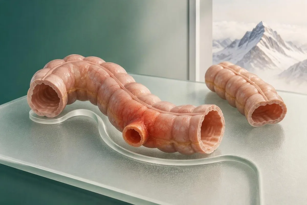
ಡೈವರ್ಟಿಕ್ಯುಲೈಟಿಸ್ ಎಂದರೆ ಡೈವರ್ಟಿಕ್ಯುಲಮ್ನಲ್ಲಿ ಉಂಟಾಗುವ ಉರಿಯೂತ, ಇದು ಕರುಳಿನ ಗೋಡೆಯ ಮೂಲಕ ಹೊರಚಾಚುವ ಸಣ್ಣ ಚೀಲ; ಚೀಲಗಳು ಇರುವುದು ಮಾತ್ರವನ್ನು ಡೈವರ್ಟಿಕ್ಯುಲೋಸಿಸ್ ಎಂದು ಕರೆಯಲಾಗುತ್ತದೆ ಮತ್ತು ಅದು ನೋವಿನ ಕಾರಣವನ್ನು ಸ್ಥಾಪಿಸುವುದಿಲ್ಲ. ಆದ್ದರಿಂದ, ಹಿಂದಿನ ಒಂದು ಸ್ಕ್ಯಾನ್ನಲ್ಲಿ ಡೈವರ್ಟಿಕ್ಯುಲೋಸಿಸ್ ಕಂಡುಬಂದಿರುವುದರಿಂದ, ಹೊಟ್ಟೆಯ ಅಸ್ವಸ್ಥತೆಯ ಹೊಸ ಪ್ರಕರಣವು ಡೈವರ್ಟಿಕ್ಯುಲೈಟಿಸ್ ಎಂದು ಸಾಬೀತುಪಡಿಸಲಾಗುವುದಿಲ್ಲ.
AGA ತಜ್ಞರ ವಿಮರ್ಶೆಯ ಪ್ರಕಾರ, ವೈದ್ಯಕೀಯ ಅನುಮಾನವೊಂದರ ಆಧಾರದ ಮೇಲೆ ಡೈವರ್ಟಿಕ್ಯುಲೈಟಿಸ್ ಅನ್ನು ಸರಿಯಾಗಿ ಗುರುತಿಸುವುದು ಸುಮಾರು ಅನುಮಾನಿತ ಪ್ರಕರಣಗಳಲ್ಲಿ 40–65% ಮಾತ್ರ, (Peery et al., 2021). ಈ ಲಕ್ಷಣಗಳ ಅತಿಕ್ರಮಣ ಮುಖ್ಯ: ಮಲಬದ್ಧತೆ, ಮೂತ್ರದ ಕಾಯಿಲೆಗಳು, ಕೊಲೈಟಿಸ್ ಮತ್ತು ಶ್ರೋಣಿಯ ಭಾಗದ ಸಮಸ್ಯೆಗಳು ಇದೇ ರೀತಿಯ ನೋವನ್ನು ಉಂಟುಮಾಡಬಹುದು; ತೊಡಕು ಕೂಡ ಆರಂಭದಲ್ಲಿ ತೊಡಕುಗಳಿಲ್ಲದ ಪ್ರಕರಣದಂತೆ ಕಾಣಿಸಬಹುದು.
ಉದಾಹರಣೆಗೆ, 18 ಗಂಟೆಗಳಿಂದ ಕ್ರಮೇಣ ಹೆಚ್ಚುತ್ತಿರುವ ಎಡ ಕೆಳ ಹೊಟ್ಟೆನೋವು, ಹಸಿವು ಕಡಿಮೆಯಾಗಿರುವುದು ಮತ್ತು ಮಲಬದ್ಧತೆ ಇರುವ 56 ವರ್ಷದ ವ್ಯಕ್ತಿಗೆ, ತಾಪಮಾನ 37.4°C ಇದ್ದರೂ ಮೌಲ್ಯಮಾಪನ ಅಗತ್ಯ. ನಡೆಯಲು ಸಾಧ್ಯವಾಗಿರುವುದು ಅಥವಾ ಪ್ರಯೋಗಾಲಯ ವರದಿ ಸಾಮಾನ್ಯವಾಗಿ ಕಂಡುಬರುವುದು ಸುರಕ್ಷತೆಯನ್ನು ಸಾಬೀತುಪಡಿಸುವುದಿಲ್ಲ; ಲಕ್ಷಣಗಳ ಬೆಳವಣಿಗೆಯ ಸ್ವರೂಪ ಮತ್ತು ಪರೀಕ್ಷೆಯೂ ಮುಖ್ಯ.
Kantesti ಒಂದು AI ರಕ್ತ ಪರೀಕ್ಷಾ ವಿಶ್ಲೇಷಕ ಇದು ಸಾಮಾನ್ಯ CRP ಫಲಿತಾಂಶವನ್ನು ಡೈವರ್ಟಿಕ್ಯುಲೈಟಿಸ್ ಇಲ್ಲ ಎಂಬುದಕ್ಕೆ ಪುರಾವೆಯೆಂದು ಪರಿಗಣಿಸದೆ, ಪ್ರಯೋಗಾಲಯದ ಫಲಿತಾಂಶಗಳನ್ನು ವಿವರಿಸುತ್ತದೆ. Kantesti LTD ಯ ಸಾಂಸ್ಥಿಕ ಮಾಹಿತಿ ನಮ್ಮ ಶೈಕ್ಷಣಿಕ ಸೇವೆಯನ್ನು ಯಾರು ಒದಗಿಸುತ್ತಾರೆ ಎಂಬುದನ್ನು ಗುರುತಿಸುತ್ತದೆ; ನಿರಂತರ ಹೊಟ್ಟೆನೋವಿನ ಮೊದಲ ಪ್ರಕರಣದ ಕುರಿತ ನಿರ್ಧಾರಗಳನ್ನು ಸ್ವಯಂಚಾಲಿತ ವ್ಯಾಖ್ಯಾನದಿಂದಲ್ಲ, ವೈದ್ಯರಿಂದ ತೆಗೆದುಕೊಳ್ಳಬೇಕು.
ಡೈವರ್ಟಿಕ್ಯುಲೈಟಿಸ್ನ ಆರಂಭಿಕ ಲಕ್ಷಣಗಳು ಯಾವುವು?
ಡೈವರ್ಟಿಕ್ಯುಲೈಟಿಸ್ನ ಆರಂಭಿಕ ಲಕ್ಷಣಗಳು ಸ್ಥಳೀಯ ಹೊಟ್ಟೆನೋವು ನಿರಂತರವಾಗುವುದು, ಹೊಸದಾಗಿ ಸ್ಪರ್ಶಸಂವೇದನೆ ಕಾಣಿಸಿಕೊಳ್ಳುವುದು, ಹಸಿವು ಕಡಿಮೆಯಾಗುವುದು ಮತ್ತು ಮಲಬದ್ಧತೆ ಅಥವಾ ಅತಿಸಾರವನ್ನು ಒಳಗೊಂಡಿರಬಹುದು. ಗಂಟೆಗಂಟೆಗೆ ಅನುಸರಿಸುವ ವಿಶ್ವಾಸಾರ್ಹ ಕ್ರಮವಿಲ್ಲ: ಲಕ್ಷಣಗಳು 1–2 ದಿನಗಳಲ್ಲಿ ಬೆಳೆಯಬಹುದು ಅಥವಾ ಹೆಚ್ಚು ಹಠಾತ್ತನೆ ಕಾಣಿಸಿಕೊಳ್ಳಬಹುದು; ಜ್ವರ ನಂತರ ಕಾಣಿಸಿಕೊಳ್ಳಬಹುದು ಅಥವಾ ಎಂದಿಗೂ ಬಾರದಿರಬಹುದು.
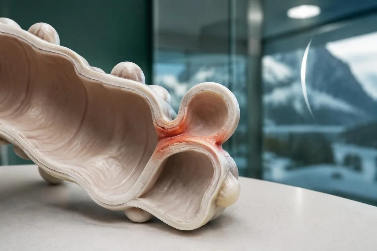
ಸ್ವಲ್ಪ ಹೊತ್ತು ಮಾತ್ರ ಇರುವ ಪ್ರತ್ಯೇಕ ಚುಚ್ಚುವ ನೋವಿಗಿಂತ, ನೋವು ಹೆಚ್ಚಾಗಿ ನಿರಂತರವಾಗುವುದು ಹೆಚ್ಚು ಕಳವಳಕಾರಿ, ಆದರೂ ನೋವಿನ ಸ್ವರೂಪದಿಂದ ಡೈವರ್ಟಿಕ್ಯುಲೈಟಿಸ್ ಅನ್ನು ರೋಗನಿರ್ಣಯ ಮಾಡಲು ಸಾಧ್ಯವಿಲ್ಲ. ಹಿಂದಿನ 24 ಗಂಟೆಗಳಲ್ಲಿ ನಿಮ್ಮ ಸಾಮಾನ್ಯ ಸ್ಥಿತಿಯೊಂದಿಗೆ ಹೋಲಿಸುವುದು ಉಪಯುಕ್ತ: ಸೆಳೆತಗಳ ನಡುವೆಯೂ ಕಡಿಮೆಯಾಗದ ಅಸ್ವಸ್ಥತೆ, ಅಥವಾ ಕುಡಿಯುವುದಕ್ಕೂ ಚಲಿಸುವುದಕ್ಕೂ ಅಡ್ಡಿಯಾಗಲು ಆರಂಭಿಸುವ ಅಸ್ವಸ್ಥತೆ ಕಂಡುಬಂದರೆ, ಇನ್ನೊಂದು ದಿನ ಗಮನಿಸುತ್ತಿರುವುದಕ್ಕಿಂತ ಮರುಮೌಲ್ಯಮಾಪನ ಮಾಡಿಸಿಕೊಳ್ಳಿ.
ಮಲವಿಸರ್ಜನೆಯಲ್ಲಿನ ಹೊಸ ಬದಲಾವಣೆಗಳು ಒಂದು ಪ್ರಕರಣವನ್ನು ವಿವರಿಸಲು ನೆರವಾಗುತ್ತವೆ; ಆದರೆ ಅದರ ಕಾರಣವನ್ನು ಗುರುತಿಸಲು ಅವು ಸಾಕಷ್ಟು ನಿರ್ದಿಷ್ಟವಲ್ಲ. ಮಲವು ಬ್ರಿಸ್ಟಲ್ ಪ್ರಕಾರ 4ರಿಂದ ಪ್ರಕಾರ 1ಕ್ಕೆ ಅಥವಾ ಪ್ರಕಾರ 6ಕ್ಕೆ ಬದಲಾಗಿದೆ ಎಂದು ದಾಖಲಿಸುವುದು, ಕೇವಲ 'ಅಸಹಜ' ಎಂದು ಬರೆಯುವುದಕ್ಕಿಂತ ಹೆಚ್ಚು ಉಪಯುಕ್ತ; ನಮ್ಮ ಮಲದ ಸಾಂದ್ರತೆಯ ಮಾರ್ಗದರ್ಶಿ ಆ ವಿವರಣೆಗಳನ್ನು ರೋಗನಿರ್ಣಯ ಮಾಡದೆ ವಿವರಿಸುತ್ತದೆ.
ಊಹಾತ್ಮಕವಾಗಿ, 44 ವರ್ಷದ ವ್ಯಕ್ತಿಗೆ ಆರಂಭದಲ್ಲಿ 6 ಗಂಟೆಗಳ ಕಾಲ ಹೊಟ್ಟೆ ಉಬ್ಬರವೆನಿಸಬಹುದು; ನಂತರ ರಾತ್ರಿ ಹೊತ್ತಿಗೆ ಎಡ ಕೆಳಭಾಗದಲ್ಲಿ ಒಂದೇ ಸ್ಥಳದಲ್ಲಿ ನೋವು ಮತ್ತು ವಾಕರಿಕೆ ಕಾಣಿಸಿಕೊಳ್ಳಬಹುದು. ಈ ಲಕ್ಷಣಗಳ ಕ್ರಮವು ಡೈವರ್ಟಿಕ್ಯುಲೈಟಿಸ್ಗೆ ಹೊಂದಿಕೆಯಾಗುತ್ತದೆ, ಆದರೆ ಇತರ ಹೊಟ್ಟೆಯ ಕಾಯಿಲೆಗಳಿಗೂ ಹೊಂದಿಕೆಯಾಗುತ್ತದೆ; ಮಲವಿಸರ್ಜನೆಯ ನಂತರ ನೋವು ಕಡಿಮೆಯಾಗುವುದರಿಂದ ಇವೆರಡರ ನಡುವಿನ ವ್ಯತ್ಯಾಸವನ್ನು ವಿಶ್ವಾಸಾರ್ಹವಾಗಿ ನಿರ್ಧರಿಸಲಾಗುವುದಿಲ್ಲ.
ಮನೆಯಲ್ಲಿ ಬಳಸಲು ಮಾನ್ಯತೆ ಪಡೆದ ಯಾವುದೇ ರೋಗಲಕ್ಷಣದ ಅಂಕಮಾಪನ ವಿಧಾನವಿಲ್ಲ. that can confirm an early episode from 2 or 3 symptoms. Before contacting a clinician, note the starting time, whether pain is spreading, your measured temperature and whether you can keep fluids down; these details often change the urgency more than the word 'cramp'.
ಡೈವರ್ಟಿಕ್ಯುಲೈಟಿಸ್ನ ನೋವು ಸಾಮಾನ್ಯವಾಗಿ ಎಲ್ಲಿ ಇರುತ್ತದೆ?
Diverticulitis pain location is usually the lower-left abdomen, because the sigmoid colon is commonly affected in Western populations. Right-sided or central pain is possible, however, and the location of 1 painful area cannot reliably distinguish diverticulitis from appendicitis, a urinary problem or another acute abdominal condition.
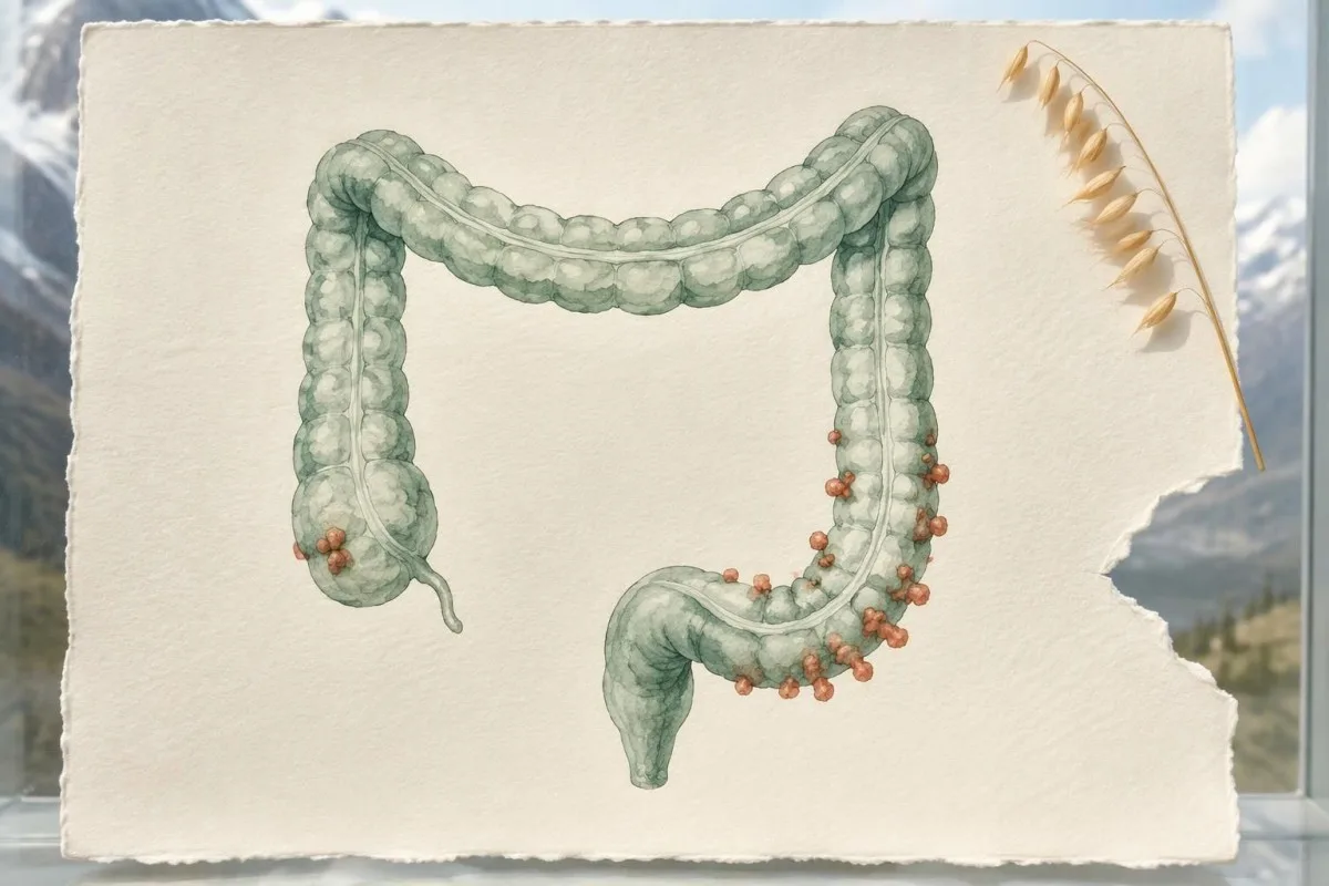
The sigmoid colon has a variable position, so pain may be felt above the pubic area rather than neatly in the left lower quadrant. A pelvic sigmoid segment can also irritate nearby structures; 2 symptoms such as suprapubic discomfort and urinary frequency therefore do not automatically identify a bladder infection.
Right-sided diverticulitis occurs more often in Asian populations than in Western populations, although individual anatomy matters more than nationality. Right-lower abdominal pain still requires assessment for appendicitis; our guide to ಅಪೆಂಡಿಸೈಟಿಸ್ನಲ್ಲಿನ ಸಾಮಾನ್ಯ ಪರೀಕ್ಷೆಗಳು explains why 1 reassuring CBC cannot safely decide between these possibilities.
Pain that spreads from 1 localized area across the abdomen raises concern for a wider process, especially when accompanied by marked tenderness or systemic illness. Clinicians assess guarding and peritoneal irritation themselves; repeatedly pressing hard on the abdomen at home adds discomfort without providing a dependable diagnostic test.
For a hypothetical 63-year-old, pain just left of the midline and a temperature of 38.2°C might suggest sigmoid diverticulitis, but a scan could reveal a different cause. Point with 1 finger to where the pain began, then describe where it is now; the change in distribution is clinically useful.
ಜ್ವರವಿಲ್ಲದೆಯೂ ಡೈವರ್ಟಿಕ್ಯುಲೈಟಿಸ್ನ ರೋಗಲಕ್ಷಣಗಳು ಕಾಣಿಸಿಕೊಳ್ಳಬಹುದೇ?
Diverticulitis symptoms without fever are possible, particularly early in an episode, in older adults or in people taking medicines that alter the immune response. A temperature below 38°C does not exclude diverticulitis, and an absent fever must not override worsening pain, inability to drink or other warning signs.
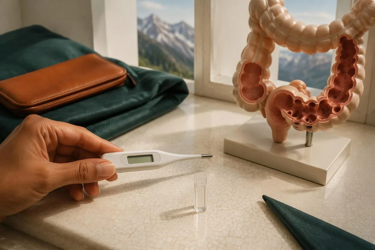
A single temperature reading is a snapshot, not a complete assessment of illness. A hypothetical reading of 36.9°C after paracetamol can coexist with clinically significant abdominal disease; tell the clinician when fever-reducing medicine was taken rather than presenting the number as an untreated baseline.
Older adults may have a less prominent fever response, while corticosteroids can reduce inflammatory symptoms and also change the white-cell count. In a person aged 75 taking prednisolone, 2 reassuring features—no fever and only modest tenderness—should not cancel concern about progressive symptoms; our ವಯಸ್ಸಾದವರಲ್ಲಿ ಔಷಧಿಗಳ ಪರಿಣಾಮಗಳ guide explains related laboratory pitfalls.
Immunosuppression lowers the threshold for imaging and treatment because symptoms can underestimate the severity of diverticulitis. The AGA review recommends a low threshold for CT in these patients (Peery et al., 2021); a transplant recipient with 1 day of new focal abdominal pain should seek prompt advice rather than wait for a temperature rise.
Feeling generally unwell without fever still matters, especially if accompanied by weakness, reduced urine output or a faster heartbeat. If a home thermometer gives an unexpected result, repeat it using the device instructions—but do not spend another 2 hours checking temperatures while severe pain or confusion is developing.
ಯಾವ ಮಲವಿಸರ್ಜನೆಯ ಬದಲಾವಣೆಗಳು ರೋಗನಿರ್ಣಯವನ್ನು ಬೆಂಬಲಿಸುತ್ತವೆ—ಅಥವಾ ಅದನ್ನು ಪ್ರಶ್ನಿಸುತ್ತವೆ?
Constipation and diarrhea can both accompany diverticulitis, and neither bowel pattern confirms the diagnosis. New mucus, urgency or urinary irritation may occur, but frequent watery stools, recent antibiotics or a shared food exposure should prompt clinicians to consider competing explanations, even when pain has lasted only 1 day.
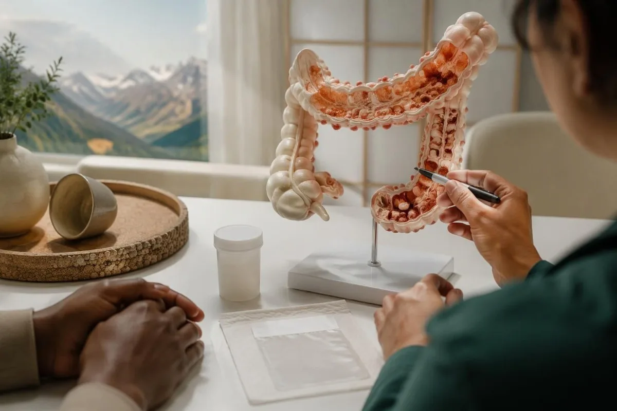
A change from your baseline is more useful than stool frequency alone: 2 bowel movements daily may be normal for one person and a major change for another. Passing a stool does not rule out diverticulitis; conversely, constipation with localized pain should not automatically be treated with a laxative before an acute abdominal problem is assessed.
Diarrhea with fever and food exposure can indicate intestinal infection, not necessarily diverticulitis. For example, a hypothetical household in which 3 people develop diarrhea after the same meal creates a different diagnostic picture; our explanation of Campylobacter stool PCR describes when organism-specific testing may clarify the cause.
Recent antibiotic use changes the workup when there are 24 ಗಂಟೆಗಳಲ್ಲಿ 3 ಅಥವಾ ಅದಕ್ಕಿಂತ ಹೆಚ್ಚು ಆಕಾರವಿಲ್ಲದ ಮಲವಿಸರ್ಜನೆಗಳು, a commonly used threshold for considering C. difficile testing in the right context. Our guide to ಹೊಂದಿಕೆಯಾಗದ C. difficile ಫಲಿತಾಂಶಗಳು explains why even a positive screening component must be interpreted alongside symptoms.
Urinary frequency or discomfort can reflect irritation near the sigmoid colon, but recurrent urinary infections, gas during urination or stool-like material in urine raise concern for a bowel–bladder connection. These are not routine early symptoms; report even 1 such episode promptly, because imaging may be needed to investigate a fistula.
ಯಾವ ಎಚ್ಚರಿಕೆಯ ಲಕ್ಷಣಗಳು ಹುಣ್ಣು, ರಂಧ್ರ ಅಥವಾ ಸೆಪ್ಸಿಸ್ ಇರುವುದನ್ನು ಸೂಚಿಸುತ್ತವೆ?
Severe or spreading abdominal pain, a rigid or markedly swollen abdomen, repeated vomiting, confusion, fainting or reduced urine output require urgent assessment. Swelling with vomiting and inability to pass stool or gas can indicate obstruction; no fever cutoff, including 38°C, is required before seeking emergency care.
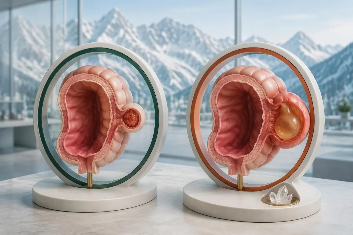
An abscess is a contained collection associated with the affected bowel, while perforation allows material to escape through the bowel wall. Neither can be reliably distinguished by pain intensity or 1 laboratory value; NICE recommends same-day hospital assessment when suspected diverticulitis is accompanied by features suggesting a complication (NICE NG147, 2019).
Sepsis can involve altered mental state, fast breathing, circulatory instability or unexpectedly low urine output, sometimes without a high temperature. Even a procalcitonin below 0.5 ng/mL cannot safely exclude a localized abdominal complication; our ಪ್ರೋಕ್ಯಾಲ್ಸಿಟೋನಿನ್ ಅರ್ಥೈಸುವ ಮಾರ್ಗದರ್ಶಿ explains why a biomarker must not replace assessment of the person.
Large-volume rectal bleeding is not a typical defining feature of diverticulitis, and diverticular bleeding is often painless. Passing blood with dizziness, fainting or weakness requires urgent care for possible significant blood loss, regardless of whether pain is present; our blood in stool guide separates color clues from emergency symptoms.
CT findings—not a home pain score—help determine how a complication is treated. NICE advises considering drainage or surgery for diverticular abscesses larger than 3 ಸೆಂ.ಮೀ when feasible, while smaller collections may be managed differently; do not interpret that size threshold as permission to wait at home with suspected complications.
ವೈದ್ಯರು ಯಾವಾಗ CT ಇಮೇಜಿಂಗ್ ಅನ್ನು ಶಿಫಾರಸು ಮಾಡುತ್ತಾರೆ?
CT is often recommended when diverticulitis has not previously been confirmed, the diagnosis is uncertain, symptoms are severe, complications are suspected or recovery is not progressing. Abdominal and pelvic CT has approximately 95% ಸಂವೇದನಾಶೀಲತೆ ಮತ್ತು ವಿಶೇಷತೆ for diverticulitis, but a scan is not automatically necessary for every familiar, mild recurrence.
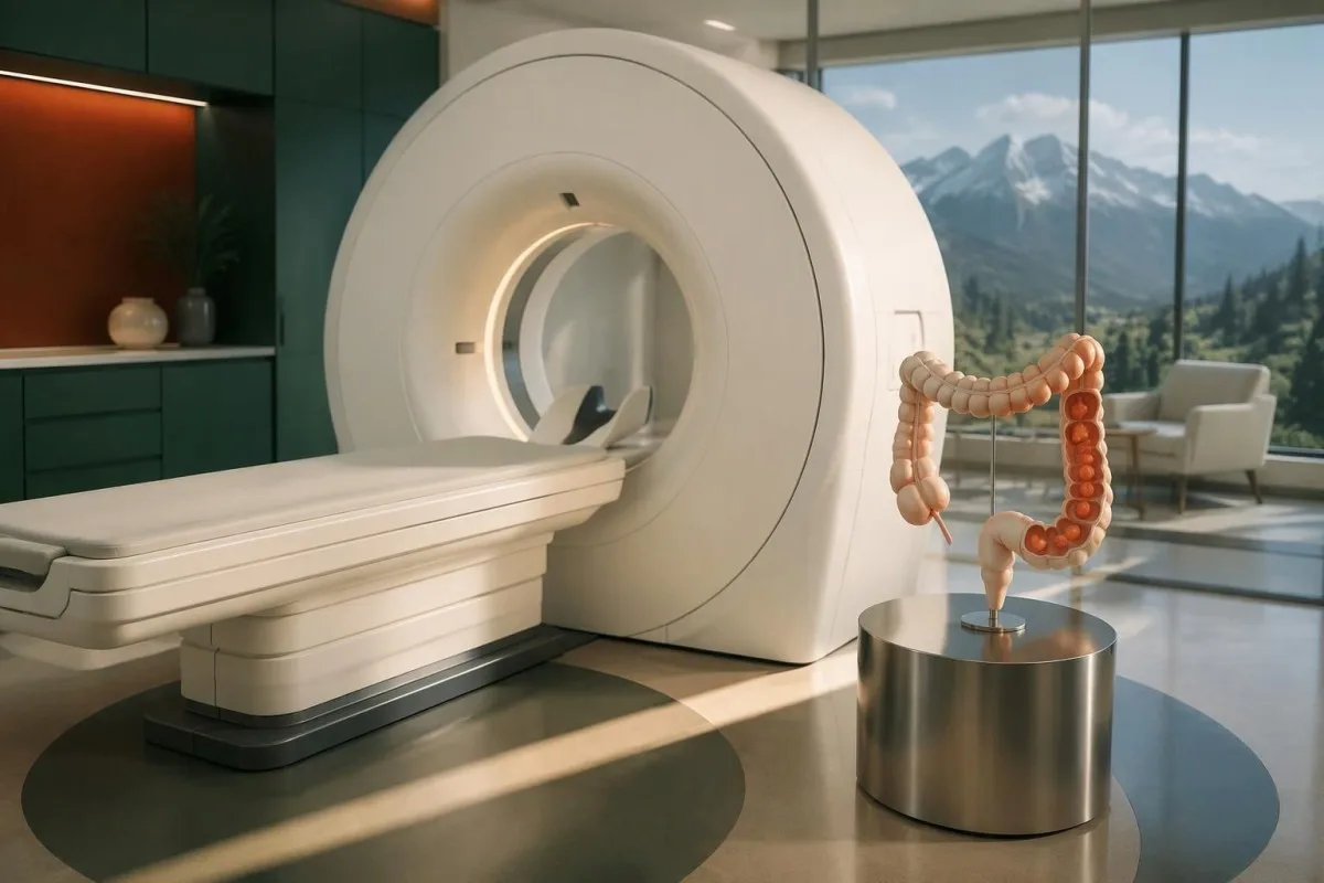
The AGA expert review emphasizes CT for patients without a prior imaging-confirmed diagnosis and for severe presentations or failure to improve (Peery et al., 2021). The ACP guideline recommends CT when there is diagnostic uncertainty (Qaseem et al., 2022); these approaches overlap but do not mean every person with 1 mild episode receives the same imaging pathway.
Suspected complicated diverticulitis is a hospital assessment problem, not an outpatient waiting-list problem. NICE recommends contrast CT within 24 hours of admission when inflammatory markers are raised and a complication is suspected; this timing follows admission and must not be misread as advice to remain at home for another day.
Intravenous contrast can improve evaluation of the bowel and surrounding tissues, while oral contrast use varies by local protocol. Kidney function and acute dehydration inform the risk–benefit discussion; an eGFR below 30 mL/min/1.73 m² usually prompts additional consideration rather than an automatic ban, as our kidney tests after dehydration guide explains.
Possible pregnancy changes imaging planning because ultrasound or MRI may be preferred where clinically suitable, although necessary emergency imaging should not be withheld reflexively. A negative urine pregnancy test taken 2 days before an expected period may be too early to settle pregnancy status; our ಆರಂಭಿಕ ಗರ್ಭಧಾರಣೆಯ ಪರೀಕ್ಷೆಯ ಮಿತಿಗಳು guide explains that uncertainty.
WBC ಮತ್ತು CRP ಫಲಿತಾಂಶಗಳು ನಿಜವಾಗಿ ಏನನ್ನು ತಿಳಿಸಬಲ್ಲವು?
WBC and CRP can support an assessment of inflammation, but neither confirms diverticulitis or reliably excludes it when normal. Many adult laboratories use a WBC interval around 4–11 × 10⁹/L and a CRP upper limit of ಸುಮಾರು; other laboratories use different intervals, including CRP below 10 mg/L.
CRP reflects an inflammatory response rather than a bowel-specific disease, and its rise can lag behind symptoms. A hypothetical CRP of 4 mg/L shortly after pain begins cannot settle the diagnosis; our WBC ಮತ್ತು CRP ಹೋಲಿಕೆ explains why early timing, medication and the clinical examination can outweigh apparent disagreement between results.
The AGA review identifies CRP above 140 mg/L ಅಥವಾ WBC above 15 × 10⁹/L as features supporting antibiotics in patients with otherwise uncomplicated diverticulitis (Peery et al., 2021). These values identify increased concern within a clinical diagnosis; they are not thresholds at which diverticulitis suddenly becomes confirmed or a complication becomes certain.
Kantesti ಒಂದು AI ರಕ್ತ ಪರೀಕ್ಷೆ ವರದಿ ಅರ್ಥ ಪ್ಲಾಟ್ಫಾರ್ಮ್ that can explain an uploaded CRP of 86 mg/L alongside the laboratory's own interval and other available results. Our interpretation can clarify that the result is elevated, but it cannot determine whether the source is diverticulitis, another infection or noninfectious inflammation without clinical assessment.
Creatinine, electrolytes and hemoglobin answer different questions: dehydration, organ function and possible anemia, rather than confirmation of diverticulitis. A hypothetical creatinine rise from 80 to 125 µmol/L during vomiting needs attention to hydration and kidney function; our BUN ಮತ್ತು ಕ್ರಿಯಾಟಿನಿನ್ ಮಾರ್ಗದರ್ಶಿ explains why trends are more informative than one ratio.
| ಮಟ್ಟ | ವ್ಯಾಪ್ತಿ | ಅದರ ಅರ್ಥ |
|---|---|---|
| Typical adult WBC interval | Approximately 4–11 × 10⁹/L | Laboratory-dependent; a result within range does not rule out diverticulitis. |
| Common CRP reference limit | Below 5 mg/L; some labs use below 10 mg/L | A normal result may be reassuring in context but can miss an early inflammatory response. |
| ಹೆಚ್ಚಿದ CRP | Above the laboratory's upper limit | Indicates inflammation without identifying the cause or locating it. |
| AGA higher-risk treatment features | CRP >140 mg/L or WBC >15 × 10⁹/L | Supports antibiotic treatment in clinically assessed uncomplicated diverticulitis; not a diagnostic rule. |
ನಿರಂತರ ಎಡ-ಕೆಳ ಹೊಟ್ಟೆ ನೋವಿಗೆ ಬೇರೆ ಯಾವ ಕಾರಣಗಳಿರಬಹುದು?
Persistent lower-left pain can come from constipation, colitis, urinary stones, urinary infection, pelvic conditions or bowel cancer—not only diverticulitis. Sudden severe pain, pain with rectal bleeding or pain during possible pregnancy changes the urgency and the diagnostic pathway, even when only 1 symptom initially seems prominent.
Abrupt one-sided pelvic pain with nausea may indicate a time-sensitive pelvic emergency, and blood inflammation markers cannot reliably exclude it. A hypothetical 29-year-old with sudden pain lasting 2 hours needs a different assessment from someone with gradual constipation; our urgent ovarian cyst symptoms guide describes rupture and torsion warning patterns.
A possible ectopic pregnancy requires prompt assessment when lower abdominal pain occurs with a missed period, vaginal bleeding, shoulder-tip pain or faintness. Having 1 early negative home test does not eliminate the possibility; clinicians may use a pregnancy test, serial hCG measurements and ultrasound according to symptoms and timing.
Urinary stones can cause pain radiating toward the groin, while urinary infection can produce frequency, burning and fever. Urine white cells or microscopic blood do not independently identify the cause of abdominal pain; our ಮೂತ್ರದ ಅವಕ್ಷೇಪದ ವ್ಯಾಖ್ಯಾನ ಮಾರ್ಗದರ್ಶಿಯಲ್ಲಿ ವಿವರಿಸಿದಂತೆ, explains why 2 abnormal urine findings can still require imaging or repeat sampling.
Sudden abdominal pain followed by rectal bleeding can indicate ischemic colitis, particularly when circulation is compromised, while persistent bowel changes and weight loss raise different concerns. A hypothetical 68-year-old with new pain and blood in stool should not assume a familiar diverticulitis recurrence merely because a scan 3 years earlier showed diverticulosis.
ತೊಂದರೆಯಿಲ್ಲದ ಡೈವರ್ಟಿಕ್ಯುಲೈಟಿಸ್ಗೆ ಯಾವಾಗಲೂ ಆ್ಯಂಟಿಬಯಾಟಿಕ್ಗಳು ಅಗತ್ಯವೇ?
Antibiotics are not required for every episode of mild, uncomplicated diverticulitis, but withholding them is a clinician-led decision for selected patients. The AGA review estimates progression to complicated disease at about 5% among patients with uncomplicated diverticulitis; immunosuppression, systemic illness, vomiting and higher inflammatory markers alter management.
NICE allows a no-antibiotic strategy for people who are systemically well, while recommending antibiotics when patients are systemically unwell, immunosuppressed or have significant comorbidity (NICE NG147, 2019). AGA guidance similarly supports selective use but highlights CRP above 140 mg/L, WBC above 15 × 10⁹/ಲೀ, frailty and refractory symptoms as reasons for treatment.
Outpatient care requires more than a reassuring scan: pain must be manageable, oral fluids tolerated and follow-up accessible. A hypothetical 58-year-old with uncomplicated CT findings who vomits after every drink over 6 hours may still need hospital care; imaging classification does not erase dehydration or practical barriers to recovery.
Antibiotic selection depends on allergies, kidney function, local resistance and interactions, not just the word 'diverticulitis' on a report. Someone reporting a rash after penicillin 20 years earlier should describe the reaction accurately; our ಪೆನಿಸಿಲಿನ್ ಅಲರ್ಜಿ ಮರುಮೌಲ್ಯಮಾಪನ ಮಾರ್ಗದರ್ಶಿ explains supervised reassessment, but acute illness is not a reason to test an allergy yourself.
Pain relief also needs context: paracetamol is commonly considered, while NICE advises avoiding NSAIDs and opioids where possible because of associated risks. If symptoms are not improving over 48–72 ಗಂಟೆಗಳು, contact the treating clinician sooner than a routine appointment; worsening pain, new vomiting or confusion requires immediate reassessment rather than waiting for that interval.
ಶಂಕಿತ ಘಟನೆಯ ಸಮಯದಲ್ಲಿ ಏನು ತಿನ್ನಬೇಕು ಮತ್ತು ಕುಡಿಯಬೇಕು?
Food and fluid advice depends on symptom severity and the clinician's assessment, not a universal fasting rule. A short clear-liquid phase may improve comfort during confirmed uncomplicated diverticulitis, but inability to tolerate fluids is a warning sign; the AGA advises reassessment if diet cannot advance after 3–5 ದಿನಗಳಲ್ಲಿ.
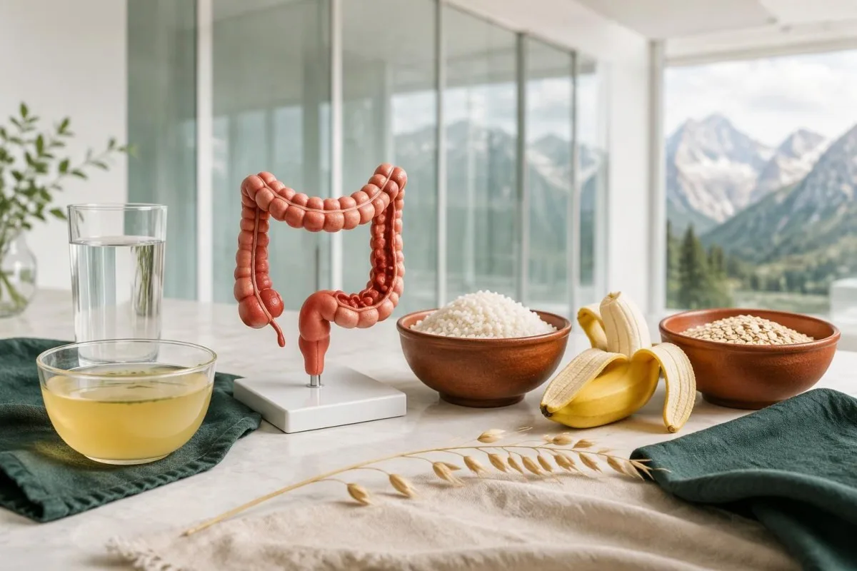
A liquid diet does not diagnose or cure diverticulitis, and prolonged restriction can reduce energy and protein intake. For a hypothetical person who has eaten little for 3 days, ongoing pain and difficulty advancing food are reasons to contact the clinical team, not evidence that stricter fasting is needed.
Small, frequent drinks may be easier to tolerate than a large glass, but there is no single fluid target suitable for everyone. A person with a prescribed 1.5-liter daily fluid restriction for heart or kidney disease needs individualized advice; vomiting, diarrhea and reduced urine output can require assessment rather than an automatic instruction to drink more.
Temporary diarrhea during recovery should be interpreted alongside pain, fever and medication exposure rather than attributed to food alone. Our ಜೀರ್ಣಕ್ರಿಯೆಯ ಲಕ್ಷಣಗಳ ಮಾರ್ಗದರ್ಶಿ discusses stool changes after restricted eating; 3 watery stools daily after antibiotics call for a different discussion from one softer stool as meals restart.
Routine avoidance of nuts, seeds and popcorn is not supported as a prevention strategy, according to the AGA review (Peery et al., 2021). After recovery, a high-quality dietary pattern and gradual return toward a usual fiber intake—often around 25–30 g/day for adults—can be discussed, but suddenly adding a large fiber supplement during acute pain is not a substitute for assessment.
ಮೌಲ್ಯಮಾಪನದ ನಂತರ ರೋಗಲಕ್ಷಣಗಳನ್ನು ಹೇಗೆ ಗಮನಿಸಬೇಕು?
Monitor pain direction, drinking tolerance, temperature, bowel function and urine output after the clinician establishes a care plan. A routine check-in over 48–72 ಗಂಟೆಗಳು may be appropriate, but deterioration at any point overrides that schedule; a falling CRP does not make worsening abdominal symptoms safe to ignore.
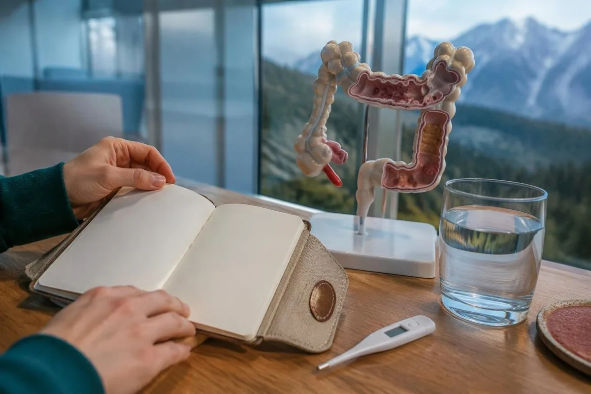
A useful home record needs only a few consistent observations, not hourly measurements of everything. For example, note pain location, measured temperature and fluid tolerance 2–3 times daily while awake, or as advised; repeated measurements without a clear action plan can increase anxiety while failing to address new warning signs.
Clinical improvement and biomarker improvement can occur at different speeds, and repeated blood testing is not mandatory when recovery is straightforward. A hypothetical CRP fall from 110 to 58 mg/L over 2 days is encouraging only if pain and general condition also improve; our ಆಸ್ಪತ್ರೆ ನಂತರದ ಪ್ರಯೋಗಾಲಯ ಬದಲಾವಣೆಗಳು guide explains why trends need clinical context.
Pain that remains severe or increasingly localized, new vomiting or inability to advance intake should trigger a fresh examination and possibly CT. Having started antibiotics 24 hours earlier does not protect against every complication; persistent symptoms may reflect an abscess, an alternative diagnosis or a treatment problem rather than simply slow recovery.
Kantesti can help explain why a CRP of 58 mg/L remains elevated after a higher previous result, but our platform is not a continuously monitored emergency service. Share meaningful changes with the treating clinician, and use local emergency services for severe deterioration; uploading another report should never delay urgent assessment.
ಡೈವರ್ಟಿಕ್ಯುಲೈಟಿಸ್ ನಂತರ ಕೊಲೊನೋಸ್ಕೋಪಿ ಯಾವಾಗ ಅಗತ್ಯ?
Colonoscopy may be recommended after recovery to exclude another bowel condition, especially following complicated diverticulitis or a first uncomplicated episode. The AGA recommends waiting 6–8 ವಾರಗಳೊಳಗೆ, or until symptoms have fully resolved if longer, and allows deferral in selected people with a recent high-quality colonoscopy within 1 year.
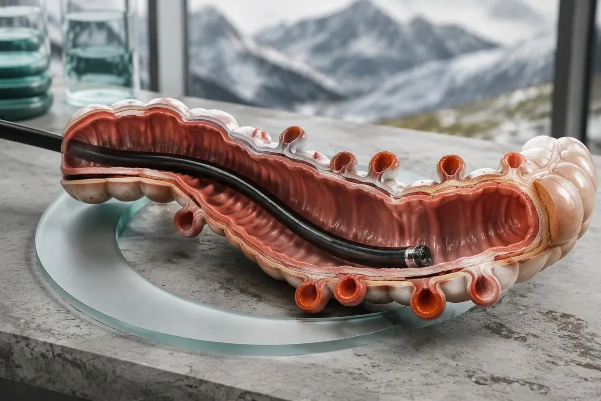
Bowel cancer can occasionally resemble diverticulitis on presentation, which is why follow-up is risk-based rather than a statement that diverticulitis causes cancer. The meta-analysis summarized by AGA found colorectal cancer in 1.9% of patients after diverticulitis, with higher prevalence after complicated episodes (7.9%) than uncomplicated episodes (1.3%) (Peery et al., 2021).
Routine colonoscopy is usually avoided during the acute painful phase, because examination can be technically harder and potentially riskier. The 6–8-week interval is a practical recovery window, not a guarantee that every bowel has healed by a calendar date; ongoing tenderness or systemic symptoms should be discussed before the procedure.
Iron-deficiency anemia, weight loss, persistent rectal bleeding or an ongoing change in bowel habits may alter follow-up urgency. A positive FIT requires its own evaluation and is not cancelled by a diverticulitis label; our ಧನಾತ್ಮಕ FIT ಮುಂದಿನ ಹಂತಗಳು guide explains why 1 positive stool screening result needs a defined diagnostic plan.
Persistent discomfort does not always mean continuing diverticulitis: the AGA review notes periodic abdominal pain in approximately 45% of patients at 1 year in a cited trial. Clinicians may investigate ongoing inflammation or another diagnosis with imaging and lower endoscopy before considering visceral hypersensitivity; recurrent severe symptoms should not be assigned that explanation without assessment.
Kantesti ಏನನ್ನು ವಿವರಿಸಬಲ್ಲದು—ಮತ್ತು ಯಾವುದನ್ನು ರೋಗನಿರ್ಣಯ ಮಾಡಲಾರದು
Kantesti ಒಂದು AI-ಚಾಲಿತ ರಕ್ತ ಪರೀಕ್ಷೆ ವಿಶ್ಲೇಷಣಾ ಸಾಧನ that can help explain CRP, WBC and other laboratory findings, but it cannot confirm diverticulitis or exclude an abscess from a report. A CRP of 3 mg/L or 150 mg/L still requires symptoms, examination and sometimes imaging to determine the cause.
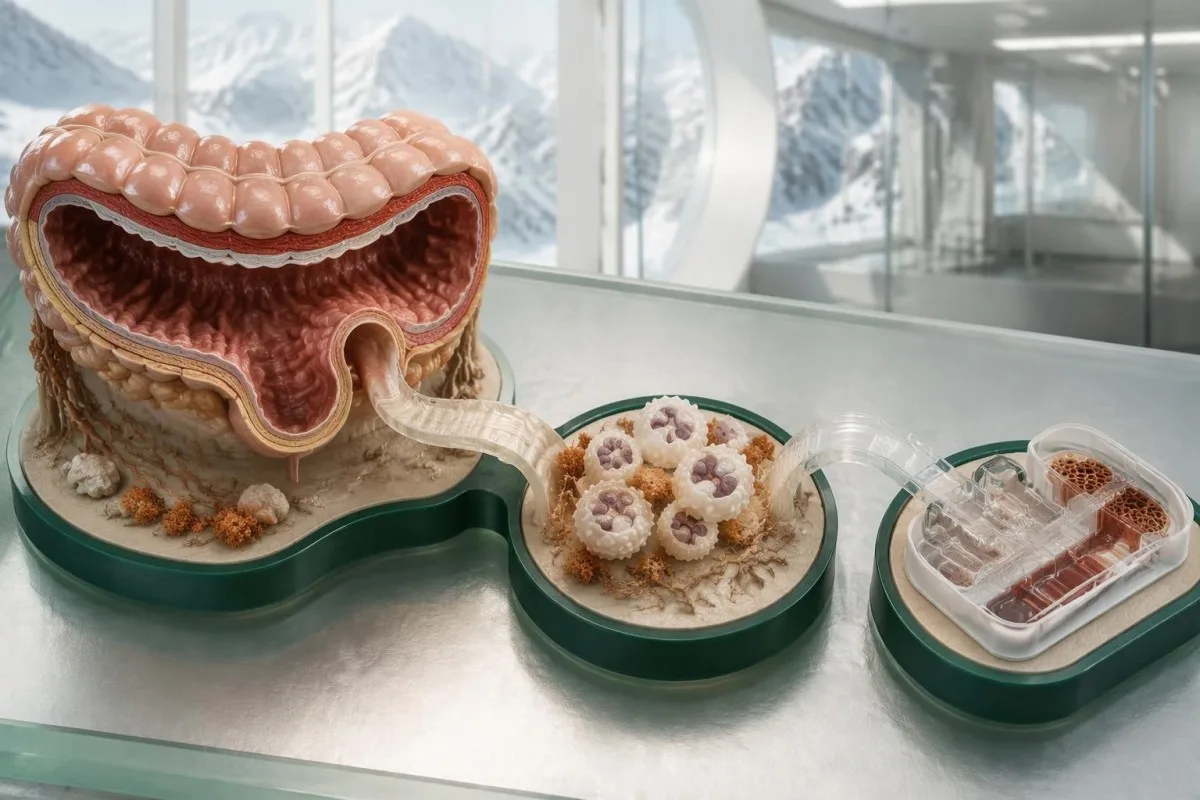
Our platform can explain units and available trends, while the limits of automated interpretation remain explicit. For example, 15 × 10⁹/ಲೀ ಮತ್ತು 15,000 cells/µL describe the same WBC count; our ತಂತ್ರಜ್ಞಾನ ವಿವರಣೆ provides context for how uploaded laboratory information is processed, not evidence that a scan is unnecessary.
Technical performance and clinical diagnosis are different claims, so a software benchmark must not be read as a diverticulitis diagnostic trial. Kantesti's ಪರಿಶೀಲನೆ ಮತ್ತು ಮೇಲ್ವಿಚಾರಣೆಯ ಮಾಹಿತಿ should be interpreted within its stated scope; no general laboratory benchmark establishes that an AI can safely exclude perforation from 1 CBC or CRP result.
The 2 Zenodo publications listed below provide platform reporting and RDW education, respectively; neither is a clinical guideline or direct evidence for diagnosing diverticulitis. Kantesti's Global Health Report describes analysis activity, while the RDW guide concerns red-cell size variation—a high RDW should not be presented as confirmation of an acute bowel disorder.
Use AI explanations to prepare questions, not to postpone care, particularly during a first episode or a worsening recurrence. Our ಜವಾಬ್ದಾರಿಯುತ AI ತತ್ವಗಳು describe these boundaries; the practical question is not whether 1 number looks reassuring, but whether your symptoms require an examination, imaging or urgent treatment.
ಆಗಾಗ್ಗೆ ಕೇಳಲಾಗುವ ಪ್ರಶ್ನೆಗಳು
ಡೈವರ್ಟಿಕ್ಯುಲೈಟಿಸ್ನ ಆರಂಭಿಕ ಲಕ್ಷಣಗಳು ಯಾವುವು?
ಆರಂಭಿಕ ಡೈವರ್ಟಿಕ್ಯುಲೈಟಿಸ್ನಿಂದ ಹೊಟ್ಟೆಯ ಕೆಳ ಎಡಭಾಗದಲ್ಲಿ ಒಂದು ನಿರ್ದಿಷ್ಟ ಪ್ರದೇಶಕ್ಕೆ ಸೀಮಿತವಾದ ನೋವು ಉಂಟಾಗಿ, ಅದು ನಿರಂತರವಾಗಬಹುದು; ಸ್ಪರ್ಶಿಸಿದಾಗ ನೋವು, ಹಸಿವು ಕಡಿಮೆಯಾಗುವುದು ಮತ್ತು ಮಲಬದ್ಧತೆ ಅಥವಾ ಅತಿಸಾರವೂ ಕಾಣಿಸಬಹುದು. ಲಕ್ಷಣಗಳು 1–2 ದಿನಗಳಲ್ಲಿ ಕಾಣಿಸಿಕೊಳ್ಳಬಹುದು, ಆದರೆ ಅವು ಕಾಣಿಸಿಕೊಳ್ಳುವ ಕ್ರಮ ಖಚಿತವಲ್ಲ ಮತ್ತು ಜ್ವರವೂ ಇಲ್ಲದಿರಬಹುದು. ಹೊಸದಾಗಿ ಕಾಣಿಸಿಕೊಂಡು ನಿರಂತರವಾಗಿರುವ ಒಂದು ನಿರ್ದಿಷ್ಟ ಸ್ಥಳದ ನೋವಿದ್ದರೆ, ಎಲ್ಲಾ ಸಾಮಾನ್ಯ ಲಕ್ಷಣಗಳು ಕಾಣಿಸಿಕೊಳ್ಳುವವರೆಗೆ ಕಾಯದೆ ಅದೇ ದಿನ ವೈದ್ಯಕೀಯ ಸಲಹೆ ಪಡೆಯಿರಿ. ತೀವ್ರವಾಗಿರುವ ಅಥವಾ ಹರಡುವ ನೋವು, ಪದೇಪದೇ ವಾಂತಿಯಾಗುವುದು, ಮೂರ್ಛೆ ಹೋಗುವುದು ಅಥವಾ ಗೊಂದಲ ಉಂಟಾದರೆ ತುರ್ತು ಚಿಕಿತ್ಸೆ ಪಡೆಯಿರಿ.
ಜ್ವರ ಇಲ್ಲದೆಯೂ ನಿಮಗೆ ಡೈವರ್ಟಿಕ್ಯುಲೈಟಿಸ್ ಇರಬಹುದೇ?
0 ಡೈವರ್ಟಿಕ್ಯುಲೈಟಿಸ್ ಜ್ವರವಿಲ್ಲದೆಯೂ ಉಂಟಾಗಬಹುದು, ಮತ್ತು 38°Cಗಿಂತ ಕಡಿಮೆ ತಾಪಮಾನವಿದ್ದರೆ ಒಂದು ಹಂತ ಅಥವಾ ತೊಡಕು ಇಲ್ಲವೆಂದು ತೀರ್ಮಾನಿಸಲಾಗುವುದಿಲ್ಲ. ಅನಾರೋಗ್ಯದ ಆರಂಭಿಕ ಹಂತದಲ್ಲಿ, ವಯಸ್ಸಾದವರಲ್ಲಿ ಅಥವಾ ಕಾರ್ಟಿಕೊಸ್ಟೆರಾಯ್ಡ್ಗಳು ಅಥವಾ ಜ್ವರ ಕಡಿಮೆ ಮಾಡುವ ಔಷಧಿಗಳ ಚಿಕಿತ್ಸೆಯ ಸಂದರ್ಭದಲ್ಲಿ ಜ್ವರದ ತೀವ್ರತೆ ಕಡಿಮೆಯಿರಬಹುದು. ಜ್ವರ ಇಲ್ಲದಿರುವುದನ್ನು ನೋವು ಹೆಚ್ಚಾಗುತ್ತಿರುವುದು, ದ್ರವಗಳನ್ನು ಕುಡಿಯುವ ಸಾಮರ್ಥ್ಯ ಮತ್ತು ವೈದ್ಯಕೀಯ ಪರೀಕ್ಷೆಯ ಫಲಿತಾಂಶಗಳೊಂದಿಗೆ ಪರಿಗಣಿಸಬೇಕು. ಥರ್ಮಾಮೀಟರ್ನ ಅಳತೆ ಸಾಮಾನ್ಯವಾಗಿದ್ದರೂ, ಲಕ್ಷಣಗಳು ಉಲ್ಬಣಿಸಿದರೆ ತಕ್ಷಣ ವೈದ್ಯಕೀಯ ಮೌಲ್ಯಮಾಪನ ಪಡೆಯಿರಿ.
ಡೈವರ್ಟಿಕ್ಯುಲೈಟಿಸ್ನ ನೋವು ಸಾಮಾನ್ಯವಾಗಿ ಎಲ್ಲಿ ಕಾಣಿಸಿಕೊಳ್ಳುತ್ತದೆ?
0 ಕೆಳಗಿನ ಎಡಭಾಗದ ಹೊಟ್ಟೆನೋವನ್ನು ಡೈವರ್ಟಿಕ್ಯುಲೈಟಿಸ್ ಸಾಮಾನ್ಯವಾಗಿ ಉಂಟುಮಾಡುತ್ತದೆ, ಏಕೆಂದರೆ ಸಿಗ್ಮಾಯ್ಡ್ ಕೊಲನ್ ಸಾಮಾನ್ಯವಾಗಿ ಇದರಿಂದ ಬಾಧಿತವಾಗುತ್ತದೆ; ಆದರೆ ನೋವು ಹೊಟ್ಟೆಯ ಮಧ್ಯಭಾಗದಲ್ಲಿ, ಶ್ರೋಣಿಯ ಭಾಗದಲ್ಲಿ ಅಥವಾ ಬಲಭಾಗದಲ್ಲಿಯೂ ಇರಬಹುದು. ನೋವಿರುವ 1 ಪ್ರದೇಶದ ಸ್ಥಾನದಿಂದ ಮಾತ್ರ ರೋಗನಿರ್ಣಯವನ್ನು ಖಚಿತಪಡಿಸಲು ಸಾಧ್ಯವಿಲ್ಲ, ಮತ್ತು ಬಲಭಾಗದ ನೋವು ಅಪೆಂಡಿಸೈಟಿಸ್ನಂತೆಯೇ ಕಾಣಿಸಬಹುದು. ಹೊಸದಾಗಿ ಕಾಣಿಸಿಕೊಳ್ಳುವ, ಹರಡುವ ಅಥವಾ ತೀವ್ರವಾಗುವ ನೋವಿಗೆ ಮರುಮೌಲ್ಯಮಾಪನ ಅಗತ್ಯ. ನೋವಿನ ಸ್ಥಳ ಮತ್ತು ಪರೀಕ್ಷೆಯಿಂದ ಕಾರಣವನ್ನು ಸ್ಪಷ್ಟವಾಗಿ ಗುರುತಿಸಲು ಸಾಧ್ಯವಾಗದಿದ್ದಾಗ ವೈದ್ಯರು ಇಮೇಜಿಂಗ್ ಪರೀಕ್ಷೆಗಳನ್ನು ಶಿಫಾರಸು ಮಾಡಬಹುದು.
ಸಾಮಾನ್ಯ ರಕ್ತ ಪರೀಕ್ಷೆಗಳು ಡೈವರ್ಟಿಕ್ಯುಲೈಟಿಸ್ ಅನ್ನು ತಳ್ಳಿಹಾಕಬಹುದೇ?
0 ಸಾಮಾನ್ಯ WBC ಮತ್ತು CRP ಫಲಿತಾಂಶಗಳು ಡೈವರ್ಟಿಕ್ಯುಲೈಟಿಸ್ ಅನ್ನು ವಿಶ್ವಾಸಾರ್ಹವಾಗಿ ತಳ್ಳಿಹಾಕಲಾರವು, ವಿಶೇಷವಾಗಿ ರೋಗದ ಆರಂಭಿಕ ಹಂತದಲ್ಲಿ. ಅನೇಕ ವಯಸ್ಕರ ಪ್ರಯೋಗಾಲಯಗಳು WBCಗೆ ಸುಮಾರು 4–11 × 10⁹/L ವ್ಯಾಪ್ತಿಯನ್ನು ಮತ್ತು CRPಗೆ 5 mg/L ಮೇಲಿನ ಮಿತಿಯನ್ನು ಬಳಸುತ್ತವೆ, ಆದರೆ ಸ್ಥಳೀಯ ವ್ಯಾಪ್ತಿಗಳು ಭಿನ್ನವಾಗಿರುತ್ತವೆ. ಉರಿಯೂತದ ಅನೇಕ ಸ್ಥಿತಿಗಳು ಈ ಸೂಚಕಗಳನ್ನು ಹೆಚ್ಚಿಸುವುದರಿಂದ, ಏರಿದ ಫಲಿತಾಂಶಗಳೂ ಡೈವರ್ಟಿಕ್ಯುಲೈಟಿಸ್ ಅನ್ನು ಖಚಿತಪಡಿಸಲಾರವು. ರೋಗನಿರ್ಣಯವನ್ನು ಸ್ಥಾಪಿಸಲು ಮತ್ತು ತೊಡಕುಗಳನ್ನು ನಿರ್ಣಯಿಸಲು ರೋಗಲಕ್ಷಣಗಳು, ಪರೀಕ್ಷೆ ಮತ್ತು ಕೆಲವೊಮ್ಮೆ CT ಅಗತ್ಯವಾಗುತ್ತವೆ.
ಎಡಭಾಗದ ಹೊಟ್ಟೆನೋವು ಯಾವಾಗ ತುರ್ತು ಪರಿಸ್ಥಿತಿಯಾಗುತ್ತದೆ?
0 ಎಡಭಾಗದ ಹೊಟ್ಟೆನೋವು ತೀವ್ರವಾಗಿದ್ದರೆ ಅಥವಾ ಹರಡುತ್ತಿದ್ದರೆ, ಅಥವಾ ಮೂರ್ಛೆ, ಗೊಂದಲ, ಪದೇಪದೇ ವಾಂತಿ ಅಥವಾ ಗಮನಾರ್ಹ ಹೊಟ್ಟೆ ಉಬ್ಬರದೊಂದಿಗೆ ಕಾಣಿಸಿಕೊಂಡರೆ ತುರ್ತು ವೈದ್ಯಕೀಯ ಮೌಲ್ಯಮಾಪನ ಅಗತ್ಯ. ಗ್ಯಾಸ್ ಅಥವಾ ಮಲ ಹೊರಹಾಕಲು ಸಾಧ್ಯವಾಗದಿರುವುದರೊಂದಿಗೆ ಹೊಟ್ಟೆ ಉಬ್ಬರವು ಕರುಳಿನ ಅಡಚಣೆಯನ್ನು ಸೂಚಿಸಬಹುದು; ದೌರ್ಬಲ್ಯದೊಂದಿಗೆ ಹೆಚ್ಚಿನ ಪ್ರಮಾಣದ ಗುದನಾಳದ ರಕ್ತಸ್ರಾವವು ಗಣನೀಯ ರಕ್ತನಷ್ಟವನ್ನು ಸೂಚಿಸಬಹುದು. ತುರ್ತು ಚಿಕಿತ್ಸೆ ಪಡೆಯುವ ಮೊದಲು 38°C ಜ್ವರ ಬರುವವರೆಗೆ ಕಾಯಬೇಕಾಗಿಲ್ಲ. ಈ ಎಚ್ಚರಿಕೆಯ ಲಕ್ಷಣಗಳು ಕಂಡುಬಂದಾಗ ರಕ್ತಪರೀಕ್ಷೆಯ ಫಲಿತಾಂಶಕ್ಕಾಗಿ ಕಾಯಬೇಡಿ.
ಡೈವರ್ಟಿಕ್ಯುಲೈಟಿಸ್ಗೆ ನಿಮಗೆ ಯಾವಾಗಲೂ CT ಸ್ಕ್ಯಾನ್ ಅಗತ್ಯವಿದೆಯೇ?
ಹಿಂದೆ ದೃಢಪಟ್ಟ ಡೈವರ್ಟಿಕ್ಯುಲೈಟಿಸ್ನ ಪ್ರತಿಯೊಂದು ಸೌಮ್ಯ ಮರುಕಳಿಕೆಗೆ CT ಅಗತ್ಯವಿಲ್ಲ, ಆದರೆ ಮೊದಲ ಬಾರಿಗೆ ಅನಿಶ್ಚಿತತೆ ಇರುವ ಪ್ರಸಂಗ, ತೀವ್ರ ಲಕ್ಷಣಗಳು, ತೊಡಕುಗಳ ಶಂಕೆ ಅಥವಾ ಸುಧಾರಣೆ ಕಾಣದಿರುವ ಸಂದರ್ಭಗಳಲ್ಲಿ ಇಮೇಜಿಂಗ್ ಅನ್ನು ಸಾಮಾನ್ಯವಾಗಿ ಶಿಫಾರಸು ಮಾಡಲಾಗುತ್ತದೆ. ಡೈವರ್ಟಿಕ್ಯುಲೈಟಿಸ್ ಪತ್ತೆಗೆ CT ಸುಮಾರು 95% ಸಂವೇದನಾಶೀಲತೆ ಮತ್ತು ನಿರ್ದಿಷ್ಟತೆಯನ್ನು ಹೊಂದಿದ್ದು, ಬಾವು ಅಥವಾ ನೋವಿಗೆ ಪರ್ಯಾಯ ಕಾರಣವನ್ನು ಗುರುತಿಸಬಲ್ಲದು. ರೋಗನಿರ್ಣಯದ ಅನಿಶ್ಚಿತತೆ ಇದ್ದಾಗ CT ಮಾಡಲು ACP ಮಾರ್ಗಸೂಚಿ ಶಿಫಾರಸು ಮಾಡುತ್ತದೆ; ಹಿಂದಿನ ಇಮೇಜಿಂಗ್ನಿಂದ ರೋಗನಿರ್ಣಯ ದೃಢಪಡಿಸದ ರೋಗಿಗಳಿಗೂ AGA ಮಾರ್ಗದರ್ಶನ ಒತ್ತು ನೀಡುತ್ತದೆ. ಗರ್ಭಧಾರಣೆ, ಮೂತ್ರಪಿಂಡದ ಕಾರ್ಯಕ್ಷಮತೆ ಮತ್ತು ಸ್ಥಳೀಯ ಪರಿಣತಿ ಇಮೇಜಿಂಗ್ ವಿಧಾನವನ್ನು ನಿರ್ಧರಿಸುತ್ತವೆ.
ಡೈವರ್ಟಿಕ್ಯುಲೈಟಿಸ್ಗೆ ಯಾವಾಗಲೂ ಪ್ರತಿಜೀವಕಗಳ ಅಗತ್ಯವಿದೆಯೇ?
Selected immunocompetent adults with mild, uncomplicated diverticulitis may be treated without antibiotics under a clinician's supervision. Antibiotics are generally recommended when systemic illness, immunosuppression, significant comorbidity or higher-risk features are present. AGA guidance identifies CRP above 140 mg/L or WBC above 15 × 10⁹/L as treatment-relevant risk features, not diagnostic cutoffs. Inability to drink, worsening pain or new emergency symptoms requires reassessment regardless of whether antibiotics have been prescribed.
ನಿಮ್ಮ ರಕ್ತ ಪರೀಕ್ಷೆ ಫಲಿತಾಂಶಗಳನ್ನು ವಿವರಿಸಿಕೊಳ್ಳಿ
ನಿಮ್ಮ ಪ್ರಯೋಗಾಲಯ ವರದಿಯನ್ನು ಅಪ್ಲೋಡ್ ಮಾಡಿ; Kantestiಯ AI ಕೆಲವೇ ಸೆಕೆಂಡುಗಳಲ್ಲಿ ಪ್ರತಿಯೊಂದು ಜೈವಿಕ ಸೂಚಕವನ್ನು ಸರಳ ಭಾಷೆಯಲ್ಲಿ ವಿವರಿಸುತ್ತದೆ. ವಿಶ್ವದಾದ್ಯಂತ 2 ದಶಲಕ್ಷಕ್ಕೂ ಹೆಚ್ಚು ಜನರು ಬಳಸಿದ್ದಾರೆ.
ಉಲ್ಲೇಖಗಳು
- Klein, T., Mitchell, S., & Weber, H. (2026). AI ರಕ್ತ ಪರೀಕ್ಷೆ ವಿಶ್ಲೇಷಕ: 2.5M ಪರೀಕ್ಷೆಗಳು ವಿಶ್ಲೇಷಿಸಲಾಗಿದೆ | ಗ್ಲೋಬಲ್ ಹೆಲ್ತ್ ರಿಪೋರ್ಟ್ 2026. Kantesti AI ವೈದ್ಯಕೀಯ ಸಂಶೋಧನೆ. ಡಿಒಐ
- Klein, T., Mitchell, S., & Weber, H. (2026). RDW ರಕ್ತ ಪರೀಕ್ಷೆ: RDW-CV, MCV ಮತ್ತು MCHC ಕುರಿತು ಸಂಪೂರ್ಣ ಮಾರ್ಗದರ್ಶಿ. Kantesti AI ವೈದ್ಯಕೀಯ ಸಂಶೋಧನೆ. ಡಿಒಐ
- National Institute for Health and Care Excellence (2019). Diverticular disease: diagnosis and management (NG147). NICE Clinical Guideline.
- Peery AF et al. (2021). AGA Clinical Practice Update on Medical Management of Colonic Diverticulitis: Expert Review. Gastroenterology. ಡಿಒಐ ಪಬ್ಮೆಡ್
- Qaseem A et al. (2022). Diagnosis and Management of Acute Left-Sided Colonic Diverticulitis: A Clinical Guideline From the American College of Physicians. Annals of Internal Medicine. ಡಿಒಐ
- 2ಮಿ+ವಿಶ್ಲೇಷಿಸಿದ ಪರೀಕ್ಷೆಗಳು
- 127+ದೇಶಗಳು
- 100+ಭಾಷೆಗಳು
ವೈದ್ಯಕೀಯ ತಂಡದ ಬಗ್ಗೆ


E-E-A-T ವಿಶ್ವಾಸ ಸಂಕೇತಗಳು
ಅನುಭವ
ಲ್ಯಾಬ್ ಅರ್ಥೈಸುವ ಕಾರ್ಯಪ್ರವಾಹಗಳ ಮೇಲೆ ವೈದ್ಯರಿಂದ ನೇತೃತ್ವದ ಕ್ಲಿನಿಕಲ್ ವಿಮರ್ಶೆ.
ಪರಿಣಿತಿ
ಕ್ಲಿನಿಕಲ್ ಸಂದರ್ಭದಲ್ಲಿನ ಬಯೋಮಾರ್ಕರ್ಗಳು ಹೇಗೆ ವರ್ತಿಸುತ್ತವೆ ಎಂಬುದರ ಮೇಲೆ ಪ್ರಯೋಗಾಲಯ ವೈದ್ಯಕೀಯದ ಕೇಂದ್ರೀಕರಣ.
ಅಧಿಕಾರಯುತತೆ
ಡಾ. ಥಾಮಸ್ ಕ್ಲೈನ್ ಅವರ ವೈದ್ಯಕೀಯ ವಿಮರ್ಶೆ; ಡಾ. ಸಾರಾ ಮಿಚೆಲ್ ಮತ್ತು ಪ್ರೊ. ಡಾ. ಹಾನ್ಸ್ ವೆಬರ್ ಅವರ ಕೊಡುಗೆಗಳೊಂದಿಗೆ.
ವಿಶ್ವಾಸಾರ್ಹತೆ
ಎಚ್ಚರಿಕೆಯನ್ನು ಕಡಿಮೆ ಮಾಡಲು ಸ್ಪಷ್ಟವಾದ ಮುಂದಿನ ಹಂತಗಳೊಂದಿಗೆ ಸಾಕ್ಷ್ಯಾಧಾರಿತ ಅರ್ಥೈಸಿಕೆ.
ವೈದ್ಯಕೀಯ ಹಕ್ಕುತ್ಯಾಗ
ಈ ಲೇಖನವು ಕೇವಲ ಶಿಕ್ಷಣ ಉದ್ದೇಶಗಳಿಗಾಗಿ이며 ವೈದ್ಯಕೀಯ ಸಲಹೆಯನ್ನು ನೀಡುವುದಿಲ್ಲ. ರೋಗನಿರ್ಣಯ ಮತ್ತು ಚಿಕಿತ್ಸೆ ನಿರ್ಧಾರಗಳಿಗಾಗಿ ಸದಾ ಅರ್ಹ ಆರೋಗ್ಯ ಸೇವಾ ಪೂರೈಕೆದಾರರನ್ನು ಸಂಪರ್ಕಿಸಿ.
ಪ್ರಕಟಿಸಲಾಗಿದೆ · ಸಂಪಾದಕೀಯ ತಂಡವನ್ನು ಸಂಪರ್ಕಿಸಿ
Kantesti Ltd ಪ್ರಕಟಿಸಿದೆ, ಇಂಗ್ಲೆಂಡ್ ಮತ್ತು ವೇಲ್ಸ್ನಲ್ಲಿ ನೋಂದಾಯಿತವಾಗಿದೆ, ಕಂಪನಿ ಸಂಖ್ಯೆ. 17090423, ಲಂಡನ್, ಯುನೈಟೆಡ್ ಕಿಂಗ್ಡಮ್.
