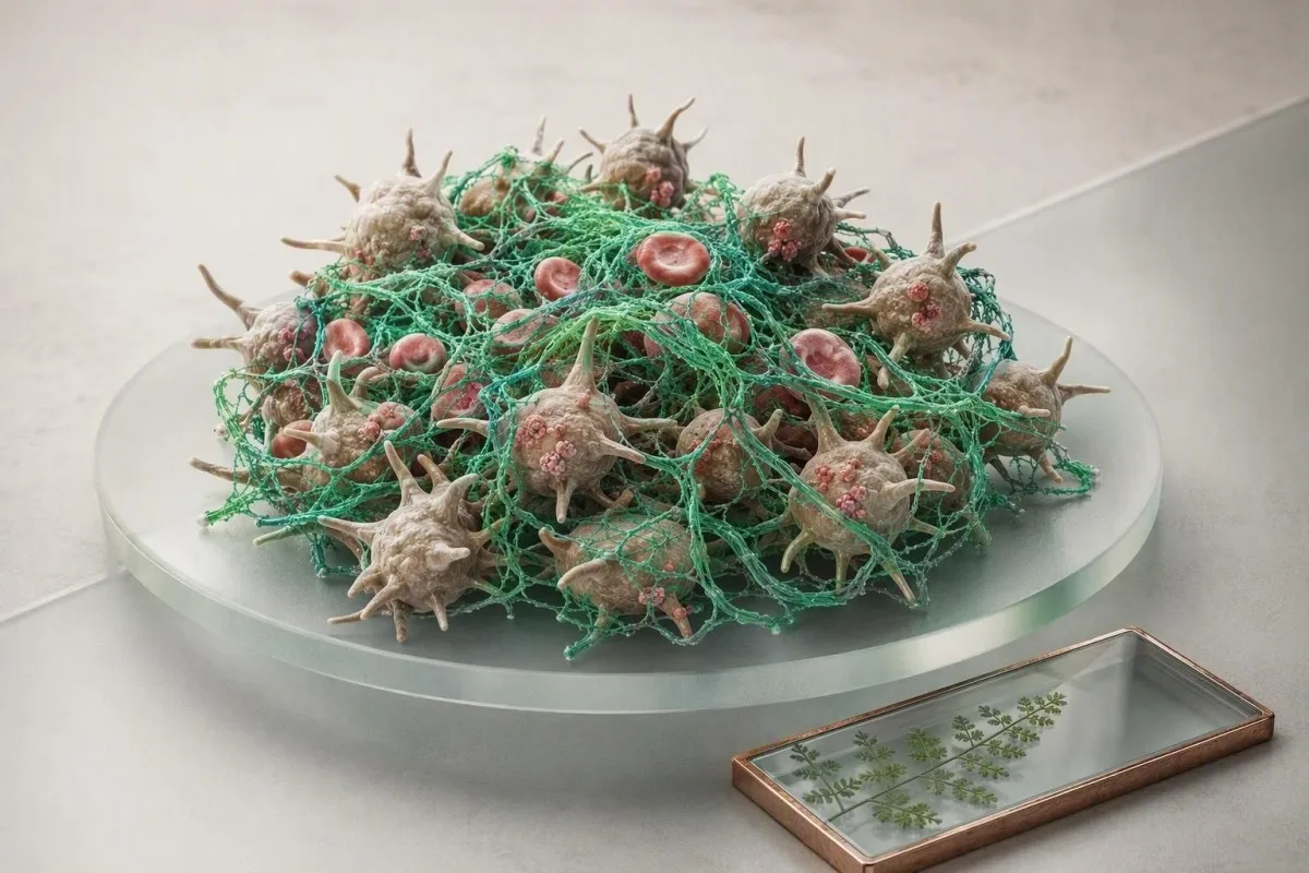
Қалыпты скрининг нәтижелері сенімді, бірақ олар гемостаздың тек таңдалған бөліктерін тексереді. Іріңдеу, қатты етеккіру, тромбтар, отбасылық тарих және дәрі-дәрмектер әлі де мақсатты тестілеуді ақтауы мүмкін.


- PT және aPTT қалыпты фон Виллебранд ауруын, тромбоциттердің қызмет ету бұзылыстарын, XIII фактор тапшылығын, фактор тапшылығының жеңіл формаларын немесе көптеген тұқым қуалайтын тромбофилияларды жоққа шығармайды.
- Әдеттегі ересек PT шамамен 10–13 секунд және INR шамамен 0,8–1,2 варфаринсіз, бірақ әр зертхана өзінің сілтеме аралығын ұсынуы керек.
- Әдеттегі aPTT жиі 25–35 секундты құрайды; қалыпты нәтиже VIII, IX немесе XI фактор тапшылығының жеңіл формасын сенімді түрде жоққа шығармайды.
- Етеккірдің мол қан кетуі сағатына 2 сағаттан артық қорғанысты сіңіретін жағдай шұғыл клиникалық бағалауды талап етеді, тіпті скрининг тесттері қалыпты болса да.
- Тромбоциттер саны санды, қызметін емес, өлшейді; 250 × 10⁹/л санауы клиникалық маңызы бар тромбоциттердің қызмет ету бұзылысымен қатар жүруі мүмкін.
- D-димер вена тромбоэмболиясын тек тест алдындағы ықтималдық төмен немесе орташа болғанда және валидацияланған жол қолданылғанда ғана жоққа шығаруға көмектеседі.
- Фон Виллебранд факторын тестілеу фактор VIII мен қайта зерттеу арқылы ең жақсы түсіндіріледі, себебі стресс, жүктілік, қабыну және эстроген нәтижелерді уақытша жоғарылатуы мүмкін.
- Шұғыл белгілер қан түкіру, кенеттен ентігу, бір жақты аяқ ісінуі, жаңа неврологиялық симптомдар, қара нәжіс немесе бақыланбайтын қан кетуді қамтиды.
Неліктен қалыпты скрининг нәтижелері зерттеуді тоқтатпайды
Қалыпты PT, INR және aPTT барлық қан кету немесе ұю бұзылуларын жоққа шығармайды. Олар негізінен таңдалған плазма ұю жолдарын бағалайды, ал тромбоциттер функциясы, фон Виллебранд факторы, фактор XIII және көптеген тромбоз тәуекелдері көрінбей қалуы мүмкін. 2026 жылғы 22 тамыздағы жағдай бойынша, бұл айырмашылық ұю панелінің нәтижелерін түсіндірудің ең пайдалы бастапқы нүктесі болып табылады.
Менің клиникалық тәжірибемде жиі кездесетін қателік - қалыпты панельді әмбебап босату сертификаты ретінде қарастыру. Олай емес. PT сыртқы және ортақ жолды сынақтан өткізеді; aPTT ішкі және ортақ жолды сынақтан өткізеді; ешқайсысы тромбоциттердің кіші ыдыстың зақымдануына қаншалықты жабысатынын өлшемейді. Доктор Томас Кляйн мұны әсіресе стоматологиялық қан кетуден немесе өмір бойы ауыр етеккірден кейін байқайды, бұл кезде зертханалық скрининг мүлдем ерекшеленбейді.
Кантести - бұл AI қан анализінің анализаторы толық қан саны, бауыр маркерлері, дәрі-дәрмектер мен хабарланған симптомдармен қатар ұю мәндерін оқиды, нәтижені жай ғана “жақсы” деп жариялаудан гөрі. Қалыпты тромбоцит саны 150–450 × 10⁹/л көптеген адамдар үшін жеткілікті тромбоциттердің бар екенін растайды, бірақ тромбоциттердің қалыпты сигнал беретінін немесе агрегацияланатынын көрсете алмайды. Біздің толық қан есептеуі нұсқаулығында санақ пен функцияның бөлек сұрақтар екенін түсіндіреді.
Пайдалы ақыл-ой моделі үш қабаттан тұрады: ыдыс қабырғасы, тромбоциттер және ұю ақуыздары. PT және aPTT негізінен үшінші қабатты тексереді. Егер біреуде 25 минутқа созылатын мұрыннан қан кету, тіс етінен қан кету, етеккірден темір тапшылығы, ата-анасында осыған ұқсас симптомдар болса, бұл тарих бір реттік қалыпты скринингтен гөрі диагностикалық салмаққа ие.
Қалыпты нәтиже іс жүзінде нені білдіреді
Қалыпты скринингтік панель өлшенген ұю уақыты сынақтың шарттарында зертхананың интервалында болғанын білдіреді. Бұл гемостаздың операция, босану, жарақат немесе антикоагулянттық әсер кезінде қалыпты болатынын дәлелдемейді.
PT, INR және aPTT шынымен не өлшейді
PT, INR және aPTT зертханалық плазманың жасанды реагенттер қосылғаннан кейін фибрин түзу уақытын өлшейді. Олар жол скринингтері болып табылады, бүкіл денені қан кету қаупін немесе адамда қауіпті тромб бар-жоғын тікелей тексеретін тесттер емес.
A PT шамамен 10–13 секунд, INR 0.8–1.2, және aPTT шамамен 25–35 секунд антикоагулянттарды қолданбайтын адамдарда жиі кездесетін ересектер интервалдары, бірақ реагенттер жеткілікті түрде ерекшеленеді, сондықтан есептің өз диапазоны жеңіп шығады. INR PT-ді негізінен варфарин бақылауы үшін стандарттайды; бұл “қанның қалыңдығының” әмбебап өлшемі емес. 1.1 INR аспириннің тромбоцит функциясын бұзғанын немесе бұзбағанын айтпайды.
PT VII фактор төмен болғанда, К дәруменінің қолжетімділігі азайғанда, варфарин белсенді болғанда немесе бауырдың синтетикалық функциясы бұзылғанда ұзартылады. aPTT гепаринмен, қызылша антикоагулянтымен және VIII, IX, XI немесе XII факторларын қамтитын тапшылықтармен ұзаруы мүмкін. Үлгісі жоғары ПТ және қалыпты aPTT мүмкіндіктерді тарылтады, бірақ қалыпты мәндер логиканы кері бұрмайды.
Мен қараған 42 жастағы пациентте процедура алдында aPTT 29 секунд болды, бірақ операциялық сайттың сәйкессіз сүзілуі пайда болды. Қайта зерттеу балалық шақтағы эпистаксис пен аналық меноррагияны анықтады; кейінгі тестілеу фон Виллебранд ауруын қолдады. Экран өз жұмысын атқарды - ол тек соңғы сұраққа жауап беруге арналмаған.
Қалыпты PT және aPTT жіберіп алуы мүмкін қан кету жағдайлары
фон Виллебранд ауруы және сапалы тромбоциттер бұзылуы - бұл қалыпты PT және aPTT кезінде шырышты-тері қан кетуінің екі ең көп таралған себебі. XIII фактор тапшылығы, жеңіл гемофилия, дәнекер тін бұзылуы және жергілікті гинекологиялық себептер - басқа мүмкіндіктер.
фон Виллебранд ауруы (VWD) клиникалық маңызы бар деңгейде шамамен 0,1% адамды құрайды, бірақ төмен VWF өлшемдері жиірек кездеседі. Әдетте бұл мұрыннан қайта-қайта қан кетуге, оңай көгеруге, ұзақ тіс қан кетуіне және ауыр етеккір қан кетуіне әкеледі. ASH/ISTH/NHF/WFH диагностикалық нұсқаулығы қан кету тарихында күдік тудырса (James et al., 2021), VWF антигенін, тромбоциттерге тәуелді VWF белсенділігін және VIII факторды тексеруді ұсынады.
Тромбоциттердің функциялық бұзылулары қалыпты PT, aPTT және тромбоциттер санын тудыруы мүмкін. Аспирин тромбоциттердің өмір сүру ұзақтығы - шамамен 7-10 күн - олардың функциясына әсер етуі мүмкін, ал ибупрофеннің әсері қысқа және айнымалы; балық майы, гинкго және жоғары дозадағы сарымсақ сияқты қоспалар процедуралар кезінде маңызды болуы мүмкін, бірақ процедураларға дейін әдеттегідей тоқтату туралы дәлелдер аралас. А тіс экстракциясы алдындағы тромбоциттер саны пайдалы, бірақ ол қан кету тарихын алмастыра алмайды.
XIII фактор PT және aPTT өлшенгеннен кейін фибринді тұрақтандырады, сондықтан ауыр тапшылық екі скринингті қалыпты қалдыруы мүмкін. Бұл сирек кездеседі, бірақ процедурадан кейінгі кідіріспен қайта-қайта қан кету, жараның нашар жазылуы немесе жаңа туған нәрестедегі кіндік бауының ерекше қан кетуі мамандардың кеңесін талап етуі керек. Сондықтан “қалыпты коагуляция панелі” ешқашан айқын фенотипті жоққа шығармауы керек.
VWF тесттерін неге кейде қайталау қажет
VWF - бұл жедел фазалық реагент: жаттығу, инфекция, мазасыздық, жүктілік, эстроген және тіпті сынама алу тәжірибесі оны жоғарылатуы мүмкін. Жедел ауру кезінде алынған шекаралық-қалыпты нәтижені клиникалық жағдайда сау кезде, әдетте гематология жоспарымен қайталау қажет болуы мүмкін.
Қан кету үлгісі PT-ден гөрі келесі тестті жақсы бағыттайды
Шырышты қабаттардың қан кетуі алдымен тромбоциттер немесе фон Виллебранд проблемаларына бағыттайды, ал терең бұлшық ет қан кетуі және буынның қайта-қайта ісінуі коагуляция факторының тапшылығын көрсетеді. Модель мінсіз емес, бірақ ол VWF, тромбоциттер функциясы, факторлар немесе анатомияға бағытталуы керек пе, соны анықтайды.
ПеТехиялар, сағыздың қан кетуі, жиі мұрыннан қан кету, тістен кейінгі дереу қан кету және ауыр етеккір әдетте бастапқы гемостаз белгілері. Стандартталған қан кетуді бағалау құралы “сіз оңай көгересіз бе?” деген сұраққа қарағанда жиірек пайдалы, өйткені іс жүзінде бәрі кейде көгереді. Менің алаңдаушылығымды өзгертетін мәлімет - бұл 5 см-ден үлкен көгерулер, анық жарақатсыз, немесе медициналық көмекті қажет ететін қан кету.
Терең гематомалар, операциядан кейінгі 24-48 сағат ішіндегі кідіріспен қан кету және гемартроз фактор тапшылығын көрсетеді. Жеңіл гемофилияда ағызғыш пен фактор деңгейіне байланысты aPTT қалыпты болуы мүмкін, әсіресе VIII немесе IX фактор белсенділігі шамамен 30-40 IU/dL жоғары болған кезде. фактор сынамасы - қайталанған жалпы скрининг емес - бұл сұраққа жауап береді.
Анемия пайдалы кросс-чекті қамтамасыз етеді. Гемоглобиннің төмендеуі, ферритиннің азаюы, тромбоциттер санының артуы және ауыр етеккір созылмалы темір жоғалуын көрсетуі мүмкін, тіпті PT және aPTT қалыпты болған кезде де; шолу әйелдердегі ферритин мәндері қан кету тарихымен қатар. Бұл комбинацияның маңыздылығы жинақталған жоғалту болып табылады: бір “қалыпты” етеккір маңызды емес, бірақ жылдар бойы жоғалтулар маңызды.
Тері және дәнекер тіндер ұю проблемасын имитациялауы мүмкін
Элерс-Данлос синдромдары, стероидті қолдану, қартаю терісі және С дәруменінің жетіспеушілігі қалыпсыз ұю сынағысыз-ақ көгеруге себеп болуы мүмкін. Клиникалық дәрігерлер әдетте буын гипермобильділігін, терінің нәзіктігін, диета тарихын және дәрі-дәрмектерді кең фактор панельдерін тапсырыс беру алдында қарайды.
Неліктен қалыпты скрининг тесттері тромбты жоққа шығармайды
Қалыпты PT, INR және aPTT терең вена тромбозын, өкпе эмболиясын, антифосфолипидтік синдромды немесе тұқым қуалайтын тромбофилияны жоққа шығармайды. Бұл сынақтар дайындалған плазмадағы ұю жылдамдығын өлшейді, аяқта, өкпеде немесе басқа қан тамырында ұйықтың пайда болғанын емес.
Қалыпты aPTT адамның тромбоздан қорғалғанын білдірмейді; шындығында, кейбір протромботикалық жағдайлар ешқандай скринингтік аномалияны тудырмайды. Фактор V Лейден, протромбин G20210A, протеин C тапшылығы, протеин S тапшылығы және антитромбин тапшылығы мақсатты тестілеуді қажет етеді, және нәтижелер жүктілік, жедел тромбоз немесе антикоагулянтты емдеу кезінде оңай бұрмаланады. Коннорс (2017) тромбофилияны таңдалмай тестілеу көбінесе күтімді өзгертпей, шатасу тудыратынын ескертеді.
Өкпе эмболиясы күдігі болған жағдайда, кенеттен себепсіз ентігу, плевральді кеуде ауыруы, қақырықпен қан жөтелу, естен тану немесе жүрек соғысының жиілеуі PT немесе aPTT-ға қарамастан, бір күндік жедел жәрдем бағалауды талап етеді. Терең вена тромбозына, бір жақты бұзау ісінуіне, жылуына немесе ауырсынуына мүмкін болған жағдайда, панельден тыныштандырудан гөрі клиникалық ықтималдық бағалау және ультрадыбыс қажет.
D-димер тест алдындағы ықтималдық төмен немесе орташа болған кезде ең пайдалы. 50 жастан асқан ересектерде көптеген валидацияланған жолдар жасқа сәйкестендірілген шекті мәнді жас × 10 нг/мл FEU қолданады; сондықтан 70 жастағы адам үшін бұл жолдарда 700 нг/мл FEU шегі бар. Біздің D-димер дәлдігі бойынша нұсқаулығымызды оқыңыз.
D-димер мен фибриноген жауап емес, қосымша белгілер береді
D-димер сезімтал, бірақ спецификалық емес, ал фибриноген қабыну кезінде жоғары болуы мүмкін және ауыр аурудың басында қалыпты болуы мүмкін. Ешбір тест симптомдардан, уақытынан, бейнелеу шешімдерінен және қалған ұю панелінен бөлек интерпретацияланбауы керек.
D-димер кіріккен фибрин ыдыраған кезде артады, сондықтан ол жасқа, жүктілікке, қатерлі ісікке, операцияға, жарақатқа, инфекцияға және ауруханаға жатқызуға байланысты артуы мүмкін - тек тромбоз ғана емес. Тізе операциясынан 10 күн өткен соң науқастағы 1200 нг/мл FEU D-димері ұйықты диагностикаламайды; симптомдар мен бейнелеу келесі қадамды анықтайды. Керісінше, төмен нәтиже антикоагулянтты алғаннан кейін немесе аурудың өте кешінде алынса, жалған тыныштандыруы мүмкін.
Фибриноген әдетте 2,0–4,0 г/л айналасында болады, бірақ әдістер әр түрлі. 1,5 г/л төменгі мәні процедуралық қан кетуге ықпал етуі мүмкін, ал белсенді қан кеткен науқаста 1,0 г/л төменгі деңгейлері көбінесе жедел ауруханада ауыстыру шешімдерін талап етеді; бұл үйде басқару шегіне қарағанда контекстке сезімтал. Біздің фибриноген талдауы жоғары деңгейлердің бастапқы кезеңдегі тұтынуды қалай жасыруы мүмкін екенін түсіндіреді.
Kantesti AI сәйкессіз үлгілерді белгілейді — мысалы, ұзартылған PT және төмен фибриноген, азаятын тромбоциттер және жедел ауру — бір санға мән беруден гөрі, жедел қарау үлгісі ретінде. Kantesti - бұл AI биомаркерлерін түсіндіру платформасы пайдаланушылар нәтижелерді күндер бойынша салыстырған кезде бұл қарым-қатынастарды сақтау үшін жасалған.
Қалыпты D-димер әмбебапты анықтамалы емес
Егер дәрігер валидацияланған жолды қолданып, адамды төмен немесе орташа тәуекелді деп бағалағаннан кейін ғана жоғары сезімтал D-димер теріс нәтижесі бейнелеу қажеттілігін қауіпсіз төмендетуі мүмкін. Жүктілік, жақында болған операция және жоғары клиникалық ықтималдық бұл теңдеуді өзгертеді.
Дәрі-дәрмектер, қоспалар және жинау қателері нәтижелерді бұрмалауы мүмкін
Антикоагулянттар, антитромбоцитарлық дәрі-дәрмектер және пре-аналитикалық қателер ұю нәтижелерін күтпеген жерден қалыпты немесе қалыпсыз көрінуіне себеп болуы мүмкін. Препарат тізімі және үлгінің қалай алынғаны сандық нәтиже сияқты ақпаратты береді.
Варфарин әдетте INR-ді арттырады, фракцияланбаған гепарин жиі aPTT-ді ұзартады, ал тікелей ауыз қуысы антикоагулянттары PT, aPTT және мамандандырылған сынақтарға әр түрлі әсер етуі мүмкін. Қалыпты PT немесе aPTT апиксабан, ривароксабан, дабигатран немесе эдоксабанның жоқтығын немесе белсенді еместігін растай алмайды. Шұғыл хирургия немесе ауыр қан кету үшін кейде арнайы калибрленген дәрі деңгейлері қажет.
Көкшіл цитрат түтігіндегі қан мен антикоагулянттың дұрыс арақатынасы болуы керек. Оны аз толтыру PT мен aPTT-ді жалған ұзартуы мүмкін, ал 55% жоғары гематокрит цитрат көлемінің реттелуін талап етуі мүмкін, себебі артық цитрат сынақ кезінде кальцийді тым көп байланыстырады. Бұл мәліметтер қажетсіз төтенше жағдайды болдырмағанша қызықсыз.
Біздің шолуларымызда біз уақытты да сұраймыз: соңғы доза, бүйрек функциясы, диарея, антибиотиктер, алкогольді ішу және жаңа шөп өнімдері. А қан сынағының уақыт кестесі нақты физиологиялық өзгерісті уақытында дұрыс өлшемеуден ажыратуға көмектеседі. Тағайындалған антикоагуляцияны зертханалық сынақ үшін дәрігердің нақты жоспарынсыз тоқтатпаңыз.
Бауыр нәтижелері неге маңызды
Бауыр көптеген ұю факторларын өндіреді, сондықтан PT-нің ауытқуы билирубин, альбумин немесе бауыр ферменттері өзгерген кезде алғашқы синтетикалық функцияның белгісі болуы мүмкін. PT-нің өзі бауыр ауруын диагностикалай алмайды, ал қалыпты PT ерте ауруды жоққа шығармайды.
Қашан симптомдар фон Виллебранд немесе тромбоциттерді тестілеуді ақтайды
PT және aPTT қалыпты болса да, қан кету қайталанатын, пропорционалды емес, отбасылық немесе темір тапшылығымен байланысты болған кезде VWF және тромбоциттерді мақсатты түрде тексеру орынды. Тестілеу кең ауқымды скрининг ретінде емес, нақты клиникалық сұрақ айналасында тағайындалғанда ең өнімді болады.
Сұрау себептеріне жатырдан шыққан ауыр етеккір, босанғаннан кейінгі қан кету, тістің тым көп қан кетуі, аптасына екі немесе одан да көп өздігінен мұрыннан қан кету, қайталанатын үлкен көгерулер немесе бірінші дәрежелі туысының қан кету бұзылысы диагнозы қойылған жағдайлар жатады. Ауыр етеккір қан кетуі репродуктивті жастағы пациенттердің 30% дейінгі бөлігіне әсер етеді, бірақ олардың тек бір бөлігінде тұқым қуалайтын гемостатикалық бұзылыс бар; тарих тексеруден кім пайда көретінін көрсетеді.
VWD бағалауы әдетте қамтиды VWF антигені, тромбоциттерге тәуелді VWF белсенділігі және фактор VIII белсенділігі. 2021 ASH/ISTH/NHF/WFH нұсқаулығы 1 типті VWD-ны қан кетуіне қарамастан растау үшін 0.30 IU/mL төмен VWF қолданады, ал ауыр қан кетуі бар адамда 0.30–0.50 IU/mL; интерпретация жергілікті сынақ тәжірибесін талап етеді (James et al., 2021).
Тромбоциттердің агрегациялық зерттеулері мамандандырылған, дайындауға сезімтал және әр көгеру үшін қолайлы бірінші сынақ емес. Олар дәрі-дәрмек әсерлері, тромбоцитопения және VWD бағаланғаннан кейін қарастырылуы мүмкін. Kantesti AI симптомдар мен нәтижелерді жинақтауды клиницист үшін ұйымдастыра алады, бірақ диагноз емдеуші маманды және жиі гемостаз зертханасын талап етеді.
Жүктілік басқаша базалық деңгейді талап етеді
Жүктілік кезінде VWF және фактор VIII әдетте артады, сондықтан тарихи төмен мән жүктіліктің соңында қалыпты болып көрінуі мүмкін. Босанғаннан кейінгі қан кету немесе бұрыннан бар VWD бар пациенттерге босанар алдында акушерлік-гематологиялық жоспар қажет, жүктіліктің үшінші триместрінде бір реттік скрининг емес.
PT немесе aPTT қалыпты болмаған кезде не болады
Араластыру зерттеуі PT немесе aPTT ұзартылған кезде ұю факторының жоқтығын ингибитордан ажыратуға көмектеседі. Пациент плазмасын қалыпты плазмамен араластырғаннан кейін түзету тапшылықты көрсетеді; тұрақты ұзару ингибиторды немесе дәрі-дәрмектің әсерін көрсетеді.
Сынақ алдамшы түрде қарапайым: зертхана науқастың және қалыпты жиналған плазманың тең бөліктерін араластырады, содан кейін бірден және кейде инкубациядан кейін ұю сынағын қайталайды. Сілтеме аралығына бағытталған түзету фактор тапшылығын қолдайды; түзетудің болмауы қызыл шаян антикоагулянтын, нақты фактор ингибиторын немесе дәрі-дәрмек әсерін көрсетуі мүмкін. Біздің араластыру зерттеуі туралы түсіндірме логиканы толығырақ қамтиды.
Қандағы антифосфолипидтік антителалар әдетте in vitro зертханалық зерттеулерде aPTT уақытын ұзартады, бірақ бұл қан кету қаупімен емес, тромбоз қаупімен байланысты. Бұл қайшылықты жағдай пациенттерді, әрине, алаңдатады. Қан кету негізінен екінші мәселе болғанда ғана алаңдатушылық тудырады — мысалы, тромбоцитопения, сирек кездесетін антифосфолипидтік синдромдағы протромбиннің төмендеуі немесе антикоагулянтты емдеу.
Факторлық тесттер белсенділікті IU/dL немесе IU/mL бірліктерінде сандық бағалайды, және тестті таңдау маңызды. Гемофилия А-ның кейбір жеңіл нұсқалары бір сатылы және хромогенді фактор VIII тесттерімен әртүрлі анықталады, сондықтан бастапқы қалыпты тест кейде мамандандырылған орталықта басқа әдіспен қайта тексерілуі мүмкін. Бұл қан ұю сынақтарының шектеулерінің әдістемелік, елестететін емес классикалық мысалы болып табылады.
Аралас зерттеулерді тек олардың нәтижелері бойынша талдамаңыз.
Аралас зерттеулердің үлгілері реагенттің сезімталдығына, инкубацияға және бұзылу дәрежесіне байланысты. Гематолог оларды қан ұю уақытымен, анти-Xa тестілеуімен, фактор деңгейлерімен және дәрі-дәрмектерге байланысты жағдайлармен біріктіріп, ауруды анықтайды.
Қашан тұқым қуалайтын тромбофилияны тестілеу пайдалы - және пайдасыз
Тұқым қуалайтын тромбофилияны тестілеу әдетте себебі түсініксіз немесе әдеттегіден тыс жерде пайда болған тромбоз, отбасында тромбоздың жиі кездесуі немесе емдеуді өзгертетін шешімдер қабылданатын жағдайларда таңдалған адамдарға ғана жүргізіледі. Қалыпты PT және aPTT тұқым қуалайтын қан ұю қаупін жоққа да шығармайды, немесе растамайды.
Егер қан ұю белгілі бір себептерден (ірі операция, ұзақ уақыт қозғалыссыз жату немесе уақытша қауіп факторы) туындаса, тест нәтижелері сирек кездеседі, өйткені емдеу ұзақтығы әдетте оқиғаға және қан кету қаупіне байланысты. Connors (2017) тұқым қуалайтын нұсқаның оң нәтижесінің алғашқы жалпы вена тромбоэмболиясынан кейін антикоагулянтты емдеу шешімдерін өзгертпейтінін атап өтті. Алайда, нәтиже отбасын жоспарлаудың немесе әдеттегіден тыс тромбоз жағдайларының мұқият таңдалған контексттерінде маңызды болуы мүмкін.
С дәрумені, S дәрумені және антитромбин деңгейлері жедел тромбоз, жүктілік, бауыр ауруы, нефроз диапазонындағы ақуыздың жоғалуы және антикоагулянттармен азаюы мүмкін. Осы кезеңдерде тестілеу адамды қате анықтауы мүмкін. Практикада гематолог әдетте жедел оқиғадан кейін және емдеуді қауіпсіз тоқтатуға болатынын қарастырғаннан кейін тестілеуді таңдайды — егер тестілеу әсерлі болса.
Антифосфолипидтік синдром басқаша: ол жүре пайда болған, кемінде 12 апта аралықта тұрақты оң антиденелерді талап етеді және емдеуді таңдауға әсер етуі мүмкін. Қалыпты aPTT оны жоққа шығармайды. Инсульт, қайталанатын жүктілікке байланысты асқынулар немесе себебі белгісіз тромбозбен ауырған адамдар үшін “осы тест менің жоспарымды өзгерте ме?” деген сұрақ қойылуы керек, “барлығын тексере аламыз ба?” деген емес.”
Отбасылық анамнезге нақтылық қажет.
“Менің әжем 82 жасында тромбоз алды” деген мәлімет 50 жасқа дейін себебі белгісіз тромбоз, қайталанатын оқиғалар немесе ми венасының тромбозы сияқты әдеттегіден тыс жерде тромбоз алған ата-анасы немесе бауыры болған жағдайдан гөрі аз алаңдатады. Нақты жас, триггерлер және диагноздар ұзақ расталмаған отбасылық тізімнен гөрі пайдалырақ.
Хирургия, стоматология немесе босану алдындағы қалыпты нәтижелер
Процедура алдындағы қалыпты PT және aPTT операция кезіндегі қалыпты қан кетуді кепілдендірмейді, сондықтан құрылымдалған қан кету анамнезі жақсы бірінші скрининг болып қала береді. Хирургиялық, операция алдындағы немесе босанғаннан кейінгі кез келген күтпеген қан кету, тіпті операция алдындағы панель қалыпты болса да, хабарлануы тиіс.
Қан кету анамнезі жоқ, төмен қауіпті адамдарда PT және aPTT-ны жүйелі түрде тестілеудің тиімділігі төмен және елемеулі ауытқулардан кейін кешігулер тудыруы мүмкін. Трансфузия, қайта операция жасау, жараны қайта таңу, босанғаннан кейінгі қан кету немесе тіс жұлудан кейінгі ұзақ қан кету анамнезі көбірек болжамды. Анестезиологтар мен хирургтер VWF тестілеуі, факторлық тесттер немесе гематологиялық кеңес беру тиімді ме екенін сонда шешеді.
Стоматологиялық процедуралар үшін әрбір антикоагулянтты тоқтату керек деп ойламаңыз. Көптеген ұсақ процедуралар жергілікті гемостатикалық шаралармен және дәрігер басқаратын жоспармен, тромбоз қаупін тудыратын бақылаусыз үзілістен гөрі қауіпсіз. 50 × 10⁹/л жоғары тромбоциттер саны көптеген инвазивті процедуралар үшін жеткілікті, бірақ процедура түрі және тромбоциттердің қызметі бұл шекті елеулі өзгерте алады.
Босану жедел физиологиялық өзгерістерді қосады: VWF деңгейлері босанғаннан кейін бірнеше күннен аптаға дейін базалық деңгейге қайта түсуі мүмкін. Бұрын ауыр босанғаннан кейінгі қан кету болған адамға босану, транексам қышқылы немесе факторлық қолдау қажет болған жағдайда жоспар, және бақылау қажет. жүктілік кезіндегі тромбоциттер диапазондары туралы ресурстарымыз пайдалы контекст береді, бірақ жоспарды алмастырмайды.
Операция алдындағы қабылдауға не әкелу керек
Операция алдындағы жазбаларды, дәл дәрі-дәрмектер мен қоспалар тізімін, отбасылық диагноздарды және бұрынғы қан кету эпизодтарының күндерін әкеліңіз. “Мен көп қан кетемін” деген жалпы сөздер түсінікті, бірақ “тіс жұлудан кейін 18 сағат бойы жараны таңу қажет болды” деген нақты сөздерден гөрі аз әсерлі.”
Клиникалық контекстте коагуляция нәтижелерін қалай оқуға болады
Қан ұю нәтижелері CBC, бауыр профилі, бүйрек функциясы, дәрі-дәрмектерге байланысты жағдайлар мен уақытты қамтитын үлгі ретінде интерпретацияланған кезде ең сенімді болады. Бір қалыпты немесе қалыпты емес қан ұю уақыты бүкіл суретті сирек көрсетеді.
Сау адамдағы жалғыз INR 1.3 көрсеткіші реактивтің өзгеруін, диетаны, К дәруменінің әсерін немесе үлгіні өңдеуді білдіруі мүмкін; дабыл алдында растауды қажет етеді. INR 1.3, сондай-ақ сарғаю, төмен альбумин, жоғары билирубин және көгеру клиникалық суретті мүлдем басқаша етеді. Қарастыру бауыр панелінің құрамдас бөліктері PT-ны жеке бауыр сынағы ретінде қарастырудың орнына.
Тромбоциттердің 50 × 10⁹/л төмен болуы көптеген процедуралар үшін қан кету қаупін арттырады, ал санының тез азаюы бұл шектен шықпай тұрып та маңызды болуы мүмкін. 48 сағат ішінде 280-ден 110 × 10⁹/л дейін төмендеген сан 110-ның бір реттік көрсеткішінен өзгеше ақпарат береді. Міне, сондықтан бағыттар, үлгі бойынша пікірлер және клиникалық жағдай маңызды.
Кантести - бұл AI арқылы қуаттандырылған қан талдауын жүргізу құралы өз зертханалық аралықтарымен және алдыңғы мәндерімен салыстыру үшін 127+ елдерінде қолданылады. Біздің қан сынағын салыстыру нұсқаулығы осы практикалық шындыққа негізделген: сандар - бұл белгілі бір сәтті көрсететін суреттер, бірақ шешімдер әдетте бағыт пен контекстке байланысты.
қалыпты нәтижені “түзетпеңіз”
К дәруменін, темірді, шөптерді мен қоспаларды, егер олар ұю санының қалыпты шегінде болса, оны жақсарту үшін ғана қабылдауға болмайды. Емдеу себепке байланысты жүргізіледі - тамақтанудың жетіспеушілігі, варфаринмен емдеу, бауыр ауруы немесе анықталған бұзушылық - оңтайландырылған ұю уақытына деген қалау емес.
Скрининг тесттері қалыпты болғанымен, шұғыл көмекті қажет ететін симптомдар
бақыланбайтын қан кету, қара немесе қанды нәжіс, қақырықта қан, күрт кеуде аурулары, бір жақты аяқ ісінуі, естен тану, немесе жаңа әлсіздік немесе сөйлеу қиындығы - PT және aPTT қалыпты болса да, шұғыл көмекке жүгініңіз. Бұл симптомдарды қарау, бейнелеу немесе қайта тексеру қажет етеді, бұл кәдімгі панель бере алмайды.
Өткір дем жетіспеушілігі, кеуде қуысының ауыруы, құлау, қақырықта қан, немесе жаңа бір жақты әлсіздік болса, шұғыл қызметтерге қоңырау шалыңыз. Бұл өкпе эмболиясын, жүрек ауруларын, инсультті немесе басқа уақытты талап ететін жағдайларды көрсетуі мүмкін, және амбулаторлық ұю нәтижелерін күту қауіпті. Өткен аптадағы қалыпты D-димер немесе aPTT бүгінгі симптомдарға автоматты түрде қолданылмайды.
15-20 минут қатты үздіксіз қысыммен тоқтамайтын қан кету, қан құсу, қара қоңыр нәжіс, зәрде көрінетін қан, бас жарақатынан кейінгі қатты бас ауруы, немесе бас айналумен болатын өте ауыр қынаптық қан кету жағдайларында сол күні шұғыл бағалау жасау ақылға қонымды. Варфарин, тікелей ауыздық антикоагулянттар, гепарин, аспирин немесе қос антиагрегантты терапияны қабылдайтын пациенттер кеңес алу үшін төменгі шекті болуы керек.
Мен сондай-ақ шаршау, бозғылттану, баспалдақтарда демнің тарылуы және ауыр қан кету бірге пайда болған кезде алаңдаймын, өйткені гемоглобин тыныш төмендеуі мүмкін. А гематокриттің төмендеуі туралы нұсқаулық зертханалық жағын түсіндіруге көмектеседі, бірақ симптомдар шұғылдықты анықтайды. Егер сіз есіңізден танып қалсаңыз, өзіңіз көлік жүргізбеңіз.
Шұғыл жәрдемге не айту керек
Симптомы басталған уақытты, антикоагулянттың атын және соңғы дозасын, жүктілік мәртебесін, бұрынғы тромб немесе қан кету тарихын, және мүм болса, нақты нәтижелерді көрсетіңіз. Бұл бес мәліметтің маргиналды зертханалық белгілерінің ұзын тізімінен гөрі, қауіпсіз триажды тездетуге болады.
Симтомға байланысты қайталама сапарға қалай дайындалуға болады
Ең пайдалы кейінгі қабылдау күні көрсетілген қан кету немесе тромб тарихын, дәрі-дәрмектің нақты уақытымен және бастапқы зертханалық есеппен біріктіреді. Бұл клиницистке жалпы ұю панелін қайталаудың орнына бірнеше жоғары тиімді сынақтарды таңдауға мүмкіндік береді.
Симптомдардың қашан басталғанын, ұзақтығын, триггерлерді, қысымның көмектескенін, емдеудің қажет болғанын және екі жағындағы отбасылық тарихын жазыңыз. Кезеңдер үшін ауыр күндердің, ағылудың, түнде өзгерудің және темірмен емдеудің санын құжаттаңыз. Бұл жадқа негізделген консультацияны VWF тестілеуін немесе жөндеуді қолдай алатын дәлелге айналдырады.
Толық есепті, оның ішінде жинау күнін, сілтеме диапазондарын, үлгі бойынша пікірлерді және барлық нәтижелерді - тек аномалия белгісін ғана емес, әкеліңіз. Мүмкін болса, соңғы CBC, ферритин, бауыр панелі және бүйрек панелі кіргізіңіз. Kantesti AI жүктелетін есептерден нақты хронологиялық қысқаша мазмұн жасай алады, ал біздің медициналық валидация стандарттары AI қолдауымен талдаудың шектеулерін және клиникалық растау неге қажет екенін сипаттайды.
Др. Томас Кляйнның практикалық ережесі қарапайым: “барлық ұю тесттерін” сұрамаңыз. Симптомдарыңызға қандай диагноз сәйкес келетінін, қандай тесттің емдеуді өзгертетінін және уақыттың оны сенімсіз ете алатынын сұраңыз. Бұл әңгіме тыныш, арзан және әдетте дәл болады.
Сұрауға тұрарлық сұрақтар
Қан кету үлгісі VWD немесе тромбоцит дисфункциясын көрсете ме, дәрі-дәрмектер оны түсіндіре ала ма, сау болған кезде қайта сынау жүргізу керек пе және болашақ процедура немесе жүктілік алдында не өзгеруі керек екенін сұраңыз. Мамандандырылған бақылау үшін оқырмандар біздің Медициналық консультативтік кеңес.
Қалыпты коагуляция панельдері үшін практикалық қорытынды
Қалыпты ұю панелі өлшенетін жолдары үшін сенімді, бірақ симптомдар немесе отбасылық тарихы күшті болған кезде істі жаппайды. Келесі дұрыс қадам - симптомдарға бағытталған тестілеу, ешқандай дүрбелең жоқ және ешқандай жоққа шығару жоқ.
Егер сізде ерекше қан кету, тромб белгілері жоқ, тиісті дәрі-дәрмектер жоқ және күшті отбасылық тарихы жоқ болса, қалыпты PT, INR және aPTT әдетте сенімді болады. Скринингтік тесттер ә acquired фактор проблемаларын, дәрі-дәрмектердің әсерін және негізгі жолдың ауытқуларын анықтайтындықтан пайдалы. Олар әр адамдағы барлық бұзылуларды табуға арналмаған.
Симптомдар сақталса, келесі тізімге CBC және жағынды, ферритин, VWF антигені және белсенділігі, VIII фактор, тромбоциттер функциясын тестілеу, фибриноген, тромбин уақыты, anti-Xa деңгейі, араластыру зерттеуі, факторлық сынақтар, D-димер немесе бейнелеу кіруі мүмкін. Тест сұраққа сәйкес келуі керек. Толық техникалық анықтама үшін біздің aPTT және ұюға арналған нұсқаулық.
Kantesti AI қан кетуді немесе тромбоздық бұзылуларды анықтамайды, сондай-ақ кез келген жеке нәтиже де анықтамайды. Біздің команданың мақсаты - есепті жақсы клиникалық әңгіме үшін түсінікті ету, әсіресе “қалыпты” сіздің денеңіздің мінез-құлқына сәйкес келмеген кезде. Бұл сәйкессіздікке елеулі қарау керек.
Соңғы қауіпсіздік пункті
Ешқашан онлайн интерпретацияның негізінде тағайындалған антикоагулянттарды, антитромбоцитарлық терапияны немесе витамин К қабылдауын реттемеңіз. Қауіпсіз жоспар дәрі-дәрмектің не үшін тағайындалғанына, бүйрек пен бауыр функциясына, процедуралық тәуекелге және тромбтың салдарына байланысты.
Жиі қойылатын сұрақтар
Менде Қалпына келтіру және ақпараттық-коммуникациялық технологиялар қалыпты болғанда қан кету бұзылысы болуы мүмкін бе?
Иә. Қалыпты PT және aPTT фон Виллебранд ауруын, тромбоциттердің сапалық бұзылуларын, фактор XIII тапшылығын, факторлардың аздаған тапшылығын, жарақаттанудың дәнекер тіндік себептерін немесе қан кетудің жергілікті себептерін жоққа шығармайды. Фон Виллебрандты тестілеу әдетте VWF антигенін, VWF белсенділігін және фактор VIII белсенділігін қамтиды, ал 0,30 ХБ/мл төменгі VWF қан кету тарихына қарамастан 1 типті VWD-ны қолдайды. Қайталанатын мұрыннан қан кету, қатты етеккір, тістен қан кетудің қалыпсыздығы немесе отбасылық тарихы скрининг нәтижелері қалыпты болса да, клиницисттің қарауын ақтайды.
Қалыпты PT INR және aPTT диапазоны қандай?
Әдеттегі ересектерге арналған сілтеме аралықтары антикоагулянттарды қолданбайтын адамдарда PT үшін шамамен 10–13 секунд, INR үшін 0.8–1.2 және aPTT үшін 25–35 секундты құрайды. Әр зертхананың реагенттері мен анализаторы өздерінің валидацияланған аралығын жасайды, сондықтан есеп беруде басылған диапазон басым болады. INR негізінен варфариннің бақылауы үшін PT стандарттауға арналған және қанның жалпы қалыңдығын өлшемейді. Қалыпты мән тромбоциттердің қызметі немесе фон Виллебранд факторының қалыпты екенін дәлелдемейді.
Қалыпты D-димер қан ұйындысын жоққа шығара ала ма?
Жоғары сезімталдықтағы D-димердің қалыпты көрсеткіші тек валидацияланған клиникалық жолберінде адамды төмен немесе орташа ықтималдылыққа жатқызған кезде ғана терең вена тромбозын немесе өкпе эмболиясын жоққа шығаруға көмектеседі. 50 жастан асқан ересектерде көптеген жолберлер жасқа сәйкес келтірілген шекті мәнді (жас × 10 нг/мл FEU) пайдаланады, мысалы, 70 жаста 700 нг/мл FEU. D-димер операциядан кейін, жүктілік кезінде, қатерлі ісік болғанда, жедел ауру кезінде немесе клиникалық ықтималдылық жоғары болғанда аз тиімді. Тіпті бұрынғы қалыпты нәтижеге қарамастан, кенеттен ентігу, кеудеде ауырсыну, естен тану немесе аяқтың бір жағының ісінуі шұғыл бағалауды қажет етеді.
Менің ПТ және аПТТ қалыпты, бірақ мен оңай көгеремін, неге?
Жеңіл жарақааттар, қалыпты PT және aPTT кезінде тромбоциттердің дисфункциясы, фон Виллебранд ауруы, аспирин сияқты дәрі-дәрмектер, стероидтерге байланысты терінің жұқаруы, дәнекер тіндердің бұзылуы, қоректік заттардың жетіспеушілігі немесе қарапайым жарақаттардан туындауы мүмкін. Тромбоциттердің саны 150 мен 450 × 10⁹/л аралығында болса, олардың санын растайды, бірақ тромбоциттердің жұмысын көрсете алмайды. Айқын жарақатсыз 5 см-ден үлкен жарақааттар, қызыл иектен немесе мұрыннан қан кету, ауыр етеккір немесе темір тапшылығы медициналық тарихты және мақсатты тестілеуді маңыздырақ етеді. Клиника маманы, әдетте, мамандандырылған сынақтарға тапсырыс бермес бұрын дәрі-дәрмектерді, CBC, ферритин мен қан кету үлгісін қарайды.
Операция алдында қалыпты коагулограмманы қайталауым керек пе?
Хирургия алдында қалыпты PT және aPTT көрсеткіштерін қайта тексеру, егер науқаста қан кету тарихы болмаса, бауыр ауруы болмаса және антикоагулянттармен емделмесе, әдетте пайдалы емес. Трансфузия қажеттілігі, қайта операцияға түсу, ұзақ уақыт тістен қан кету, босанғаннан кейінгі қан кету немесе диагнозы қойылған қан кету бұзылуы бар туысының болуы қайталанған скринингке қарағанда маңыздырақ. Қан кету анамнезі болған жағдайда VWF антигенін, белсенділігін және фактор VIII-ді тестілеу дұрысырақ болуы мүмкін. Тағайындаған дәрігердің процедураға арналған нақты жоспарынсыз ешқашан варфарин, тікелей ауыз арқылы әсер ететін антикоагулянт, аспирин немесе басқа да тағайындалған антитромботикалық дәрі-дәрмекті тоқтатпаңыз.
Қалыпты aPTT жүйелік қызыл жегі қоздырғышын немесе антифосфолипидті синдромды жоққа шығара ма?
Жоқ. Қалыпты aPTT (зертханалық қан ұюының белгілі бір уақытын анықтайтын зерттеу) lupus anticoagulant (қысқаша LA) немесе antiphospholipid syndrome (қысқаша APS) дегенді жоққа шығармайды, өйткені реагенттердің сезімталдығы әр түрлі және кейбір науқастарда қалыпты скринингтік зерттеулер болуы мүмкін. Antiphospholipid syndrome клиникалық критерийлерді, мысалы, тромбозды немесе жүктілік кезіндегі ерекше асқынуларды, сондай-ақ кем дегенде 12 апта айырмашылығымен жасалған сынақтарда тұрақты түрде позитивті антиденелерді талап етеді. Lupus anticoagulant зертханада aPTT-ні ұзартуы мүмкін, бірақ бұл қан кетумен емес, қан ұюымен байланысты болуы мүмкін. Егер тромбоз жас кезінде пайда болса, қайталанса немесе ерекше жерде пайда болса, маманның интерпретациясы әсіресе маңызды.
Бүгін AI арқылы қан талдауы нәтижесін алыңыз
Kantesti-ті лезде әрі дәл зертханалық талдау үшін сенетін бүкіл әлем бойынша 2 миллионнан астам пайдаланушыға қосылыңыз. Қан анализінің нәтижелерін ж.үктеп, бірнеше секунд ішінде 15,000+ биомаркерлерінің жан-жақты түсіндірмесін алыңыз.
📚 Сілтеме жасалған ғылыми жарияланымдар
Кляйн, Т., Митчелл, С., & Вебер, Х. (2026). Зәрдегі уробилиногенді тестілеу: Толық зәр талдауы нұсқаулығы 2026. (2026). Zenodo. https://doi.org/10.5281/zenodo.18226379. ResearchGate: https://www.researchgate.net/; Academia.edu: https://www.academia.edu/. Kantesti AI медициналық зерттеулер.
Кляйн, Т., Митчелл, С., & Вебер, Х. (2026). Темірді зерттеу нұсқаулығы: TIBC, темірмен қанығу және байланыстыру сыйымдылығы. (2026). Zenodo. https://doi.org/10.5281/zenodo.18248745. ResearchGate: https://www.researchgate.net/; Academia.edu: https://www.academia.edu/. Kantesti AI медициналық зерттеулер.
📖 Сыртқы медициналық сілтемелер
⚕️ Медициналық ескерту
Бұл мақала тек білім беру мақсатында ғана және медициналық кеңес болып табылмайды. Диагноз және ем туралы шешімдер үшін әрдайым білікті медицина қызметкеріне жүгініңіз.
E-E-A-T сенім сигналдары
Тәжірибе
Дәрігер басқаратын зертханалық талдауды түсіндіру жұмыс ағындарын клиникалық шолу.
Сараптама
Биомаркерлердің клиникалық контексте қалай әрекет ететініне зертханалық медицина фокусы.
Билік
Доктор Томас Клейн жазған, доктор Сара Митчелл және профессор доктор Ханс Вебер шолған.
Сенімділік
Дабыл қаупін азайту үшін айқын бақылау жолдарымен дәлелге негізделген түсіндіру.