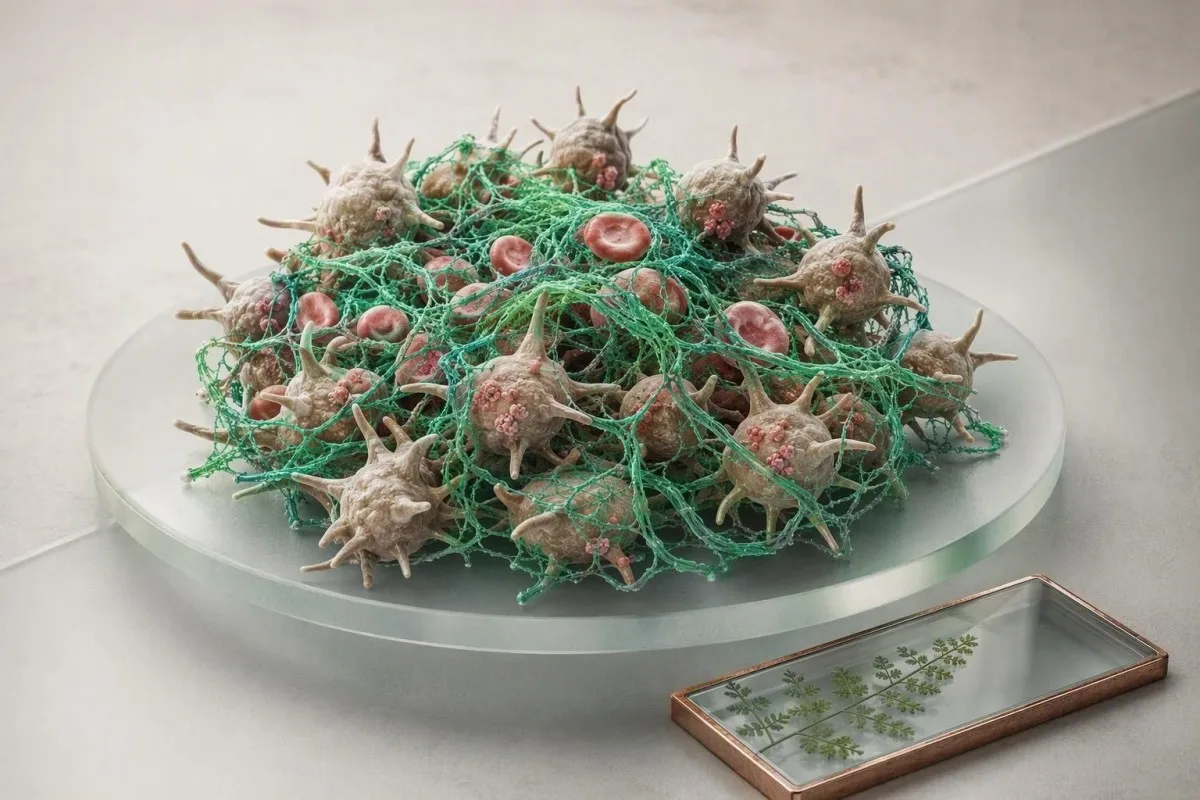
Tavaline sõeltestide tulemused on rahustavad, kuid need uurivad ainult hemostaasi valitud osi. Verevalumid, rohke menstruatsioon, verehüübed, perekondlik anamnees ja ravimid võivad siiski õigustada sihipärast testimist.


- Normaalne PT ja aPTT ei välista von Willebrand'i haigust, trombotsüütide funktsionaalseid häireid, faktori XIII puudulikkust, kergeid faktori puudulikkusi ega enamikku päritavatest trombofiiliatest.
- Tüüpiline täiskasvanu PT on umbes 10–13 sekundit ja INR umbes 0,8–1,2 ilma varfariinita, kuid iga labor peab esitama oma referentsvahemiku.
- Tüüpiline aPTT on sageli 25–35 sekundit; tavaline tulemus ei välista usaldusväärselt kerget faktori VIII, IX või XI puudulikkust.
- Tugev menstruaalverejooks mis imab kaitset iga tund rohkem kui 2 tundi järjest, vajab kiiret kliinilist hindamist isegi siis, kui sõeltestid on tavalised.
- Trombotsüütide arv mõõdab kogust, mitte toimivust; 250 × 10⁹/L loenduse korral võib esineda kliiniliselt oluline trombotsüütide funktsionaalne häire.
- D-dimeer aitab välistada venoosset tromboembooliat ainult siis, kui testi-eelne tõenäosus on madal või keskmine ja kasutatakse valideeritud teed.
- von Willebrand'i faktori testimine parim tõlgendada faktori VIII ja korduvate proovide võtmisega, kuna stress, rasedus, põletik ja östrogeen võivad tulemusi ajutiselt tõsta.
- Kiireloomulised sümptomid hõlmavad vere köhimist, äkilist hingeldust, ühepoolset jalgade turset, uusi neuroloogilisi sümptomeid, musti väljaheiteid või kontrollimatut veritsust.
Miks tavalised sõeltestide tulemused ei lõpeta uurimist
Tavalised PT, INR ja aPTT ei välista igasugust verejooksu või vere hüübimise häiret. Need hindavad peamiselt valitud vereplasma hüübimissüsteeme, samas kui trombotsüütide funktsioon, von Willebrandi tegur, faktor XIII ja paljud tromboosiriskid võivad jääda nähtamatuks. Alates 22. augustist 2026 on see erinevus kõige kasulikum lähtepunkt hüübimispaneeli tulemuste selgitamiseks.
Minu kliinilises praktikas on tavaline viga pidada tavalist paneeli universaalseks puhastussertifikaadiks. See nii ei ole. PT uurib välist ja ühist rada; aPTT uurib sisemist ja ühist rada; kumbki ei mõõda, kui hästi trombotsüüdid väikese veresoone vigastuse korral kinnituvad. Dr Thomas Klein näeb seda eriti pärast hambaoperatsioonist tingitud verejooksu või eluaegset tugevat menstruatsiooni, kui labori sõeltest näib täiesti tavaline.
Kantesti on AI vereanalüüsi analüsaator mis loeb hüübimisväärtusi koos täisvereanalüüsiga, maksa näitajate, ravimite ja esitatud sümptomitega, mitte ei deklareeri tulemust lihtsalt “hea”. Tavaline trombotsüütide arv 150–450 × 10⁹/l kinnitab, et ringlevaid trombotsüüte on enamiku inimeste jaoks piisavalt, kuid see ei näita, kas need trombotsüüdid signaale edastavad ja normaalselt agregaatuvad. Meie täieliku vereanalüüsi juhendis selgitab, miks arv ja funktsioon on eraldi küsimused.
Kasulik mentaalne mudel on kolm kihti: veresoone sein, trombotsüüdid ja hüübimisvalgud. PT ja aPTT uurivad peamiselt kolmandat kihti. Kui kellelgi on 25 minutit kestvad ninverejooksud, igemete veritsus, rauavaegus menstruatsioonist ja vanemal sarnased sümptomid, siis see anamnees on diagnostiliselt kaalukam kui üks tavaline sõeltesti tulemus.
Mida normaalne tulemus tegelikult tähendab
Tavaline sõeltesti paneeli tulemus tähendab, et mõõdetud hüübimisaeg langes selle labori intervalli piiresse vastava analüüsi tingimustes. See ei tõesta, et hemostaas on normaalne operatsiooni, sünnituse, trauma või antikoagulantide kasutamise ajal.
Mida PT, INR ja aPTT tegelikult mõõdavad
PT, INR ja aPTT mõõdavad aega, mis kulub labori plasmas fibriini moodustumiseks pärast kunstlike reaktiivide lisamist. Need on rajauuringud, mitte otsesed testid kogu keha veritsusriski või ohtliku verehüübe olemasolu kohta.
A PT umbes 10–13 sekundit, INR 0,8–1,2ja aPTT umbes 25–35 sekundit on tavalised täiskasvanute intervallid inimestel, kes ei kasuta antikoagulantravi, kuigi reaktiivid erinevad piisavalt, et aruande enda vahemik loeb. INR standardiseerib PT peamiselt varfariini jälgimiseks; see ei ole vere “paksuse” universaalne mõõdik. 1,1 INR ei ütle meile, kas aspiriin on trombotsüütide funktsiooni kahjustanud.
PT pikeneb, kui tegur VII on madal, K-vitamiini on vähe saadaval, varfariin on aktiivne või sünteetiline maksafunktsioon on hädaline. aPTT võib pikeneda hepariini, luupusantikoagulandi ja tegurite VIII, IX, XI või XII defitsiidi korral. kõrge PT ja normaalse aPTT korral ahendab võimalusi, kuid normaalsed väärtused ei muuda loogikat.
Ühe 42-aastase patsiendi puhul, keda ma üle vaatasin, oli aPTT enne protseduuri 29 sekundit, kuid tekkis ebaproportsionaalne kirurgilise saidi nõrgumine. Korduv anamneesi läbivaatus paljastas lapsepõlve ninverejooksu ja ema menorraagia; hilisemad testid toetasid von Willebrandi haigust. Sõeltest oli oma töö teinud – see lihtsalt ei olnud loodud lõpliku küsimuse vastamiseks.
Verejooksuhäired, mida tavalised PT ja aPTT võivad jätta märkamata
Von Willebrandi tõbi ja kvalitatiivsed trombotsüütide häired on kaks kõige sagedasemat põhjust limaskesta-nahalisele verejooksule normaalse PT ja aPTT korral. Faktor XIII puudulikkus, kerge hemofiilia, sidekoehaigused ja kohalikud günekoloogilised põhjused on muud võimalused.
Von Willebrandi tõbi (VWD) mõjutab umbes 0,1% inimestest kliiniliselt olulisel tasemel, kuigi madalad VWF tasemed on sagedasemad. See põhjustab tavaliselt korduvaid ninverejookse, kerget verevalumite teket, pikaajalist veritsust pärast hammaste eemaldamist ja tugevat menstruaalveritsust. ASH/ISTH/NHF/WFH diagnostilised juhised soovitavad VWF antigeeni, trombotsüütide-sõltuva VWF aktiivsuse ja faktori VIII määramist, kui anamneesis esinev verejooksufoon tekitab kahtlust (James et al., 2021).
Trombotsüütide funktsiooni häired võivad põhjustada normaalse PT, aPTT ja trombotsüütide arvu. Aspirin võib mõjutada trombotsüütide funktsiooni kogu trombotsüütide eluea jooksul – umbes 7–10 päeva – samal ajal kui ibuprofeeni toime on lühem ja varieeruvam; toidulisandid nagu kalaõli, ginkgo ja suurtes annustes küüslauk võivad olla olulised protseduuride ümber, kuigi tõendid rutiinse katkestamise kohta on segased. A trombotsüütide arv enne hammaste eemaldamist on kasulik, kuid see ei asenda anamneesi.
Faktor XIII stabiliseerib fibriini pärast PT ja aPTT poolt mõõdetud lõpp-punkti, seega raske puudulikkus võib jätta mõlemad sõeltestid normaalseks. See on haruldane, kuid korduv hiline verejooks pärast protseduuri, halb haavade paranemine või ebatavaline nabanööri verejooks vastsündinul peaks kutsuma esile spetsialisti hinnangu. Sellepärast ei tohiks “normaalne koagulatsioonipaneel” kunagi kaaluda üles silmapaistvat fenotüüpi.
Miks VWF teste tuleb mõnikord korrata
VWF on ägeda faasi reaktant: treening, infektsioon, ärevus, rasedus, östrogeen ja isegi proovivõtmise kogemus võivad seda tõsta. Piiripealne normaalne tulemus ägeda haiguse ajal võib vajada kordamist, kui patsient on kliiniliselt hea tervise juures, tavaliselt hematoloogilise plaani järgi.
Verejooksumuster suunab järgmist testi sageli paremini kui PT
Limaskesta verejooks viitab esmalt trombotsüütide või von Willebrandi probleemidele, samas kui sügavad lihasverevalumid ja korduvad liigesepõletikud viitavad koagulatsioonifaktori puudulikkusele. Muster ei ole täiuslik, kuid see määrab, kas jälgimine peaks keskenduma VWF-ile, trombotsüütide funktsioonile, faktori tasemele või anatoomiale.
Petehhiad, igemete veritsus, sagedased ninverejooksud, vahetult pärast hamba väljatõmbamist tekkinud veritsus ja tugevad menstruatsioonid on klassikaliselt primaarse hemostaasi vihjed. Standardiseeritud verejooksu hindamise tööriist on sageli informatiivsem kui küsimus “kas teil tekivad kergesti verevalumid?”, sest peaaegu kõigil tekib aeg-ajalt. Detail, mis suurendab minu muret, on verevalumid, mis on suuremad kui 5 cm ilma selge traumata, või verejooks, mis vajab meditsiinilist abi.
Sügavad hematoomid, hilinenud verejooks 24–48 tundi pärast operatsiooni ja hemartroosid viitavad pigem faktori puudulikkusele. Kerge hemofiilia võib siiski sõltuvalt testist ja faktori tasemest omada normaalselt aPTT-d, eriti kui faktori VIII või IX aktiivsus on üle umbes 30–40 IU/dL. Faktori määramise test – mitte korduv üldine sõeltest – annab sellele küsimusele vastuse.
Anemia pakub kasulikku ristkontrolli. Langev hemoglobiini, madal ferritiini tase, tõusev trombotsüütide arv ja rohke menstruatsiooniverejooks võivad peegeldada kroonilist rauakaotust ka siis, kui PT ja aPTT on normaalsed; vaadake üle naiste ferritiini väärtused koos verejooksu anamneesiga. Põhjus, miks see kombinatsioon oluline on, on kumulatiivne kaotus: üks “normaalne” menstruatsioon pole oluline, kuid aastatepikkune kaotus on.
Nahk ja sidekude võivad jäljendada vere hüübimisprobleemi
Ehlers-Danlose sündroomid, steroidide kasutamine, vananev nahk ja C-vitamiini puudus võivad põhjustada verevalumeid ilma kõrvalekalleteta vere hüübimise analüüsis. Kliinikud otsivad tavaliselt liigeseliikuvust, naha fragiilsust, toitumisanamneesi ja ravimeid enne ulatuslike faktoripaneelide määramist.
Miks tavalised sõeltestid ei välista verehüübimist
Normaalne PT, INR ja aPTT ei välista süvaveenide tromboosi, kopsuembooliat, antifosfolipiidide sündroomi ega kaasasündinud trombofiiliat. Need analüüsid mõõdavad vere hüübimiskiirust valmistatud plasmas, mitte seda, kas veri on jalas, kopsus või muus veresoones hüübinud.
Normaalne aPTT ei tähenda, et inimene on tromboosi eest kaitstud; tegelikult ei tekita mõned protrombootilised seisundid üldse sõeluuringu kõrvalekaldeid. Factor V Leiden, protrombiin G20210A, valgukao C puudulikkus, valgukao S puudulikkus ja antitrombiini puudulikkus nõuavad sihipärast testimist ning tulemused on kergesti moonutatud raseduse, ägeda tromboosi või antikoagulantravi ajal. Connors (2017) hoiatab, et ükskõikne trombofiiliatestide tegemine tekitab sageli segadust ilma ravi muutmata.
Kahtlustatava kopsuemboolia korral, ootamatu seletamatu hingeldus, pleuriidivalu rinnus, verine köha, minestus või kiire pulss nõuab samal päeval erakorralist hindamist, olenemata PT või aPTT tulemusest. Süvaveenide tromboosi kahtluse korral vajab ühepoolne sääre turse, soojus või valu kliinilist tõenäosusskoorimist ja ultraheli, mitte kindlustunde otsimist paneelist.
D-dimer on kõige kasulikum, kui eeltesti tõenäosus on madal või keskmine. Üle 50-aastastel täiskasvanutel kasutavad paljud valideeritud protseduurid vanusest sõltuvat läveks vanus × 10 ng/mL FEU; 70-aastasel on seega nendes protseduurides 700 ng/mL FEU lävi. Lugege nüansse meie D-dimeri täpsuse juhendis.
D-dimeer ja fibriinogeeni lisamine annavad vihjeid, mitte lõplikke vastuseid
D-dimer on tundlik, kuid mittespetsiifiline, samas kui fibrinogeen võib põletiku ajal olla kõrge ja raske haiguse alguses normaalne. Ühtki testi ei tohiks tõlgendada eraldi sümptomitest, ajastusest, pildiuuringutest ja ülejäänud hüübimispaneelist.
D-dimer tõuseb, kui ristseotud fibrinogeeni laguneb, seega võib see suureneda vanuse, raseduse, vähi, operatsiooni, trauma, infektsiooni ja haiglasse vastuvõtmisega – mitte ainult tromboosiga. D-dimer 1200 ng/mL FEU patsiendil 10 päeva pärast põlveoperatsiooni ei diagnoosi hüübimist; sümptomid ja pildiuuringud määravad järgmised sammud. Vastupidi, madal tulemus võib olla valesti rahustav, kui see on võetud pärast antikoagulatsiooni või väga hilja haiguse käigus.
Fibrinogeen tavaliselt vahemikus 2,0–4,0 g/L, kuid meetodid erinevad. Alla 1,5 g/L väärtus võib põhjustada protseduuril verejooksu ja alla 1,0 g/L tasemed aktiivselt veritseval patsiendil nõuavad sageli kiiret haiglapõhist asendamisotsust; see on kontekstitundlik, mitte koduse ravi lävi. Meie fibrinogeeni testijuhend selgitab, miks kõrged tasemed võivad alguses tarbimist varjata.
Kantesti AI märgib vastuolulised mustrid – näiteks pikaajaline PT madala fibrinogeenitaseme, langevate trombotsüütide ja ägeda haigusega – kui kiire ülevaatamise muster, selle asemel et omistada tähendust ühele numbrile. Kantesti on AI biomarkeri tõlgendamise platvorm mõeldud nende suhete säilitamiseks, kui kasutajad võrdlevad tulemusi erinevate kuupäevade vahel.
Normaalne D-dimer ei ole universaalselt lõplik
Negatiivne kõrge tundlikkusega D-dimer võib ohutult vähendada pildiuuringute vajadust ainult pärast seda, kui arst on hinnanud inimese madalaks või keskmise riskiks, kasutades valideeritud protseduuri. Rasedus, hiljutine operatsioon ja kõrge kliiniline tõenäosus muudavad seda võrrandit.
Ravimid, toidulisandid ja võtmisvead võivad tulemusi moonutada
Antikoagulandid, antiagregandid ja preanalüütilised vead võivad põhjustada vere hüübimise tulemuste ootamatult normaalse või ebanormaalse ilmnemise. Ravimi loetelu ja proovivõtu viis on sageli sama informatiivsed kui numbriline tulemus.
Vartariin tõstab tavaliselt INR-i, mittفرادineeritud hepariin pikendab sageli aPTT-d ja otsesed suukaudsed antikoagulandid võivad mõjutada PT-d, aPTT-d ja spetsiifilisi teste. Tavalised PT või aPTT tulemused ei saa kinnitada apiksabaani, rivaroksabaani, dabigatraani või edoksabaani puudumist või inaktiivsust. Spetsiifilised kalibreeritud ravimitasemed on mõnikord vajalikud enne kiiret operatsiooni või suure verejooksu korral.
Kahvatussinine tsitraadituub peab olema õiges vere-antikoagulandi suhtes. Selle alatäitmine võib valesti pikendada PT-d ja aPTT-d, ning hematokrit üle 55% võib vajada kohandatud tsitraadi mahtu, kuna liigne tsitraat seob testimise ajal liiga palju kaltsiumi. Need üksikasjad on igavad, kuni need takistavad tarbetut erakorralist uurimist.
Meie ülevaadetes küsime ka manustamise aja kohta: viimane annus, neerufunktsioon, kõhulahtisus, antibiootikumid, alkoholi tarbimine ja uued taimsed tooted. vereanalüüsi ajaskaala aitab eristada tegelikku füsioloogilist muutust halvasti ajastatud mõõtmisest. Ärge lõpetage ettenähtud antikoagulatsiooni laboriuuringu jaoks ilma arsti selgesõnalise plaanita.
Miks maksa leiud on olulised
Maks toodab enamikku hüübimisfaktoritest, nii et kõrvalekalletega PT võib olla varajane sünteetilise funktsiooni vihje, kui ka bilirubiin, albumiin või maksaensüümid on muutunud. PT üksi ei saa diagnoosida maksahaigust ja tavaline PT ei välista varajast haigust.
Millal sümptomid õigustavad von Willebrand'i või trombotsüütide testimist
Sihtotstarbeline VWF ja trombotsüütide testimine on põhjendatud, kui verejooks on korduv, ebaproportsionaalne, perekondlik või seotud rauavaegusega – isegi normaalse PT ja aPTT korral. Testimine on kõige produktiivsem, kui see on suunatud selgele kliinilisele küsimusele, mitte laialdaseks sõeltestiks.
Põhjused küsimiseks hõlmavad tugevat menstruatsiooni alates menarhest, sünnitusjärgset veritsust, liigset veritsust hammastest, kahte või enamat spontaanset ninatemist nädalas, korduvaid suuri verevalumeid või esimese astme sugulast, kellel on diagnoositud verehüübimishäire. Tugev menstruaalveritsus mõjutab kuni 30% reproduktiivses eas patsientidest, kuid ainult osa neist kannatab päriliku hemostaatilise häire all; anamnees tuvastab, kes saab uurimisest kasu.
VWD hindamine hõlmab tavaliselt VWF antigeeni, trombotsüütidest sõltuvat VWF aktiivsust ja faktori VIII aktiivsust. 2021. aasta ASH/ISTH/NHF/WFH juhendis kasutatakse VWF-i alla 0,30 IU/mL tüübi 1 VWD kinnitamiseks sõltumata veritsusest ja 0,30–0,50 IU/mL abnormaliseerunud veritsusega inimesel; tõlgendamine vajab siiski kohalikku analüüsi ekspertiisi (James et al., 2021).
Trombotsüütide agregatsiooni uuringud on spetsiifilised, ettevalmistustundlikud ja mitte mõistlik esimene test iga verevalumi korral. Neid võib kaaluda pärast ravimite mõju, trombotsütopeenia ja VWD hindamist. Kantesti AI saab korraldada sümptomite ja tulemuste kokkuvõtte arstile, kuid diagnoosimine nõuab raviarsti ja sageli hemostaasi laborit.
Rasedus nõuab erinevat baastaset
VWF ja faktor VIII suurenevad raseduse ajal normaalselt, nii et varasem madal väärtus võib hilise gestatsiooniga normaalseks muutuda. Varase sünnitusjärgse verejooksu või väljakujunenud VWD-ga patsientidel vajavad nad enne sünnitust obstetriku-hematoloogia plaani, mitte ühte kolmanda trimestri sõeltesti.
Mis juhtub, kui PT või aPTT on ebatavaline
Segamisuuring aitab eristada puuduvat hüübimisfaktorit inhibiitori korral, kui PT või aPTT on pikenenud. Korrektsioon pärast patsiendi plasma segamist normaalse plasmaga viitab puudulikkusele; püsiv pikenemine viitab inhibiitorile või ravimi toimele.
Test on petlikult lihtne: labor ühendab võrdsed osad patsiendi ja normaalse kogutud plasma, seejärel kordab hüübimisanalüüsi kohe ja mõnikord pärast inkubatsiooni. Korrektsioon viiteintervalli poole toetab faktori puudulikkust; korrektsiooni ebaõnnestumine võib viidata lupus antikoagulandile, spetsiifilisele faktorinhibiitorile või ravimi toimele. Meie segamisuuringute selgitaja käsitleb loogikat üksikasjalikumalt.
Lupuse antikoagulant pikendab tavaliselt in vitro aPTT-d, kuid on seotud trombiohu, mitte verejooksuga. See näiline vastuolu teeb patsientidele mõistetavalt muret. Verejooks muutub probleemiks peamiselt siis, kui esineb teine probleem – näiteks trombotsütopeenia, harvaesineva lupuse hüpoprotrombiineemia korral madal protrombiinitase või antikoagulantravi.
Faktorite määramised kvantifitseerivad aktiivsust ühikutes IU/dL või IU/mL ja meetodi valik on oluline. Mõnda kerget hemofiilia A varianti leitakse ühefaasiliste ja kromogeensete VIII faktori määramistega erinevalt, seega võib normaalne esialgne määramine mõnikord spetsialiseerunud keskuses teise meetodiga korduda. See on klassikaline näide hüübimiskatsete piirangutest mis on metodoloogilised, mitte kujuteldavad.
Ärge tõlgendage segamisuuringuid üksi
Segamisuuringute mustrid sõltuvad reaktiivi tundlikkusest, inkubatsiooniajast ja kõrvalekalde määrast. Hematoloog integreerib need trombiiniaja, anti-Xa määramise, faktoritasemete ja ravimiallergiaga enne haiguse nimetamist.
Millal päritavate trombofiiliate testimine on kasulik – ja millal mitte
Päriliku tromboosiohu testimine on tavaliselt reserveeritud valitud inimestele, kellel on põhjustamata või ebatavalise asukohaga tromb, tugev perekonnamuster või otsused, kus tulemus muudaks ravi. Normaalne PT ja aPTT ei välista ega kinnita pärilikku trombiohtu.
Testimine harva aitab pärast selgelt põhjustatud trombi suure operatsiooni, pikaajalise liikumatusvõime või ajutise riskiteguri tõttu, kuna ravi kestus sõltub tavaliselt juhtumist ja verejooksude riskist. Connors (2017) rõhutas, et positiivne pärilik variant ei muuda sageli antikoagulatsiooni otsuseid pärast esimest tavalist venoosset tromboemboliat. Tulemus võib siiski olla oluline hoolikalt valitud pereplaneerimise või ebatavaliste tromboosijuhtumite korral.
C-valgu, S-valgu ja antitrombiini tasemed võivad ägeda tromboosi, raseduse, maksahaiguse, nefrootilise taseme valgu kadu ja antikoagulantravi tõttu väheneda. Nendel perioodidel testimine võib inimese valesti märgistada. Praktikas valib hematoloog tavaliselt aja pärast ägedat juhtumit ja pärast seda, kui on läbi vaadanud, kas ravi saab ohutult katkestada – kui testimine on üldse kasulik.
Antifosfolipiidsündroom on erinev: see on omandatud, nõuab püsivaid positiivseid antikehi vähemalt 12-nädalase intervalliga ja võib mõjutada ravi valikut. Normaalne aPTT ei välista seda. Insuldi, korduvate rasedusprobleemide või põhjustamata trompide korral peaks küsimus olema “kas see test muudab minu plaani?” pigem kui “kas me saame kõike testida?”.”
Perekonnaseis vajab täpsust
“Minu vanaemal oli tromboos 82-aastaselt” on vähem murettekitav kui vanema või õe või venna põhjustamata tromboos enne 50. eluaastat, korduvad juhtumid või ebatavaline asukoht, nagu tserebraalne venoosne tromb. Täpsed vanused, käivitajad ja diagnoosid on kasulikumad kui pikk kontrollimata perekonnaprobleemide loetelu.
Tavalised tulemused enne operatsiooni, hambaravi või sünnitust
Normaalne PT ja aPTT enne protseduuri ei taga normaalset kirurgilist verejooksu, seega on struktureeritud verejooksu ajalugu parem esimene sõel. Ootamatu varasem hambaprotees, operatiivne või sünnitusjärgne verejooks tuleb avalikustada isegi siis, kui operatsioonieelne paneel on normaalne.
Rutiinne PT ja aPTT testimine madala riskiga inimestel ilma verejooksu ajalooga on väheproduktiivne ja võib põhjustada viivitusi tühiste kõrvalekallete tõttu. Siirduse, operatsioonisaali naasmise, haava uuesti sidumise, sünnitusjärgse verejooksu või pikaajalise verejooksu pärast hambaväljatõmbamist ajalugu on ennustavam. Anestesioloogid ja kirurgid otsustavad seejärel, kas VWF-i testimine, faktorite määramised või hematoloogia arvamus on sobiv.
Hambaprotseduuride puhul ärge eeldage, et iga antikoagulant tuleb peatada. Paljud väikesed protseduurid on ohutumad kohalike hemostaatiliste meetmete ja retsepti alusel koostatud plaaniga kui järelevalveta katkestamisega, mis tekitab trombiohtu. Trombotsüütide arv üle 50 × 10⁹/L on sageli piisav paljude invasiivsete protseduuride jaoks, kuid protseduuri tüüp ja trombotsüütide funktsioon võivad seda läve oluliselt muuta.
Sünnitus põhjustab kiiret füsioloogilist muutust: VWF-i tasemed võivad sünnitusjärgselt päevade kuni nädalate jooksul tagasi baastasemeni langeda. Keegi, kellel on olnud varem raske sünnitusjärgne verejooks, vajab dokumenteeritud plaani sünnituseks, traneksaamhappe või faktori toetuse korral, kus see on näidustatud, ja jälgimist. raseduse trombotsüütide vahemikud pakuvad kasulikku konteksti, kuid ei asenda seda plaani.
Mida kaasa võtta operatsioonieelsele visiidile
Võtke kaasa varasemad operatsioonimärkused, kui need on saadaval, täpne ravimite ja toidulisandite loetelu, perekonnadiagnoosid ja varasemate verejooksude kuupäevad. Ebamäärane “ma veritsen palju” on mõistetav, kuid vähem toimiv kui “hambaväljatõmbamise järel oli sidumist vaja 18 tundi.”
Kuidas lugeda koagulatsiooni tulemusi kliinilises kontekstis
Koagulatsioonitulemused on kõige usaldusväärsemad, kui neid tõlgendatakse mustrina kogu CBC, maksa profiili, neerufunktsiooni, ravimiallergiate ja ajastuse kaudu. Üksik normaalne või kõrvalekaldeline hüübimisaeg räägib harva kogu loo.
Isolaatne INR 1,3 tervel inimesel võib peegeldada reaktiivi variatsiooni, dieeti, varajast K-vitamiini toimet või proovi käitlemist; see väärib enne häirekella andmist kinnitust. INR 1,3 koos ikteruse, madala albumiini, kõrge bilirubiini ja verevalumitega on väga erinev kliiniline pilt. Läbivaatus maksa profiili komponendid selle asemel, et käsitleda PT-d eraldiseisva maksatestina.
Trombotsüüdid alla 50 × 10⁹/L suurendavad veritsusriski paljude protseduuride korral, samas kui kiiresti langev näit võib olla oluline isegi enne selle piiri ületamist. Näit, mis langeb 48 tunni jooksul 280-lt 110 × 10⁹/L-ni, sisaldab teavet, mida ühekordsel 110-l pole. Sellepärast on suundumused, proovikommentaarid ja kliiniline seisund olulised.
Kantesti on AI-põhine vereanalüüsi analüüsitööriist kasutatakse 127+ riigis, et võrrelda tulemusi nende enda labori intervallide ja varasemate väärtustega. Meie vereanalüüsi võrdlusjuhend põhineb sellel praktilisel reaalsusel: numbrid on hetktõmmised, kuid otsused sõltuvad tavaliselt suunast ja kontekstist.
Ärge “parandage” normaalseid tulemusi
K-vitamiini, rauda, taimeteesid ja toidulisandeid ei tohiks alustada lihtsalt selleks, et parandada koagulatsiooninäitu, mis on juba vahemikus. Ravi järgib põhjust – toitumispuudust, varfariini juhtimist, maksahaigust või tuvastatud häiret – mitte soovi optimeeritud hüübimisaja järele.
Sümptomid, mis vajavad kiiret ravi hoolimata tavalistest sõeltestide tulemustest
Otsige erakorralist abi kontrollimatu verejooksu, mustade või veriste väljaheidete, vere köhimise, äkiliste rindkere sümptomite, ühepoolse jalgade turse, minestamise või uue nõrkuse või kõnedefitsiidi korral – isegi kui PT ja aPTT on normaalsed. Need sümptomid vajavad uurimist, kuvamist või kordustestimist, mida rutiinne paneel ei saa pakkuda.
Helistage hädaabiteenistusse äkilise hingeldamise, valu rinnus, kokkuvarisemise, vere köhimise või uue ühepoolse nõrkuse korral. Need võivad viidata kopsuembooliale, südamehaigusele, insuldile või muudele ajatundlikele seisunditele ning ambulatoorsete koagulatsioonitulemuste ootamine on ohtlik. Normaalne D-dimeer või aPTT eelmisest nädalast ei kehti automaatselt tänaste sümptomite kohta.
Kiire sama päeva hindamine on mõistlik verejooksu korral, mis ei peatu pärast 15–20 minutit tugevat pidevat survet, vere oksendamise, mustade tõrvaste väljaheidete, uriinis nähtava vere, tugeva peavalu pärast peavigastust või väga tugeva tupeveritsuse korral koos pearinglusega. Varfariini, otseseid suukaudseid antikoagulante, hepariini, aspiriini või kahekordset trombotsüütide agregatsiooni inhibeerivat ravi võtvatel patsientidel peaks olema madalam soovituspiir.
Samuti muretsen, kui väsimus, kahvatus, hingeldus treppidel ja tugev verejooks esinevad koos, kuna hemoglobiin võib vaikselt väheneda. A madala hematokriti juhend aitab selgitada laboripoolt, kuid sümptomid määravad kiireloomulisuse. Ärge sõitke ise autoga, kui tunnete end pearinglusena.
Mida öelda erakorralises vastuvõtus
Märkige sümptomite alguse aeg, antikoagulandi nimi ja viimane annus, raseduse staatus, varasem trombi või verejooksu anamnees ja täpsed tulemused, kui need on saadaval. Need viis üksikasja võivad kiirendada ohutut triaaži rohkem kui pikk loetelu marginaalsetest laborilippudest.
Kuidas valmistuda sümptomipõhiseks järelvisiidiks
Kõige kasulikum järelkohtumine hõlmab kuupäevastatud verejooksu või trombi anamneesi täpse ravimite ajastuse ja algse laboriaruandega. See võimaldab arstil valida väikese arvu kõrge saagikusega teste, selle asemel et korrata üldist koagulatsioonipaneeli.
Salvestage, millal sümptomid algasid, kestus, päästikud, kas surve toimis, kas ravi oli vajalik ja mõlema poole perekondlik anamnees. Perioodide puhul dokumenteerige raskete päevade, üleujutuste, öiste muutuste ja rauarohtude arv. See muudab mälupõhise konsultatsiooni tõendiks, mis võib toetada VWF-testimist või suunamist.
Tooge täielik aruanne, sealhulgas kogumise kuupäev, referentsvahemikud, proovikommentaarid ja kõik tulemused – mitte ainult ebanormaalsed lipud. Kui võimalik, lisage hiljutine CBC, ferritiin, maksa- ja neerunäitajate paneel. Kantesti AI saab üles laaditud aruannetest luua selge kronoloogilise kokkuvõtte, samas kui meie meditsiinilise valideerimise standardid kirjeldavad AI-toega tõlgenduse piiranguid ja miks kliiniline kinnitus on endiselt vajalik.
Dr Thomas Kleini praktiline reegel on lihtne: ärge küsige “kõiki verehüübimiskatseid”. Küsige, milline diagnoos teie sümptomitele kõige paremini sobib, milline test muudaks ravi, ja kas ajastus võib selle ebausaldusväärseks muuta. See vestlus on rahulikum, odavam ja tavaliselt täpsem.
Küsimused, mida tasub küsida
Küsige, kas teie verejooksu muster viitab VWD või trombotsüütide düsfunktsioonile, kas ravimid võivad seda selgitada, kas korduv testimine peaks toimuma tervena olles ja mida tuleks enne tulevast protseduuri või rasedust muuta. Spetsialistide järelevalveks saavad lugejad üle vaadata meie Meditsiininõukogu.
Tavaliste koagulatsioonipaneelide praktiline kokkuvõte
Tavaline koagulatsioonipaneel on mõõdetavate teede osas rahustav, kuid see ei lõpeta juhtumit, kui sümptomid või perekonnas esinev anamnees on veenvad. Õige järgmine samm on sümptomipõhine testimine, mitte paanika ja mitte eiramine.
Kui teil ei esine ebatavalist verejooksu, verehüüvete sümptomeid, ei võta te ravimeid ega esine tugevat perekondlikku anamneesi, on tavapärane PT, INR ja aPTT üldiselt rahustav. Sõeltestid on kasulikud just seetõttu, et need tuvastavad paljusid omandatud faktori probleeme, ravimite mõju ja peamisi teede kõrvalekaldeid. Need ei ole mõeldud iga häire tuvastamiseks iga inimese puhul.
Kui sümptomid püsivad, võib järelmeetmete nimekiri sisaldada CBC ja määret, ferriiti, VWF antigeeni ja aktiivsust, faktorit VIII, trombotsüütide funktsiooni testimist, fibrinogeeni, trombiiniaega, anti-Xa taset, segamisuuringut, faktori määramist, D-dimeeri või kujutist. Test peaks vastama küsimusele. Üksikasjaliku tehnilise viite saamiseks vaadake meie aPTT ja hüübimise juhend.
Kantesti AI ei diagnoosi verejooksu või trombootilisi häireid, samuti ei tee seda ükski isoleeritud tulemus. Meie meeskonna eesmärk on muuta aruanne piisavalt loetavaks parema kliinikute vestluse jaoks – eriti siis, kui “tavaline” ei vasta teie keha käitumisele. Sellele mittevastavusele tasub tõsiselt läheneda.
Lõplik ohutuspunkt
Ärge kunagi muutke ettenähtud antikoagulantravi, antiagregantravi või K-vitamiini tarbimist ainult veebipõhise tõlgenduse tõttu. Ohutu plaan sõltub sellest, miks ravim välja kirjutati, neerude ja maksa funktsioonist, protseduuri riskist ja verehüüvete tagajärgedest.
Korduma kippuvad küsimused
Kas mul võib esineda vere hüübimishäire, mille korral PT ja aPTT on normaalsed?
Jah. Tavaline protrombiiniaeg (PT) ja aktiveeritud osaline tromboplastiiniaeg (aPTT) ei välista von Willebrandi tõbe, kvalitatiivseid trombotsüütide häireid, faktori XIII puudulikkust, kergeid faktori puudulikkusi, sidekoe põhjustatud verevalumeid ega lokaalseid veritsuse põhjuseid. Von Willebrandi testimine hõlmab tavaliselt VWF-antigeeni, VWF-aktiivsuse ja faktori VIII aktiivsuse määramist ning VWF-i taseme < 0,30 IU/mL korral toetab see 1. tüüpi von Willebrandi tõbe, olenemata veritsusajaloost. Korduvad ninaveenid, tugevad menstruatsioonid, ebanormaalne verejooks pärast hamba väljatõmbamist või perekondlik anamnees õigustavad kliinikujuhitud ülevaatust, hoolimata normaalsest sõeluuringu tulemustest.
Mis on normaalne PT INR ja aPTT vahemik?
Tavaline täiskasvanud võrdlusvahemikud on umbes 10–13 sekundit PT korral, 0,8–1,2 INR korral ja 25–35 sekundit aPTT korral inimestel, kes antikoagulante ei kasuta. Iga labori reaktiivid ja analüsaator loovad oma valideeritud vahemiku, seega eelistatakse aruandel trükitud vahemikku. INR on loodud peamiselt PT standardiseerimiseks varfariini jälgimiseks ega mõõda vere üldist paksust. Tavalise väärtuse olemasolu ei tõenda, et trombotsüütide funktsioon või von Willebrandi tegur on normaalne.
Kas normalne stężenie D-dimeru wyklucza zakrzep?
Normaalselt kõrge tundlikkusega D-dimeeri test võib välistada süvaveenitromboosi või kopsuemboolia ainult juhul, kui valideeritud kliiniline tee hindab isiku madalaks või keskmise tõenäosusega. Üle 50-aastastel täiskasvanutel kasutavad paljud teed vanusest sõltuvat piirmäära vanus × 10 ng/mL FEU, näiteks 700 ng/mL FEU 70-aastasel. D-dimeer on vähem kasulik pärast operatsiooni, raseduse ajal, vähi korral, ägeda haiguse ajal või kui kliiniline tõenäosus on kõrge. Äkiline hingeldus, valu rinnus, minestamine või ühepoolne jala turse vajab kiireloomulist hindamist, olenemata varasemast normaalsest tulemusest.
Miks on minu PT ja aPTT normaalsed, aga ma väärtan kergesti?
Lihtne verevalumite teke normaalse PT ja aPTT korral võib olla tingitud trombotsüütide düsfunktsioonist, von Willebrandi tõvest, ravimitest nagu aspiriin, steroididest tingitud naha õhenemisest, sidekoehaigustest, toitumispuudusest või tavalisest traumast. Trombotsüütide arv vahemikus 150–450 × 10⁹/L kinnitab kogust, kuid ei näita trombotsüütide töövõimet. 5 cm suuremad verevalumid ilma selge vigastuseta, igemete või ninaverejooks, rohke menstruatsioon või rauavaegus muudavad anamneesi ja sihipärase testimise otstarbekamaks. Arst vaatab tavaliselt enne spetsiifiliste analüüside määramist üle ravimid, CBC, ferritiini ja verejooksumustri.
Kas ma peaksin enne operatsiooni kordama tavalist vere hüübimise paneeli?
Kordubluse vere hüübimise aja (PT) ja aktiveeritud osalise tromboplastiini aja (aPTT) korduvat määramist enne operatsiooni ei peeta rutiinselt kasulikuks inimestel, kellel pole verejooksu anamneesi, maksahaigust ega kokkupuudet antikoagulantidega. Enne vereülekande vajadus, korduv operatsioon, pikaajaline veritsus hambaeemaldusel, sünnitusjärgne verejooks või veritsushäirega sugulane on olulisem kui korduv vere hüübimise aja uuring. VWF antigeeni, aktiivsuse ja faktori VIII määramine võib olla sobivam, kui selline anamnees esineb. Ärge kunagi lõpetage varfariini, otsese toimega suukaudse antikoagulandi, aspiriini või muu ettenähtud antitrombootilise ravimi võtmist ilma retsepti väljakirjutaja poolt protseduurispetsiifilise plaanita.
Kas tavaline aPTT välistab luupusantikoagulandi või antifosfolipiidsündroomi?
Ei. Tavalik aPTT ei välista erütematoosset luupust ega antifosfolipiidide sündroomi, kuna reaktiivide tundlikkus varieerub ja mõned haiged inimesed saavad normaalseid sõeluuringuid. Antifosfolipiidide sündroom eeldab kliinilisi kriteeriume, nagu tromboos või spetsiifilised raseduse kahjustused, pluss püsivad positiivsed antikehad testidel, mis tehakse vähemalt 12-nädalaste intervallidega. Luupusantikoagulant võib laboratoorselt pikendada aPTT-d, kuid see on seotud pigem hüübimise kui verejooksuga. Spetsialisti tõlgendus on eriti oluline, kui tromboos tekkis noores eas, kordub või esineb ebatavalises kohas.
Hangi AI-toega vereanalüüsi analüüs juba täna
Liitu enam kui 2 miljoni kasutajaga üle maailma, kes usaldavad Kantesti-d kohese ja täpse laborianalüüsi jaoks. Laadi üles oma vereanalüüsi tulemused ja saad põhjaliku tõlgenduse 15,000+ biomarkerite kohta sekunditega.
📚 Viidatud teaduspublikatsioonid
Klein, T., Mitchell, S., & Weber, H. (2026). Urobilinogeen uriinianalüüsis: täielik uriinianalüüsi juhend 2026. (2026). Zenodo. https://doi.org/10.5281/zenodo.18226379. ResearchGate: https://www.researchgate.net/; Academia.edu: https://www.academia.edu/. Kantesti AI Medical Research.
Klein, T., Mitchell, S., & Weber, H. (2026). Raua uuringute juhend: TIBC, raua küllastus ja sidumismahutavus. (2026). Zenodo. https://doi.org/10.5281/zenodo.18248745. ResearchGate: https://www.researchgate.net/; Academia.edu: https://www.academia.edu/. Kantesti AI Medical Research.
📖 Välised meditsiinilised viited
⚕️ Meditsiiniline lahtiütlus
Käesolev artikkel on mõeldud üksnes hariduslikel eesmärkidel ega kujuta endast meditsiinilist nõuannet. Diagnoosi ja ravivalikute otsuste tegemiseks konsulteeri alati kvalifitseeritud tervishoiutöötajaga.
E-E-A-T usaldussignaalid
Kogemus
Arsti juhitud kliiniline ülevaade labori tõlgendamise töövoogudest.
Ekspertiis
Laborimeditsiin keskendub sellele, kuidas biomarkerid käituvad kliinilises kontekstis.
Autoriteetsus
Kirjutanud dr Thomas Klein, ülevaade: dr Sarah Mitchell ja prof dr Hans Weber.
Usaldusväärsus
Tõenduspõhine tõlgendus selgete edasiste sammudega, et vähendada ärevust.