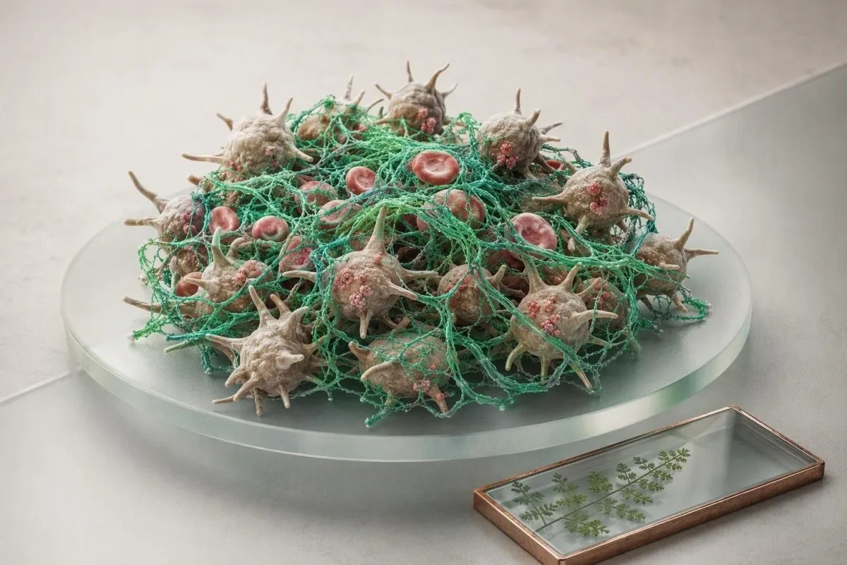
Normale siftingsresultate is gerusstellend, maar dit toets slegs geselekteerde dele van haemostase. Kneusing, swaar periodes, bloedklonte, familiegeskiedenis en medisyne kan steeds gemikte toetsing regverdig.


- Normale PT en aPTT sluit nie von Willebrand-siekte, bloedplaatjie-funksie afwykings, faktor XIII tekort, ligte faktor tekorte, of meeste oorgeërfde trombofilieë uit nie.
- Tipiese volwasse PT is ongeveer 10–13 sekondes en INR ongeveer 0.8–1.2 sonder warfarin, maar elke laboratorium moet sy eie verwysingsinterval verskaf.
- Tipiese aPTT is dikwels 25–35 sekondes; 'n normale resultaat sluit nie ligte faktor VIII, IX, of XI tekort betroubaar uit nie.
- Swaar menstruele bloeding wat beskerming elke uur vir meer as 2 opeenvolgende ure week, verdien dringende kliniese assessering selfs wanneer siftings toetse normaal is.
- Bloedplaatjietelling meet hoeveelheid, nie prestasie nie; 'n telling van 250 × 10⁹/L kan saam voorkom met 'n klinies beduidende bloedplaatjie-funksie afwyking.
- D-dimeer help om veneuse trombo-embolisme uit te sluit slegs wanneer pre-toets waarskynlikheid laag of intermediêr is en 'n gevalideerde pad gebruik word.
- Von Willebrand-faktor toetsing word die beste interpretasie met faktor VIII en herhalende monsters omdat stres, swangerskap, inflammasie en estrogeen resultate tydelik kan verhoog.
- Dringende simptome sluit bloed hoes, skielike kortasem, een-sided been swelling, nuwe neurologiese simptome, swart stoelgange, of ongekontroleerde bloeding in.
Hoekom normale siftingsresultate nie die ondersoek beëindig nie
Normale PT, INR en aPTT sluit nie elke bloeding- of stollingversteuring uit nie. Hulle assesseer hoofsaaklik geselekteerde plasma stollingspaaie, terwyl plaatjie funksie, von Willebrand faktor, faktor XIII en baie trombose risiko's onsigbaar kan bly. Vanaf 22 Augustus 2026 is daardie onderskeid die nuttigste beginpunt vir koagulasiestelresultate wat verduidelik word.
In my kliniese praktyk is die algemene fout om 'n normale paneel as 'n universele vrystelling sertifikaat te behandel. Dit is nie. PT monster die eksterne en algemene pad; aPTT monster die interne en algemene pad; geen meet hoe goed plaatjies kleef by 'n klein-vatsel besering nie. Dr Thomas Klein sien dit veral na tandheelkundige bloeding of lewenslange swaar periodes, wanneer die laboratorium skerm heeltemal onopmerklik voorkom.
Kantesti is 'n KI-bloedtoetsontleder wat koagulasiewaardes langs die volledige bloedtelling, lewermerkers, medisyne en gerapporteerde simptome lees eerder as om “n resultaat eenvoudig ”reg" te verklaar. 'n Normale plaatjie telling van 150–450 × 10⁹/L bevestig dat daar genoeg sirkulerende plaatjies is vir die meeste mense, maar dit kan nie wys of daardie plaatjies normaal sein en aggregeer nie. Ons volledige bloedtelling-gids verduidelik hoekom telling en funksie aparte vrae is.
'n Nuttige denkmodel is drie lae: vaatwand, plaatjies en stollingproteïene. PT en aPTT ondersoek meestal die derde laag. As iemand neusbloeding het wat 25 minute duur, tandvleisbloeding, ystertekort van periodes, en 'n ouer met soortgelyke simptome, dra daardie geskiedenis meer diagnostiese gewig as een normale siftings tekening.
Wat 'n normale resultaat eintlik beteken
'n Normale siftings paneel beteken dat die gemete stolling tyd binne daardie laboratorium se interval geval het onder daardie toets se toestande. Dit bewys nie dat hemostase normaal sal wees tydens chirurgie, kind geboorte, trauma, of antikoagulant blootstelling nie.
Wat PT, INR en aPTT werklik meet
PT, INR en aPTT meet die tyd wat dit neem vir laboratorium plasma om fibrin te vorm nadat kunsmatige reagense bygevoeg is. Hulle is pad siftings, nie direkte toetse van hele-liggaam bloeding risiko of of 'n persoon 'n gevaarlike klont het nie.
A PT van ongeveer 10–13 sekondes, INR 0.8–1.2, en aPTT van ongeveer 25–35 sekondes is algemene volwasse intervalle by mense wat nie antikoagulante gebruik nie, hoewel reagense genoeg verskil dat die verslag se eie reeks wen. INR standaardiseer PT hoofsaaklik vir warfarin monitering; dit is nie “n universele maatstaf van ”bloed dikte" nie. 'n 1.1 INR vertel ons nie of aspirien plaatjie funksie benadeel het nie.
PT word verleng wanneer faktor VII laag is, vitamien K beskikbaarheid verminder word, warfarin aktief is, of sintetiese lewer funksie benadeel is. aPTT kan verleng met heparien, lupus antikoagulant, en tekorte wat faktore VIII, IX, XI of XII behels. Die patroon van hoë PT met normale aPTT vernou moontlikhede, maar normale waardes keer die logika om nie.
'n 42-jarige pasiënt wat ek hersien het, het aPTT 29 sekondes gehad voor 'n prosedure, maar het buitensporige chirurgiese wond dreining ontwikkel. Herhaalde geskiedenis het kinderlike neusbloeding en moederlike menopouse bloeding onthul; daaropvolgende toetsing het von Willebrand siekte ondersteun. Die siftings het sy werk gedoen—dit was net nie ontwerp om die finale vraag te beantwoord nie.
Bloedingstoestande wat normale PT en aPTT kan mis
Von Willebrand-siekte en kwalitatiewe bloedplaatjie-afwykings is die twee algemeenste verklarings vir mukokutane bloeding met normale PT en aPTT. Faktor XIII-tekort, ligte hemofilie, bindweefselafwykings en plaaslike ginekologiese oorsake is ander moontlikhede.
Von Willebrand-siekte (VWD) raak ongeveer 0.1% van mense op 'n klinies betekenisvolle vlak, hoewel lae VWF-metings meer algemeen is. Dit veroorsaak tipies herhalende neusbloeding, maklike kneusing, verlengde tandebloeding en swaar menstruele bloeding. Die ASH/ISTH/NHF/WFH diagnostiese riglyn beveel VWF-antigeen-, bloedplaatjie-afhanklike VWF-aktiwiteit en faktor VIII-toetsing aan wanneer bloedingsgeskiedenis verdagte wees (James et al., 2021).
Bloedplaatjie-funksieafwykings kan normale PT, aPTT en bloedplaatjietelling produseer. Aspirien kan bloedplaatjie-funksie beïnvloed vir die bloedplaatjie-lewe – ongeveer 7–10 dae – terwyl ibuprofen se effek korter en veranderlik is; aanvullings soos visolie, ginkgo en hoë-dosis knoffel kan belangrik wees rondom prosedures, hoewel bewyse vir roetine-staking gemeng is. 'n Bloedplaatjie-telling voor tandekstraksie is nuttig, maar dit kan nie 'n bloedingsgeskiedenis vervang nie.
Faktor XIII stabiliseer fibrin na die eindpunt wat deur PT en aPTT gemeet word, so ernstige tekort kan beide skerms normaal laat. Dit is skaars, maar herhalende vertraagde post-prosedure bloeding, swak wondgenesing, of ongewone naelstring-bloeding by “n pasgeborene moet spesialistoorsig aanmoedig. Dit is hoekom ”n "normale koagulasiepaneel" nooit 'n treffende fenotipe moet omverwerp nie.
Hoekom VWF-toetse soms herhaal moet word
VWF is 'n akute-fase reaktant: oefening, infeksie, angs, swangerskap, estrogeen en selfs die monstername-ervaring kan dit verhoog. 'n Grens-normale resultaat verkry tydens akute siekte mag herhaal moet word wanneer klinies gesond, gewoonlik met 'n hematologieplan.
Bloedingspatroon rig dikwels die volgende toets beter as PT
Mukosale bloedingspunte wys eers na bloedplaatjie- of Von Willebrand-probleme, terwyl diep spierbloeding en herhalende gewrigswelling dui op koagulasiefaktor-tekort. Die patroon is nie perfek nie, maar dit bepaal of opvolg op VWF, bloedplaatjie-funksie, faktore of anatomie moet fokus.
Petechiae, tandvleisbloeding, gereelde neusbloeding, onmiddellike post-tandebloeding en swaar periodes is klassiek primêre hemostase leidrade. “n Gestandaardiseerde bloedingsbeoordelingsinstrument is dikwels meer onthullend as om te vra ”kneus jy maklik?" omdat byna almal dit af en toe doen. Die detail wat my kommer verander, is kneusing groter as 5 cm sonder duidelike trauma, of bloeding wat mediese sorg benodig.
Diep hematomas, vertraagde bloeding 24–48 uur na chirurgie, en hemartroses is meer suggestief van faktor tekort. Lig hemofilie kan steeds 'n normale aPTT hê afhangende van die toets en faktor vlak, veral wanneer faktor VIII of IX aktiwiteit bo ongeveer 30–40 IU/dL is. 'n Faktor toets – nie 'n herhaalde generiese toets nie – beantwoord daardie vraag.
Anemie bied 'n nuttige dwarstoets. Dalende hemoglobien, lae feritien, stygende bloedplaatjiegetal en swaar periodes kan chroniese ysterverlies weerspieël selfs wanneer PT en aPTT normaal is; hersien feritienwaardes by vroue langs die bloedingstorie. Die rede waarom hierdie kombinasie saak maak, is kumulatiewe verlies: een “normale” periode is irrelevant, maar jare se verlies is nie.
Vel en bindweefsel kan 'n stolprobleem naboots
Ehlers-Danlos-sindrom, steroïedblootstelling, verouderende vel en vitamien C-tekort kan kneusplekke veroorsaak sonder 'n abnormale stollingstoets. Klinici soek gewoonlik na gesamentlike hipermobiliteit, velvrugtheid, dieetgeskiedenis en medisyne voordat breë faktorpanele bestel word.
Hoekom normale siftings toetse nie 'n bloedklont uitsluit nie
Normale PT, INR en aPTT sluit nie diepvenetrombose, pulmonêre embolisme, antifosfolipied-sindroom, of oorerflike trombofilie uit nie. Hierdie toetse meet stollingspoed in voorbereide plasma, nie of 'n stolsel in 'n been, long of ander bloedvat gevorm het nie.
'n Normale aPTT beteken nie dat 'n persoon teen trombose beskerm is nie; trouens, sommige protrombotiese toestande lewer geen siftingsafwyking hoegenaamd nie. Faktor V Leiden, protrombien G20210A, proteïen C-tekort, proteïen S-tekort en antitorombien-tekort vereis geteikende toetsing, en resultate word maklik verdraai tydens swangerskap, akute trombose of antikoagulante behandeling. Connors (2017) waarsku dat onoordeelkundige trombofilie-toetsing dikwels verwarring skep sonder om sorg te verander.
Vir vermoedelike pulmonêre embolisme, skielike onverklaarbare kortasemigheid, pleuritis-borspyn, bloedhoes, flouval, of 'n vinnige pols regverdig dieselfde dag noodbeoordeling, ongeag PT of aPTT. Vir moontlike diepvenetrombose, benodig eenzydige kuitswelling, warmte of pyn kliniese waarskynlikheidtelling en ultraklank eerder as gerusstelling van 'n paneel.
D-dimeer is die nuttigste wanneer voor-toets-waarskynlikheid laag of intermediêr is. By volwassenes ouer as 50 jaar, gebruik baie gevalideerde weë 'n ouderdom-aangepaste drempel van ouderdom × 10 ng/mL FEU; 'n 70-jarige het dus 'n 700 ng/mL FEU drempel in daardie weë. Lees die nuanses in ons D-dimeer akkurate gids.
D-dimer en fibrinogeen voeg leidrade by, nie finale antwoorde nie
D-dimeer is sensitief maar onsipies, terwyl fibrinogeen hoog kan wees tydens inflammasie en normaal vroeg in ernstige siekte. Geen toets moet geïnterpreteer word apart van simptome, tydsberekening, beeldvormingsbesluite en die res van die stollingpaneel nie.
D-dimeer styg wanneer gekruisde fibrin afgebreek word, so dit kan toeneem met ouderdom, swangerskap, kanker, chirurgie, trauma, infeksie en hospitaalopname—nie net trombose nie. 'n D-dimeer van 1 200 ng/mL FEU by 'n pasiënt 10 dae na knie-operasie is nie diagnosties van 'n stolsel nie; simptome en beeldvorming dryf die volgende stap. Omgekeerd kan 'n lae resultaat vals gerusstellend wees indien dit getrek is na antikoagulasie of baie laat in die siekte.
Fibrinogeen sit gewoonlik rondom 2.0–4.0 g/L, maar metodes verskil. 'n Waarde onder 1.5 g/L kan bydra tot prosedurele bloeding, en vlakke onder 1.0 g/L by 'n pasiënt wat aktief bloei, vereis dikwels dringende hospitaal-geleide vervangingsbesluite; dit is kontekssensitief eerder as 'n tuisbestuursdrempel. Ons fibrinogeen-toetsriglyn verduidelik waarom hoë vlakke verbruik vroegtydig kan maskeer.
Kantesti AI merk diskordante patrone uit—byvoorbeeld, 'n verlengde PT met lae fibrinogeen, dalende bloedplaatjies en akute siekte—as 'n dringende hersieningspatroon eerder as om betekenis aan een nommer te heg. Kantesti is 'n KI-biomerkers-interpretasieplatform ontwerp om hierdie verhoudings te bewaar wanneer gebruikers resultate oor datums vergelyk.
'n Normale D-dimeer is nie universeel definitief nie
'n Negatiewe hoë-sensitiwiteit D-dimeer kan veilig die behoefte aan beeldvorming verminder slegs nadat 'n klinikus die persoon as lae of intermediêre risiko beoordeel het met behulp van 'n gevalideerde pad. Swangerskap, onlangse chirurgie en hoë kliniese waarskynlikheid verander daardie vergelyking.
Medisyne, aanvullings en versamelingsfoute kan resultate verdraai
Antikoagulante, antiplaatjie-medikasie en pre-analitiese foute kan stollingresultate onverwags normaal of abnormaal laat lyk. Die medikasielys en die manier waarop 'n monster versamel is, is dikwels net so insiggewend as die numeriese resultaat.
Warfarin verhoog gewoonlik INR, ongefraksioneerde heparien verleng dikwels aPTT, en direkte orale antikoagulante kan PT, aPTT en gespesialiseerde toetse wisselend beïnvloed. 'n Normale PT of aPTT kan nie bevestig dat apixaban, rivaroxaban, dabigatran of edoxaban afwesig of onaktief is nie. Spesifieke gekalibreerde geneesmiddelvlakke is soms nodig voor dringende chirurgie of vir ernstige bloeding.
Die ligblou sitraatbuisie moet die korrekte bloed-tot-antikoagulant-verhouding hê. Onder-vulling daarvan kan PT en aPTT vals verleng, en 'n hematokrit bo 55% mag aangepaste sitraatvolume vereis omdat oortollige sitraat te veel kalsium bind tydens toetsing. Hierdie besonderhede is vervelig totdat dit 'n onnodige noodopvolging voorkom.
In ons oorsigte vra ons ook oor tydsberekening: laaste dosis, nierfunksie, diarree, antibiotika, alkoholgebruik en nuwe kruieprodukte. A bloedtoets-tydlyn help om 'n werklike fisiologiese verandering te skei van 'n swak getimede meting. Moenie voorgeskrewe antikoagulasie stop vir 'n laboratoriumtoets sonder die voorskrywer se eksplisiete plan nie.
Hoekom lewer bevindinge saak maak
Die lewer produseer die meeste stolling faktore, so 'n abnormale PT kan 'n vroeë sintetiese-funksie wenk wees wanneer bilirubien, albumien of lewer ensieme ook verander is. PT alleen kan nie lewersiekte diagnoseer nie, en normale PT sluit vroeë siekte nie uit nie.
Wanneer simptome von Willebrand of bloedplaatjies toetsing regverdig
Geteikende VWF en bloedplaatjie toetsing is redelik wanneer bloeding herhalend, onevenredig, familiaal, of geassosieer met ystertekort is—selfs met normale PT en aPTT. Toetsing is die mees produktief wanneer dit rondom 'n duidelike kliniese vraag bestel word eerder as 'n breë siftingsondersoek.
Redes om te vra sluit in swaar periodes van menarche, postpartumbloeding, oormatige tandebloeding, twee of meer spontane neusbloedinge per week, herhalende groot kneusplekke, of 'n eerste-graad familielid met 'n gediagnoseerde bloedingsversteuring. Swak menstruele bloeding affekteer tot 30% van pasiënte van reproduktiewe ouderdom, maar slegs 'n subset het 'n oorgeërfde hemostatiese versteuring; die geskiedenis identifiseer wie baat vind by opvolging.
'n VWD evaluasie sluit gewoonlik in VWF antigeen, bloedplaatjie-afhanklike VWF aktiwiteit en faktor VIII aktiwiteit. Die 2021 ASH/ISTH/NHF/WFH riglyn gebruik VWF onder 0.30 IU/mL om tipe 1 VWD te bevestig, ongeag bloeding, en 0.30–0.50 IU/mL by 'n persoon met abnormale bloeding; interpretasie benodig steeds plaaslike toets-kundigheid (James et al., 2021).
Bloedplaatjie-aggregasiestudies is gespesialiseerd, voorbereidings-sensitief en nie 'n verstandige eerste toets vir elke kneusplek nie. Hulle mag oorweeg word nadat medikasie-effekte, trombositopenie en VWD beoordeel is. Kantesti AI kan die simptoom-en-resultaat opsomming vir 'n klinikus organiseer, maar diagnose vereis 'n behandelende professionele persoon en, dikwels, 'n hemostase laboratorium.
Swangerskap vereis 'n ander basislyn
VWF en faktor VIII neem normaalweg toe tydens swangerskap, so 'n historiese lae waarde mag laat in die dragtigheid normaal lyk. Pasiënte met vorige postpartumbloeding of gevestigde VWD benodig 'n obstetriese-hematologiese plan voor bevalling, nie 'n enkele derde-trimester siftingsondersoek nie.
Wat gebeur wanneer PT of aPTT abnormaal is
'n Mengstudie help om 'n ontbrekende stolling faktor te onderskei van 'n inhibeerder wanneer PT of aPTT verleng is. Korreksie na die vermenging van pasiënt plasma met normale plasma dui op tekort; aanhoudende verlenging dui op 'n inhibeerder of medikasie effek.
Die toets is misleidend eenvoudig: die laboratorium kombineer gelyke dele pasiënt en normale gepoolde plasma, en herhaal dan die stollingstoets onmiddellik en soms na inkubasie. Korreksie na die verwysingsinterval ondersteun 'n faktor tekort; mislukking om te korrigeer mag 'n lupus antikoagulant, 'n spesifieke faktor inhibeerder, of 'n geneesmiddel effek suggereer. Ons mengstudie verduideliker dek die logika in meer detail.
'n Lupus-antikoagulant verleng die aPTT dikwels in vitro, maar word eerder met 'n klontrisiko as met bloeding geassosieer. Hierdie oënskynlike teenstrydigheid ontstel pasiënte, verstaanbaar. Bloeding word hoofsaaklik 'n bekommernis wanneer daar 'n tweede probleem is – soos trombositopenie, lae protrombien in seldsame lupus hipoprotrombinemie, of antikoagulant-behandeling.
Faktor-toetse kwantifiseer aktiwiteit as IU/dL of IU/mL, en die keuse van toets is belangrik. Sommige ligte tipe A-hemofilie variante word anders opgespoor deur eenstadium- teenoor chromogeniese faktor VIII-toetse, dus kan 'n normale aanvanklike toets soms met 'n ander metode by 'n spesialissentrum herhaal word. Dit is 'n klassieke voorbeeld van klonttoetslimitasies wat metodologies is, nie vergesog nie.
Moenie mengstudies alleen interpreteer nie
Mengstudie-patrone hang af van reagenssensitiwiteit, inkubasie en die graad van abnormaliteit. 'n Hematoloog integreer dit met trombientyd, anti-Xa-toetsing, faktervlakke en medikasie-blootstelling voordat 'n afwyking benoem word.
Wanneer oorgeërfde trombofilie toetsing nuttig is—en wanneer dit nie is nie
Toetsing vir oorerflike trombofilie word gewoonlik gereserveer vir geselekteerde persone met onuitgelokte of ongewone-plek trombose, sterk familiepatrone, of besluite waar 'n resultaat bestuur sou verander. Normale PT en aPTT sluit nie oorerflike klontrisiko uit of bepaal dit nie.
Toetsing help selde na 'n duidelik uitgelokte klont van groot chirurgie, langdurige onbeweeglikheid of 'n tydelike risikofaktor, omdat die behandelingstydperk gewoonlik deur die gebeurtenis en bloedingsrisiko bepaal word. Connors (2017) het beklemtoon dat 'n positiewe oorerflike variant dikwels nie antikoagulasiebesluite verander na 'n eerste algemene veneuse trombo-embolisme nie. Die resultaat mag egter belangrik wees in noukeurig gekose gesinsbeplanning of ongewone-trombose konteks.
Proteïen C, proteïen S en antitrombien waardes kan verminder word deur akute trombose, swangerskap, lewersiekte, nefrotiese-reeks proteïenverlies en antikoagulante. Toetsing gedurende daardie periodes kan iemand verkeerdelik etiketteer. In die praktyk kies 'n hematoloog dikwels die tydsberekening na die akute gebeurtenis en na die oorsig of behandeling veilig onderbreek kan word – indien toetsing selfs die moeite werd is.
Antifosfolipied sindroom is anders: dit is verkry, vereis aanhoudende positiewe teenliggame op minstens 12 weke uitmekaar, en kan behandelingkeuse beïnvloed. “n Normale aPTT sluit dit nie uit nie. Vir persone met beroerte, herhaaldelike swangerskapmorbiditeit of onuitgelokte klonte, moet die vraag wees ”sal hierdie toets my plan verander?“ eerder as ”kan ons alles toets?".”
Familiegeskiedenis benodig presisie
“”n Grootjie het op 82 'n klont gehad" is minder kommerwekkend as 'n ouer of broer/suster met 'n onuitgelokte klont voor die ouderdom van 50, herhaaldelike gebeurtenisse, of 'n ongewone plek soos serebrale veneuse trombose. Presiese ouderdomme, snellers en diagnoses is meer nuttig as 'n lang onverifieerde familie lys.
Normale resultate voor chirurgie, tandheelkunde of geboorte
Normale PT en aPTT voor 'n prosedure waarborg nie normale chirurgiese bloeding nie, dus bly 'n gestruktureerde bloedingsgeskiedenis die beter eerste siftingsinstrument. Onverwags vorige tandheelkundige, operatiewe of postpartum bloeding moet verklaar word, selfs al is 'n pre-operatiewe paneel normaal.
Roetine PT en aPTT toetsing by lae-risiko persone sonder 'n bloedingsgeskiedenis het 'n swak opbrengs en kan vertragings veroorsaak van geringe abnormaliteite. 'n Geskiedenis van transfusie, terugkeer na teater, wond-herverpakking, postpartum bloeding, of langdurige bloeding na tandekstraksie is meer voorspellend. Narkotiseurs en chirurge sal dan besluit of VWF-toetsing, faktor-toetse of 'n hematologie-opinie gepas is.
Vir tandheelkundige prosedures, moenie aanvaar dat elke antikoagulant gestop moet word nie. Baie geringe prosedures is veiliger met plaaslike hemostatiese maatreëls en 'n voorskrif-geleide plan as met 'n onbewaakte onderbreking wat klontrisiko skep. 'n Plaatjie telling bo 50 × 10⁹/L is dikwels voldoende vir baie indringende prosedures, maar die prosedure tipe en plaatjie funksie kan daardie drempel aansienlik verander.
Kindgeboorte voeg vinnige fisiologiese verandering by: VWF-vlakke kan binne dae tot weke na geboorte terugval na basislyn. Iemand met 'n vorige ernstige postpartum bloeding benodig 'n gedokumenteerde plan vir geboorte, traneksamiensuur of faktor-ondersteuning waar aangedui, en opvolg. Die swangerskapplaatjie-reekse bied nuttige konteks maar vervang nie daardie plan nie.
Wat om mee te bring na 'n pre-operatiewe besoek
Bring vorige operatiewe notas indien beskikbaar, die presiese medisyne en aanvullende lys, familie diagnoses, en datums van vorige bloedingsepisodes. “n Vage ”ek bloei baie“ is verstaanbaar maar minder uitvoerbaar as ”verband was nodig na ekstraksie vir 18 uur."
Hoe om koagulasieresultate in 'n kliniese konteks te lees
Koagulasieresultate is die mees betroubare wanneer dit geïnterpreteer word as 'n patroon oor die CBC, lewerprofiel, nierfunksie, medikasie-blootstelling en tydsberekening. 'n Enkele normale of abnormale stollingstyd vertel selde die hele storie.
'n Geïsoleerde INR van 1.3 by 'n gesonde persoon kan weerspieël: reagent variasie, dieet, vroeë vitamien K-effek of monsterhantering; dit verdien bevestiging voor alarm. INR 1.3 plus geelsug, lae albumien, hoë bilirubien en kneusing is 'n baie ander kliniese beeld. Hersien lewerskermkomponente in plaas daarvan om PT as 'n alleenstaande lewer toets te behandel.
Bloedplaatjies onder 50 × 10⁹/L verhoog bloedingrisiko vir baie prosedures, terwyl 'n vinnig dalende telling selfs kan saak maak voordat dit daardie lyn oorskry. 'n Telling wat van 280 tot 110 × 10⁹/L oor 48 uur val, het inligting wat 'n eenmalige 110 nie het nie. Dit is hoekom tendense, spesimenopmerkings en kliniese status saak maak.
Kantesti is 'n KI-aangedrewe bloedtoets-analisehulpmiddel gebruik regoor 127+ lande om resultate met hul eie laboratoriumintervalle en vorige waardes te vergelyk. Ons bloedtoets-vergelykingsgids is gebou rondom daardie praktiese realiteit: getalle is oomblikopnames, maar besluite hang gewoonlik af van rigting en konteks.
Moenie “n normale resultaat ”korrigeer" nie
Vitamien K, yster, kruie en aanvullings moet nie begin word bloot om 'n koagulasiegetal te verbeter wat reeds binne die omvang is nie. Behandeling volg 'n oorsaak - dieettekort, warfarienbestuur, lewersiekte of 'n geïdentifiseerde versteuring - nie die begeerte vir 'n geoptimaliseerde stollingstyd nie.
Simptome wat dringende sorg benodig ten spyte van normale siftings toetse
Soek noodbeoordeling vir ongekontroleerde bloeding, swart of bloedige stoelgange, bloedhoes, skielike borsimptome, een-sided been swelling, floute, of nuwe swakheid of spraak probleme - selfs al is PT en aPTT normaal. Hierdie simptome benodig ondersoek, beeldvorming of herhalende toetse wat 'n roetinepaneel nie kan voorsien nie.
Bel nooddienste vir skielike kortasem, borspyn, ineenstorting, bloedhoes, of nuwe een-sided swakheid. Hierdie kan pulmonale embolisme, hartsiekte, beroerte of ander tydsensitiewe toestande aandui, en wag vir buitepasiënt koagulasie resultate is onveilig. 'n Normale D-dimer of aPTT van verlede week is nie outomaties van toepassing op simptome van vandag nie.
Dringende dieselfde-dag beoordeling is sinvol vir bloeding wat nie met 15-20 minute van stewige deurlopende druk stop nie, bloed braak, swart teeragtige stoelgange, sigbare bloed in urine, erge hoofpyn na kopbesering, of baie swaar vaginale bloeding met duiseligheid. Pasiënte wat warfarien, direkte orale antikoagulante, heparien, aspirien of dubbele antiplaatjie terapie neem, moet 'n laer drempel vir advies hê.
Ek bekommer my ook wanneer moegheid, bleekheid, kortasem op trappe en swaar bloeding saam voorkom omdat hemoglobien stil kan afneem. 'n lae hematokriet gids help om die laboratoriumkant te verduidelik, maar simptome bepaal die dringendheid. Moenie jouself bestuur as jy flou voel nie.
Wat om te sê by noodhulp
Gee die tyd van simptoom aanvang, antikoagulant naam en laaste dosis, swangerskap status, vorige stolling of bloeding geskiedenis, en die presiese resultate indien beskikbaar. Hierdie vyf besonderhede kan veilige triage versnel meer as 'n lang lys van marginale laboratorium vlae.
Hoe om voor te berei vir 'n simptoom-gedrewe opvolgbesoek
Die mees nuttige opvolgafstaan kombineer 'n gedateerde bloeding of stolling geskiedenis met presiese medikasie tydsberekening en die oorspronklike laboratorium verslag. Dit laat die klinikus toe om 'n klein aantal hoë-opbrengs toetse te kies in plaas daarvan om 'n generiese koagulasie paneel te herhaal.
Teken aan wanneer simptome begin het, duur, snellers, of druk gewerk het, of behandeling nodig was, en familiegeskiedenis aan beide kante. Vir periodes, dokumenteer die aantal swaar dae, vloede, oornag veranderinge en yster behandeling. Dit verander 'n geheue-gebaseerde konsultasie in getuienis wat VWF toetse of verwysing kan ondersteun.
Bring die volledige verslag, insluitend versamelingsdatum, verwysingsreekse, monsteropmerkings en alle resultate - nie net die abnormale vlag nie. Indien moontlik, sluit onlangse CBC, feritien, lewerpaneel en nierpaneel in. Kantesti AI kan 'n duidelike chronologiese opsomming uit opgelaaide verslae skep, terwyl ons mediese valideringstandaarde beskryf die beperkings van KI-ondersteunde interpretasie en waarom kliniese bevestiging noodsaaklik bly.
Dr Thomas Klein se praktiese reël is eenvoudig: vra nie vir “elke stollings toets nie.” Vra watter diagnose die beste by u simptome pas, watter toets die sorg sou verander, en of tydsberekening dit onbetroubaar sou kon maak. Daardie gesprek is kalmer, goedkoper en gewoonlik meer akkuraat.
Vrae wat die moeite werd is om te vra
Vra of u bloedingspatroon VWD of bloedplaatjie-disfunksie voorstel, of medisyne dit kon verklaar, of herhaalde toetsing moet plaasvind wanneer u gesond is, en wat moet verander voor 'n toekomstige prosedure of swangerskap. Vir spesialis toesig, kan lesers ons oorsig oorweeg Mediese Adviesraad.
Die praktiese kernpunt vir normale koagulasiepanele
'n Normale koagulasiepaneel is gerusstellend vir die weë wat dit meet, maar dit sluit die saak nie as die simptome of familiegeskiedenis oortuigend is nie. Die regte volgende stap is simptoom-gedrewe toetsing, nie paniek nie en nie verwerping nie.
As u geen ongewone bloeding het nie, geen bloedklont simptome nie, geen relevante medisyne nie en geen sterk familiegeskiedenis nie, is normale PT, INR en aPTT oor die algemeen gerusstellend. Siftingstoetse is nuttig juis omdat dit baie verworwe faktor probleme, medikasie-effekte en groot weë-abnormaliteite vang. Hulle is nie bedoel om elke versteuring by elke persoon te vind nie.
As simptome voortduur, kan die opvolg lys CBC en smear, feritien, VWF-antigeen en aktiwiteit, faktor VIII, bloedplaatjie-funksie toetsing, fibrinogeen, trombietoets, anti-Xa vlak, vermengingstudie, faktor-assays, D-dimer of beeldvorming insluit. Die toets moet by die vraag pas. Vir 'n gedetailleerde tegniese verwysing, sien ons aPTT en stollingsgids.
Kantesti AI diagnoseer nie bloeding of bloedklont versteurings nie, en ook nie enige geïsoleerde resultaat nie. Wat ons span beoog om te doen is om die verslag leesbaar genoeg te maak vir “n beter klinikusgesprek—veral wanneer ”normaal” nie ooreenstem met hoe u liggaam opgetree het nie. Daardie wanpassing is die moeite werd om ernstig op te neem.
'n Finale veiligheidspunt
Moet nooit voorgeskrewe antikoagulante, antiplaatjie-terapie of vitamien K-inname aanpas slegs op grond van 'n aanlyn interpretasie nie. Die veilige plan hang af van waarom die medisyne voorgeskryf is, nier- en lewerfunksie, prosedure risiko en die gevolge van 'n bloedklont.
Gereelde vrae
Kan ek 'n bloedingsteuring hê met normale PT en aPTT?
Ja. Normale PT en aPTT sluit von Willebrand siekte, kwalitatiewe bloedplaatjie afwykings, faktor XIII tekortkoming, ligte faktor tekortkominge, bindweefsel oorsake van kneusing, of plaaslike oorsake van bloeding nie uit nie. Von Willebrand toetsing sluit gewoonlik VWF-antigeen, VWF-aktiwiteit en faktor VIII-aktiwiteit in, en VWF onder 0.30 IE/mL ondersteun tipe 1 VWD ongeag bloedingsgeskiedenis. Herhalende neusbloeding, swaar periodes, abnormale tandheelkundige bloeding of 'n familiegeskiedenis regverdig 'n klinikus-gedrewe oorsig ondanks normale siftingsresultate.
Wat is 'n normale PT INR en aPTT-reeks?
Tipiese verwysingsintervalle vir volwassenes is ongeveer 10–13 sekondes vir PT, 0.8–1.2 vir INR, en 25–35 sekondes vir aPTT by mense wat nie antikoagulante gebruik nie. Elke laboratorium se reagense en ontleder skep sy eie gevalideerde interval, so die reeks wat op die verslag gedruk word, geniet voorkeur. INR is hoofsaaklik ontwerp om PT vir warfarinmonitering te standaardiseer en meet nie algehele bloedviskositeit nie. 'n Normale waarde bewys nie dat plaatjie funksie of von Willebrand faktor normaal is nie.
Kan 'n normale D-dimer 'n bloedklont uitsluit?
'n Normale hoë-sensitiwiteits D-dimer kan die uitsluiting van diepveneuse trombose of longembolisme slegs help wanneer 'n gevalideerde kliniese pad die persoon as lae of intermediêre waarskynlikheid klassifiseer. By volwassenes ouer as 50, gebruik baie bane 'n ouderdom-aangepaste afsnydingspunt van ouderdom × 10 ng/mL FEU, soos 700 ng/mL FEU by ouderdom 70. D-dimer is minder nuttig na chirurgie, tydens swangerskap, met kanker, tydens akute siekte, of wanneer kliniese waarskynlikheid hoog is. Skielike kortasem, borspyn, flouval of eensydige been swelling benodig dringend ondersoek, ongeag 'n vorige normale resultaat.
Hoekom is my PT en aPTT normaal, maar ek kneus maklik?
Maklike kneusing met normale PT en aPTT kan voortspruit uit bloedplaatjie-disfunksie, von Willebrand-siekte, medisyne soos aspirien, steroïed-verwante velduntdheid, bindweefselafwykings, voedingstekorte, of gewone trauma. Bloedplaatjiegetalle tussen 150 en 450 × 10⁹/L bevestig die hoeveelheid, maar toon nie die bloedplaatjie se prestasie nie. Kneuse groter as 5 cm sonder duidelike besering, bloeding uit die tandvleis of neus, swaar periodes, of ystertekort maak 'n mediese geskiedenis en gerigte toetsing meer die moeite werd. 'n Klinikus sal gewoonlik medisyne, CBC, ferritien en bloedingspatroon oorsien voordat spesialisondersoeke aangevra word.
Moet ek 'n normale stolpaneel voor 'n operasie herhaal?
Om 'n normale PT en aPTT voor die operasie te herhaal, is nie roetine-nuttig by 'n persoon sonder 'n bloedinggeskiedenis, geen lewersiekte en geen antikoagulerende blootstelling nie. 'n Vorige behoefte aan bloedoortapping, terugkeer na teater, langdurige tandheelkundige bloeding, postpartumbloeding of 'n familielid met 'n gediagnoseerde bloedingstoornis is meer relevant as 'n herhaalde toets. VWF-antigeen, -aktiwiteit en faktor VIII-toetsing mag meer gepas wees wanneer daardie geskiedenis teenwoordig is. Hou nooit op met warfarin, 'n direkte orale antikoagulant, aspirien of ander voorgeskrewe antitrombotiese medisyne sonder 'n prosedurespesifieke plan van die voorskrywer nie.
Sluit 'n normale aPTT 'n lupus-antikoagulant of antifosfolipiedsindroom uit?
Nee. 'n Normale aPTT sluit lupus antikoagulans of antifosfolipied sindroom nie uit nie omdat reagensgevoeligheid wissel en sommige geaffekteerde persone normale siftingsassays het. Antifosfolipied sindroom vereis kliniese kriteria soos trombose of spesifieke swangerskap morbiditeit plus aanhoudende positiewe teenliggaampies op toetse wat minstens 12 weke uitmekaar uitgevoer is. Lupus antikoagulans kan 'n aPTT in die laboratorium verleng, maar wel geassosieer word met stolling eerder as bloeding. Spesialisinterpretasie is veral belangrik indien trombose op 'n jong ouderdom voorgekom het, herhaal word, of 'n ongewone area behels.
Kry vandag KI-aangedrewe bloedtoets-analise
Sluit aan by meer as 2 miljoen gebruikers wêreldwyd wat Kantesti vertrou vir onmiddellike, akkurate laboratoriumtoetsanalise. Laai jou bloedtoetsresultate op en ontvang omvattende interpretasie van 15,000+-biomerkers binne sekondes.
📚 Verwysde navorsingspublikasies
Klein, T., Mitchell, S., & Weber, H. (2026). Urobilinogeen in Urine Toets: Volledige Urinale Analise Gids 2026. (2026). Zenodo. https://doi.org/10.5281/zenodo.18226379. ResearchGate: https://www.researchgate.net/; Academia.edu: https://www.academia.edu/. Kantesti KI Mediese Navorsing.
Klein, T., Mitchell, S., & Weber, H. (2026). Ysterstudies Gids: TIBC, Ysterversadiging & Bindingskapasiteit. (2026). Zenodo. https://doi.org/10.5281/zenodo.18248745. ResearchGate: https://www.researchgate.net/; Academia.edu: https://www.academia.edu/. Kantesti KI Mediese Navorsing.
📖 Eksterne mediese verwysings
⚕️ Mediese Vrywaring
Hierdie artikel is slegs vir opvoedkundige doeleindes en vorm nie mediese advies nie. Raadpleeg altyd ’n gekwalifiseerde gesondheidsorgverskaffer vir besluite oor diagnose en behandeling.
E-E-A-T Vertrouenseine
Ervaring
Kliniese oorsig gelei deur ’n geneesheer van laboratorium-interpretasie-werksvloei.
Kundigheid
Laboratoriumgeneeskunde fokus op hoe biomerkers in ’n kliniese konteks optree.
Gesagsvermoë
Geskryf deur dr. Thomas Klein met hersiening deur dr. Sarah Mitchell en prof. dr. Hans Weber.
Betroubaarheid
Bewysgebaseerde interpretasie met duidelike opvolgpaaie om alarm te verminder.