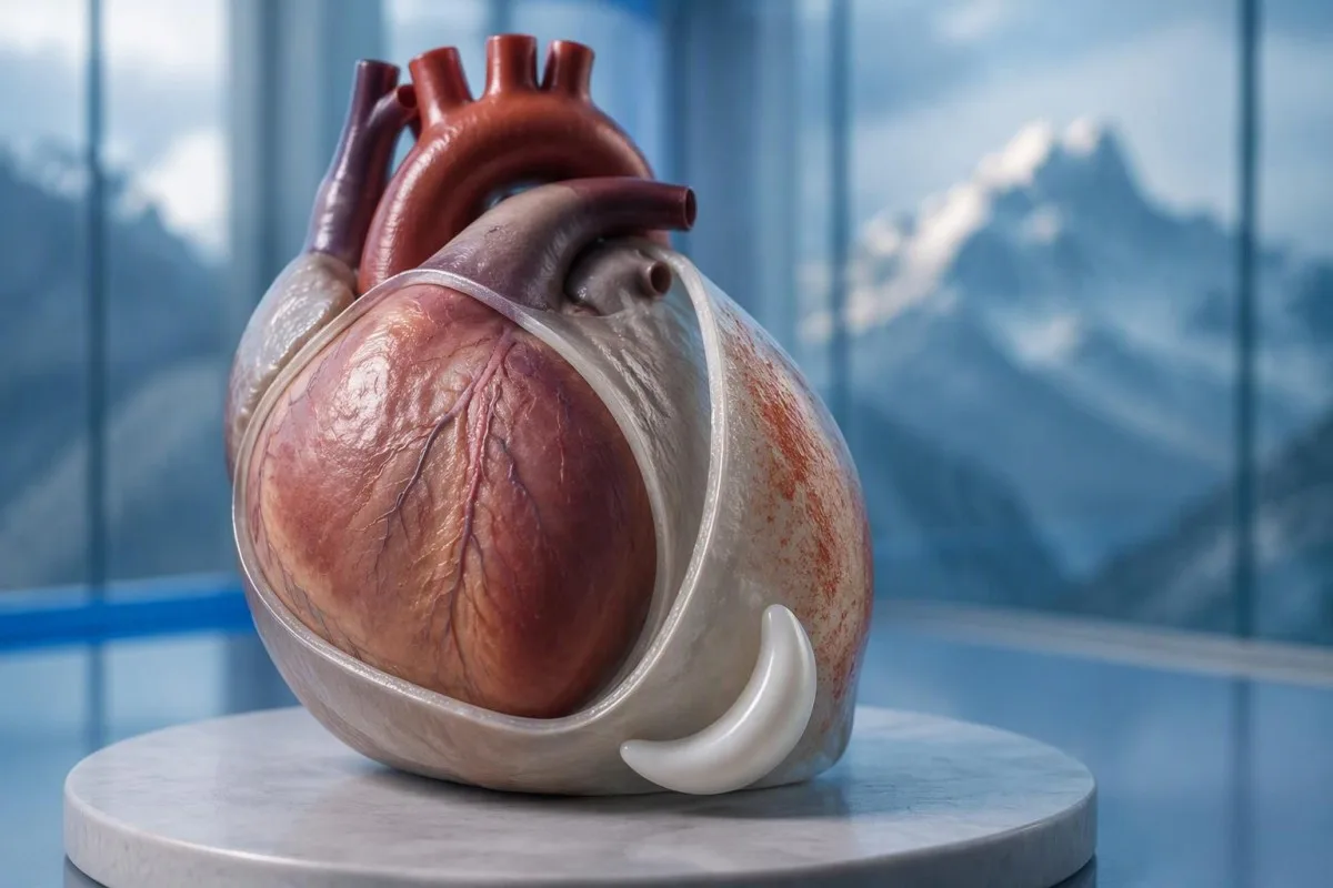
დიახ — პერიკარდიტის ტკივილი გულმკერდის არეში ხშირად ძლიერდება წოლისას და უმჯობესდება ჯდომისას ან წინ დახრისას. მაგრამ ეს ნიმუში არ გამორიცხავს გულის შეტევას; ახალი, აუხსნელი ტკივილი გულმკერდის არეში საჭიროებს სასწრაფო სამედიცინო შეფასებას.


- პოზიციური ტკივილი ხშირად ძლიერდება წოლისას და უმჯობესდება წინ დახრით, მაგრამ არც ერთი პასუხი არ ადასტურებს პერიკარდიტს და არ გამორიცხავს გულის შეტევას.
- სუნთქვის ტკივილი შეიძლება გაუარესდეს ღრმა ჩასუნთქვით ან ხველებით; ფილტვის ემბოლიას და ფილტვის სხვა დაავადებებს შეუძლიათ იგივე ნიმუშის გამოწვევა.
- გადაუდებელი სიმპტომები მოიცავს ძლიერ ან მუდმივ ტკივილს გულმკერდის არეში, ქოშინს, გონების დაკარგვას, ცივ ოფლს, ან ტკივილს, რომელიც ვრცელდება მკლავზე, ყბაზე ან ზურგზე. დაუყოვნებლივ დარეკეთ თქვენს ადგილობრივ სასწრაფო დახმარების ნომერზე, ყველა სიმპტომის მოლოდინის გარეშე.
- ეკგ ცვლილებები მწვავე პერიკარდიტისთვის დამახასიათებელი დაფიქსირდა 60% შემთხვევამდე 2015 წლის ESC სახელმძღვანელოში; ნორმალური ეკგ არ გამორიცხავს დაავადებას.
- ექოკარდიოგრაფია აფასებს სითხეს გულის ირგვლივ და მის გავლენას შევსებაზე. 20 მმ-ზე მეტი გამონაჟონი არის მაღალი რისკის ფაქტორი, არა ის ზღვარი, რომლის ქვემოთაც ყველა სითხე უვნებელია.
- ტროპონინი ასეი-სპეციფიკური 99-ე პერცენტილის ზემოთ მიუთითებს გულის კუნთის დაზიანებაზე; მხოლოდ შედეგი არ შეუძლია გულის შეტევის გარჩევა მიოკარდიტისგან.
- ანთების ტესტები როგორიცაა CRP და ESR, ხელს უწყობს შეფასებას და თვალყურს, მაგრამ ნორმალური შედეგები არ შეიძლება დამოუკიდებლად გამორიცხავდეს პერიკარდიტს.
- შემდგომი დაკვირვება ჩვეულებრივ ვითარდება დაახლოებით 1 კვირის განმავლობაში სათანადოდ შერჩეული ამბულატორიული პაციენტებისთვის; სიმპტომების გაუარესება მოითხოვს უფრო ადრეულ ხელახალ შეფასებას.
რატომ შეიძლება გაუარესდეს პერიკარდიტის ტკივილი გულმკერდის არეში წოლისას?
პერიკარდიტით გამოწვეული ტკივილი გულმკერდის არეში ხშირად ძლიერდება დაწოლისას და ნაკლებად ძლიერდება ჯდომისას ან წინ დახრისას. პოზის ცვლილებები ცვლის გაღიზიანებული პერიკარდიუმისა და მიმდებარე სტრუქტურების მექანიკურ ურთიერთობას, მაგრამ შვება ერთ მდგომარეობაში არის დიაგნოსტიკური მინიშნება - არა მტკიცებულება იმისა, რომ გულის შეტევა შეუძლებელია.
The პერიკარდიუმი არის გულის გარშემორტმული 2-ფენიანი პარკი, რომლის გარეთა გარსი შეიცავს ტკივილზე მგრძნობიარე კავშირებს, რომლებიც თავად გულის კუნთს არ აქვს ანალოგიურად. ნაცნობი ახსნა, რომ ფენები უბრალოდ “ერთმანეთს ეხება”, არასრული: მოძრაობა, მიმდებარე ქსოვილების გაღიზიანება და ინდივიდუალური სენსორული რეაქციები ასევე გავლენას ახდენს ტკივილზე.
ჰიპოთეტურ 42-წლის მამაკაცს, რომელსაც მწვავე ტკივილი უვითარდება რესპირატორული დაავადების შემდეგ და არ შეუძლია კომფორტულად ძილი ბრტყელად, აქვს პერიკარდიტის თავსებადი ნიმუში. მე მაინც ვიკითხავ ზუსტ დაწყებაზე, დატვირთვაზე, ქოშინზე და გულ-სისხლძარღვთა ისტორიაზე დიაგნოზის დასმამდე; ასაკი და ცოტა ხნის წინანდელი გაციება არ აუქმებს კორონარული დაავადების ან ფილტვის ემბოლიის რისკს.
კანტესტი არის AI სისხლის ტესტის ანალიზატორი რაც შეიძლება დაეხმაროს CRP და ტროპონინის შედეგების ახსნას კლინიკური შეფასების შემდეგ; ეს არ შეიძლება გადაწყვიტოს, არის თუ არა პოზიციური ტკივილი გულმკერდის არეში უსაფრთხო. როგორც თომას კლაინი, მე ამ განსხვავებას მარტივად ვფორმულირებ: 1 დამაიმედებელი სიმპტომი არასდროს არ უნდა გადაწონოს აუხსნელი მწვავე ტკივილი გულმკერდის არეში; ინფორმაცია ორგანიზაციაში ხსნის ჩვენს საგანმანათლებლო როლს.
უარესდება თუ არა პერიკარდიტის ტკივილი ღრმა ჩასუნთქვით?
პერიკარდიტით გამოწვეული ტკივილი ჩვეულებრივ ძლიერდება ღრმა ჩასუნთქვისას, ხველისას ან ზოგჯერ ყლაპვისას. კლინიცისტები სუნთქვაზე დაკავშირებულ ტკივილს პლევრიტულს უწოდებენ, მაგრამ ეს აღწერა განსაზღვრავს ტკივილის ნიმუშს და არა 1 კონკრეტულ დაავადებას; ფილტვებს, გულმკერდის კედელს და პერიკარდიუმს შეუძლიათ მისი წარმოქმნა.
ღრმა შთაგონება მოძრაობს დიაფრაგმას და ცვლის ურთიერთობებს გულმკერდის შიგნით, რაც შეიძლება გაამწვავოს გაღიზიანებული პერიკარდიუმის გარსი. კავშირები, რომლებიც მოიცავს ფრენიკულ ნერვებს, წარმოქმნილი ძირითადად C3–C5, ხელს უწყობს იმის ახსნას, თუ რატომ გრძნობენ ზოგიერთი პაციენტი რეფერალურ დისკომფორტს მხრის ან ტრაპეციის ქედის მახლობლად, ნაცვლად მხოლოდ მკერდის ძვლის უკან.
ყოველ ამოსუნთქვასთან ერთად გაჩერებული ტკივილი საჭიროებს უფრო ფართო შეფასებას, როდესაც ის მოულოდნელად იწყება, განსაკუთრებით ქოშინთან ან შეშუპებულ ფეხთან ერთად. ფილტვის ემბოლიას შეუძლია წარმოქმნას იგივე ჩივილი კლასიკური წნევის შეგრძნების გარეშე; თუნდაც ჟანგბადის გაჯერების 98% არ შეიძლება დამოუკიდებლად გამორიცხოს იგი.
ტკივილი, როდესაც კლინიცისტი აჭერს გულმკერდის 1 ნაწილს, უფრო სავარაუდოს ხდის გულმკერდის კედლის წყაროს, მაგრამ ეს არ იძლევა აბსოლუტურ კორონარული გამორიცხვის საშუალებას. ჩვენი ახსნა ტესტები აუხსნელი ტკივილისთვის განასხვავებს დამხმარე ლაბორატორიულ მინიშნებებს იმ მდგომარეობებისგან, რომლებიც საჭიროებენ გამოკვლევას და გამოსახულებას.
პერიკარდიტის რომელი სხვა სიმპტომებია მნიშვნელოვანი?
პერიკარდიტის სიმპტომები შეიძლება შეიცავდეს ცხელებას, დაღლილობას, ქოშინს და გულისცემის გახშირებას, ასევე ტკივილს გულმკერდის არეში. არცერთი არ არის სავალდებულო და მათი კომბინაცია უფრო მნიშვნელოვანია, ვიდრე ნებისმიერი 1 სიმპტომი; ძლიერი სისუსტე, გულის წასვლა ან სუნთქვის გაუარესება ცვლის შეფასების სასწრაფოობას.
ტემპერატურა ზემოთ 38°C is a higher-risk feature in the 2015 ESC pericardial guideline, particularly when the cause is uncertain. A patient with 37.4°C and considerable pain may still need urgent assessment, however; temperature thresholds help clinicians organize risk rather than define a safe home-care boundary.
A fast pulse can reflect pain, fever, dehydration, an arrhythmia, or impaired cardiac filling. I would interpret a resting pulse of 110 beats/min differently in someone comfortable and well hydrated than in someone pale and faint; our პალპიტაციების ტესტირების სახელმძღვანელო explains why laboratory results are only part of that assessment.
Breathlessness when lying flat is not automatically the same thing as chest pain when lying flat. Someone who needs 3 pillows because they cannot breathe comfortably may have congestion, an effusion, or another cardiopulmonary problem; the distinction is covered in our ქოშინის შედეგები გიდასტურებთ, რა ლაბორატორიულ დასკვნებს შეუძლია და რა არ შეუძლია გამორიცხოს., but active breathing difficulty should not wait for online interpretation.
როდის საჭიროებს პოზიციური ტკივილი გულმკერდის არეში სასწრაფო დახმარებას?
Call emergency services for severe, persistent, or rapidly worsening chest pain, especially with breathlessness, fainting, cold sweating, or pain spreading to the arm, jaw, or back. In the UK call 999 or 112; elsewhere use your local emergency number, and do not drive yourself.
Sudden maximal pain, collapse, new confusion, or coughing up blood also warrants emergency assessment because the differential includes pulmonary embolism and aortic emergencies. Do not wait 5 or 15 minutes to see whether leaning forward fixes the problem; concerning accompanying symptoms make a waiting experiment unsafe.
New unexplained chest pain that is not severe still deserves prompt, usually same-day, medical assessment rather than a routine appointment several weeks away. The 2021 AHA/ACC chest pain guideline emphasizes structured assessment and discourages using supposedly “atypical” symptoms to dismiss acute coronary syndrome (Gulati et al., 2021).
Stop exertion while arranging help, and follow the emergency dispatcher’s instructions about any medication rather than starting treatment for presumed pericarditis. Our გულმკერდის ტკივილის სასწრაფო სისხლის ანალიზების შესახებ article explains why 1 laboratory result cannot replace an emergency ECG, examination, and appropriately timed testing.
როგორ აფასებენ კლიнициტები საეჭვო პერიკარდიტს?
Clinicians assess suspected pericarditis by combining the history, examination, ECG, echocardiography, and selected blood tests. The widely taught 2015 ESC framework requires at least 2 of 4 findings: typical pain, a pericardial rub, characteristic ECG changes, or a new or worsening pericardial effusion (Adler et al., 2015).
Sharp pain, pain worse lying down, and relief leaning forward are not 3 independent diagnostic criteria; they are different features of the same symptom. This distinction prevents a persuasive story from being mistaken for confirmation, especially when the ECG and imaging have not yet been assessed.
A pericardial friction rub may be transient, so a clinician may listen again after an initial examination rather than treating 1 quiet examination as decisive. Vital signs, signs of poor circulation, heart failure, and the history of recent procedures or systemic illness can matter more urgently than whether the rub is audible.
The 2025 ESC myocarditis and pericarditis guideline gives contemporary imaging and overlapping myocardial involvement a central place in assessment, so the older checklist is not a complete care pathway (Schulz-Menger et al., 2025). Our clinical standards information should be read as documentation about laboratory interpretation—not evidence that software can reproduce this bedside evaluation.
რას აჩვენებს ეკგ პერიკარდიტის დროს?
An ECG may show widespread ST-segment elevation and PR-segment depression in acute pericarditis, but these changes are not always present. The 2015 ESC guideline reports characteristic ECG changes in up to 60% of cases, so a normal tracing does not exclude pericarditis (Adler et al., 2015).
A 12-წამყვანიანი ეკგ (12-lead ECG) records electrical activity from several perspectives; clinicians assess which leads are affected, reciprocal changes, rhythm, and comparison with previous tracings. “The ST segment looks elevated” is not enough to separate pericarditis, early repolarization, myocardial infarction, and other causes without the clinical setting.
A heart attack more often produces changes corresponding to a coronary territory, whereas pericarditis may produce a broader pattern—but neither rule is absolute. ECG abnormalities can evolve over საათების, and repeating the tracing during recurrent or changing symptoms may reveal information absent from the first recording.
Electrolyte disturbances can add another layer of confusion, particularly when kidney disease or medication effects are present. A potassium concentration around 6.0 მმოლ/ლ-ს can be clinically urgent depending on context, and our potassium and ECG guide explains why ECG interpretation should never be detached from the rest of the assessment.
რატომ გამოიყენება ექოკარდიოგრაფია პერიკარდიტის დროს?
Echocardiography checks for fluid around the heart, ventricular function, and signs that fluid is impairing cardiac filling. A normal echocardiogram does not exclude uncomplicated pericarditis; an effusion greater than 20 mm is a recognized high-risk feature, not the definition of every dangerous effusion.
The 2015 ESC guideline describes effusions as small below 10 მმ, moderate at 10–20 mm, and large above 20 mm, using the echo-free space on imaging. These measurements help communicate size, but the distribution of fluid and its hemodynamic effects still require interpretation rather than a number-only decision.
Cardiac tamponade occurs when pericardial pressure compromises filling; how quickly fluid accumulates can matter more than its absolute volume. A collection that develops over საათების may be less well tolerated than a larger, slowly developing collection, and the classic low-pressure, raised-neck-vein, quiet-heart-sound triad is not reliably present in every patient.
The 2025 ESC guideline supports integrating echocardiography with other imaging when the diagnosis or extent of involvement remains uncertain (Schulz-Menger et al., 2025). An elevated NT-proBNP may add information about cardiac strain, but 1 value cannot diagnose tamponade; our NT-proBNP interpretation guide explains several alternative reasons for elevation.
შეუძლია თუ არა ტროპონინს განასხვავოს პერიკარდიტი გულის შეტევისგან?
Troponin identifies heart muscle injury, but an elevated result alone cannot distinguish a heart attack from myocarditis or overlapping myopericarditis. The relevant threshold is the assay-specific 99-ე პროცენტილის ზედა საცნობარო ზღვარს ზემოთ ნებისმიერი მაჩვენებელი, interpreted alongside symptoms, ECG findings, serial measurements, and sometimes imaging.
There is no universal normal troponin number for every laboratory: ტროპონინ I-სა და ტროპონინ T-ს შორის use different assays, and some laboratories apply sex-specific reference limits. If a hypothetical report lists an upper limit of 14 ნგ/ლ, a result of 20 ng/L exceeds that particular limit; it does not independently identify the cause, as our troponin I versus T explanation details.
An early normal troponin may precede a detectable rise, which is why emergency departments use validated serial pathways, often with repeat sampling at 1 or 2 hours for suitable high-sensitivity assays. Selected patients can meet validated single-sample rule-out criteria, but that decision depends on assay, symptom timing, ECG, and risk—not a home interpretation of “normal.”
Pericardial symptoms plus raised troponin prompt assessment for myocardial involvement, especially with arrhythmia, breathlessness, or reduced ventricular function. A 30-year-old with a recent viral illness still needs a proper differential; our myocarditis troponin guide explains why neither youth nor the size of the rise settles the diagnosis.
რას მატებს CRP, ESR და სისხლის ანალიზები?
CRP, ESR, and a full blood count can support evidence of inflammation, but they do not confirm pericarditis or exclude a heart attack. Many laboratories use a standard CRP upper reference limit around 5 მგ/ლ-ზე ქვემოთ, while others use different limits; the printed laboratory range takes precedence.
CRP-ის მაჩვენებელი 45 მგ/ლ indicates a substantial inflammatory response in a laboratory with an upper limit of 5 mg/L, but it cannot say whether the source is pericardial, pulmonary, autoimmune, or elsewhere. High-sensitivity CRP measures the same protein at low concentrations; acute illness values should not be interpreted using stable cardiovascular-risk categories.
კანტესტი არის AI სისხლის ანალიზის განმარტების პლატფორმა that can explain a CRP result against the supplied reference range and help organize its trend alongside other reported results. If CRP changes from 45 to 12 mg/L, the direction may support improvement, but persistent symptoms still matter; our WBC and CRP comparison discusses why these markers can disagree.
ESR often changes more slowly than CRP and is influenced by age, anemia, pregnancy, and protein concentrations, so comparing 2 different markers as though they were interchangeable can mislead follow-up. The 2015 ESC guideline treats inflammatory tests as supportive evidence rather than standalone diagnostic criteria (Adler et al., 2015); our causes of elevated ESR adds context for an unexpectedly persistent elevation.
რომელი მიზეზები და რისკის ფაქტორები ცვლის მკურნალობის გეგმას?
Pericarditis may follow a viral illness, occur after cardiac injury, or accompany autoimmune disease, severe kidney disease, tuberculosis, or other conditions. სიცხე 38°C-ზე ზემოთ 38°C, a large effusion, tamponade, or failure to improve with initial treatment are among the features that can justify hospital assessment and a broader search for causes.
The 2015 ESC guideline lists lack of response to anti-inflammatory treatment within 7 დღე as a major adverse prognostic feature, alongside subacute onset and the imaging findings above (Adler et al., 2015). This is a clinician’s reassessment trigger—not permission to wait a week through worsening pain, fever, or breathlessness.
Autoimmune testing is most useful when the history supplies a reason: recurrent episodes, joint symptoms, rashes, mouth ulcers, or known systemic disease. 1 positive antibody does not prove autoimmune pericarditis; our anti-dsDNA შედეგის გიდი explains why antibody findings need compatible clinical evidence.
Severe kidney dysfunction can change both the cause of pericarditis and medication safety; an eGFR below 15 mL/min/1.73 m² identifies kidney failure-range function but does not itself diagnose uremic pericarditis. Our urgent low-eGFR symptoms guide is relevant to that background, while chest pain in this setting requires direct medical assessment.
ადასტურებს თუ არა მედიკამენტებით გაუმჯობესება პერიკარდიტს?
Improvement with ibuprofen, aspirin, or a change in posture does not confirm pericarditis. For appropriately diagnosed uncomplicated cases, clinicians commonly use an anti-inflammatory medicine with colchicine, often for approximately 3 თვე after a first episode, but the regimen must account for kidney function, interactions, and the underlying cause.
A reduction in pain from 8 out of 10 to 3 out of 10 is useful clinical information, but several chest-pain causes can respond to analgesia. Do not use medication response as a reason to cancel assessment, and do not start high-dose anti-inflammatory treatment for undiagnosed chest pain without professional advice.
Colchicine requires particular care with kidney or liver impairment and certain interacting medicines, including some antibiotics; taking 2 medicines together can alter exposure even when each prescription is familiar. Corticosteroids are not routine first-line treatment for every uncomplicated case, and selected bacterial or systemic causes need a different strategy rather than simply more pain relief.
Kantesti can help organize laboratory follow-up for a clinician through our მედიკამენტების უსაფრთხოების ტენდენციის გზამკვლევი, but it should not select the treatment or tapering schedule. A plan that includes 1 named prescriber, clear interaction checks, and instructions for worsening symptoms is more useful than a generic dosing chart.
როდის არის უსაფრთხო ვარჯიში პერიკარდიტის სიმპტომების გაუმჯობესების შემდეგ?
Exercise should be restricted during active pericarditis, and return should follow clinical reassessment rather than pain relief alone. The decision changes substantially if troponin, rhythm testing, or imaging suggests myocardial involvement; 1 symptom-free day is not evidence that strenuous activity is safe.
The older 2015 ESC guideline used a minimum ნიშნულზე ან მის შემდეგ restriction for athletes with isolated pericarditis, while contemporary guidance places greater emphasis on individualized remission and risk assessment. Patients should not borrow an athlete’s timetable—or a myocarditis timetable—without knowing which diagnosis their clinician has established.
For a hypothetical 28-year-old runner, the difference between isolated pericarditis and myocardial involvement can change follow-up testing and return-to-training decisions. I would ask whether symptoms, inflammatory markers, ventricular function, and any relevant rhythm concerns have resolved; a normal resting pulse of 65 beats/min is only one small part of that picture.
Daily activity, physical work, and competitive sport are 3 different workloads, so a written return plan should specify what the patient can actually do. Our სისხლის ბიომარკერების სახელმძღვანელო can explain reported measurements, but no biomarker page, wearable reading, or AI result constitutes exercise clearance.
რა მოხდება, თუ გულმკერდის არეში ტკივილი დაბრუნდება ან ტესტები ნორმალურია?
Persistent or recurrent chest pain needs reassessment even when an earlier ECG, echocardiogram, or CRP result was normal. Appropriately selected low-risk outpatients are commonly reviewed within about 1 კვირის განმავლობაში., but recurrence with faintness, breathlessness, or worsening pain warrants earlier—and sometimes emergency—assessment.
Residual discomfort, recurrent inflammation, chest-wall pain, and a new unrelated problem can feel similar to the patient, so 2 episodes should not automatically receive the same explanation. Cardiac MRI can sometimes identify pericardial or myocardial involvement when first-line tests leave uncertainty; it is selected for a clinical question, not ordered for every ache.
A follow-up note should record symptom timing, temperature, activity tolerance, medication changes, and the actual laboratory reference ranges. CRP values of 8 and 6 mg/L from different laboratories may not represent a meaningful improvement without knowing the methods and context; our post-discharge result changes guide explains why small numerical changes deserve restraint.
კანტესტი არის AI-ზე დაფუძნებული სისხლის ანალიზის (blood test analysis) ხელსაწყო that can help organize reported results for discussion, but it cannot verify the cause of renewed chest pain from a PDF. My advice here, as Thomas Klein, is to agree on 3 things before leaving follow-up: the next review date, permitted activity, and the symptoms that require immediate help.
კლინიკური საზღვრები და დაკავშირებული კვლევითი პუბლიკაციები
Symptoms and laboratory interpretation cannot safely replace an acute chest-pain assessment. როგორც 3 ოქტომბერი, 2026, the clinical references below support the distinction between pericardial clues, myocardial injury, and emergency risk; the 2 Zenodo resources are background laboratory guides, not trials validating a pericarditis diagnosis.
Kantesti-ის medical advisory information describes the professional expertise behind our educational work, but readers should not equate a medical title with an examination of their own chest pain. A personal care plan still needs 1 accountable treating clinician or team who can examine the patient, review the ECG, and act on imaging or troponin findings.
The aPTT-ის ნორმალური დიაპაზონი: D-დიმერი, ცილა C სისხლის შედედების სახელმძღვანელო is available as a Zenodo educational publication with DOI 10.5281/ზენოდო.18262555. Its associated clotting-test interpretation guide is relevant when clinicians consider coagulation questions, but a normal aPTT cannot exclude pulmonary embolism and D-dimer must be used within an appropriate clinical probability pathway.
The შრატის ცილების სახელმძღვანელო: გლობულინების, ალბუმინის და A/G თანაფარდობის სისხლის ანალიზი is a second Zenodo educational publication with DOI 10.5281/ზენოდო.18316300. The associated serum protein interpretation guide supplies background on systemic laboratory findings; neither albumin nor the A/G ratio independently diagnoses pericarditis, and the discovery links below are searches rather than verified copies of these publications.
ხშირად დასმული კითხვები
პერიკარდიტით გამოწვეული ტკივილი მკერდის არეში უარესდება თუ დაწოლა?
პერიკარდიტის დროს მკერდის ტკივილი ხშირად ძლიერდება მწოლიარე მდგომარეობაში და მცირდება მჯდომარე ან წინ დახრილი მდგომარეობისას. ეს ნიმუში სავარაუდოა, თუმცა ის კლინიკური შეფასების მხოლოდ 1 კომპონენტს წარმოადგენს და არ შეიძლება გულის შეტევის გამორიცხვა. ექიმები აერთიანებენ ანამნეზს გამოკვლევასთან, ეკგ-სთან, ექოკარდიოგრაფიასთან და შერჩეულ სისხლის ანალიზებთან. ახალი აუხსნელი მკერდის ტკივილი საჭიროებს დაუყოვნებელ შეფასებას, სასწრაფო დახმარებით ძლიერი ტკივილის, ქოშინის, გულის წასვლის ან ცივი ოფლიანობის შემთხვევაში.
მკერდის ტკივილი, რომელიც გაყუჩდება წინ გადახრისას, გულის შეტევას გამორიცხავს?
გულმკერდის ტკივილი, რომელიც გაუმჯობესებულია წინ გადახრით, არ გამორიცხავს გულის შეტევას. 2021 წლის AHA/ACC-ის გულმკერდის ტკივილის სახელმძღვანელო პრინციპები მხარს უჭერს სტრუქტურირებულ შეფასებას სიმპტომების უარყოფის ნაცვლად, იმის გამო, რომ ისინი არაკლასიკურად ჟღერს. შესაძლოა საჭირო გახდეს ეკგ და შესაბამისად დროული მაღალი მგრძნობელობის ტროპონინის ტესტირება, ხოლო ადრეული ნორმალური შედეგი არ არის ავტომატურად დამაჯერებელი. გამოიძახეთ სასწრაფო დახმარება მწვავე ან მუდმივი ტკივილისთვის ან თანმხლები ქოშინის, გულის წასვლის ან ცივი ოფლიანობისთვის.
შეიძლება თუ არა პერიკარდიტი ნორმალური ეკგ-ის დროს?
დიახ, პერიკარდიტი შეიძლება განვითარდეს ნორმალური ეკგ-ით. 2015 წლის ESC-ის პერიკარდიუმის სახელმძღვანელომ დაფიქსირდა დამახასიათრებელი ეკგ ცვლილებები მწვავე შემთხვევების 60%-მდე, რაც ნიშნავს, რომ მნიშვნელოვან ნაწილს არ ჰქონდა კლასიკური ნიმუში. ეკგ-ის მონაცემები ასევე შეიძლება შეიცვალოს, ამიტომ კლინიცისტებმა შეიძლება გაიმეორონ 12-გადამყვანი ეკგ, როდესაც სიმპტომები იცვლება. მარტო ნორმალურ ეკგ-ს არ შეუძლია გამოირიცხოს პერიკარდიტი ან გულის ყოველი შეტევა.
აჩვენებს თუ არა ყოველთვის გულის ექოკარდიოგრაფია პერიკარდიტს?
ექოკარდიოგრამა ყოველთვის ვერ ავლენს გართულებების გარეშე მიმდინარე პერიკარდიტს, რადგან ანთება შესაძლოა მოხდეს სითხის ხილული დაგროვების გარეშე. ექოკარდიოგრაფია განსაკუთრებით სასარგებლოა პერიკარდიუმის სითხის გასაზომად, პარკუჭოვანი ფუნქციის შესაფასებლად და შევსების დარღვევის აღმოსაჩენად. 2015 წლის ESC კლასიფიკაციით, 20 მმ-ზე მეტი გამონაჟონი ითვლება დიდ სითხე-კრებად, თუმცა მცირე, სწრაფად დაგროვებული სითხეც შეიძლება იყოს საშიში. კლინიკური კითხვის გაურკვევლობის შემთხვევაში შესაძლოა დამატებითი ვიზუალიზაცია განიხილებოდეს.
ნორმალური ტროპონინი ნიშნავს, რომ გულმკერდის ტკივილი არ არის სერიოზული?
ნორმალური ტროპონინის შედეგი არ ნიშნავს ავტომატურად, რომ გულმკერდის ტკივილი უვნებელია. დრო, ანალიზის სპეციფიკური 99-ე პროცენტილის ლიმიტი, ეკგ-ს მონაცემები და რისკის მთლიანი შეფასება განსაზღვრავს, რას შეუძლია გამორიცხოს შედეგი. დადასტურებული მაღალი მგრძნობელობის ტროპონინის სქემები ხშირად იყენებენ განმეორებით გაზომვებს 1 ან 2 საათის შემდეგ, თუმცა შერჩეულ პაციენტებს შეიძლება ჰქონდეთ უფლება ერთჯერადი ნიმუშის გამომრიცხავი სქემისთვის. ტროპონინი ასევე არ გამორიცხავს დამოუკიდებლად ფილტვის ემბოლიას, აორტის გადაუდებელ მდგომარეობებს ან პერიკარდიტს.
რამდენი ხანი გრძელდება პერიკარდიტის სიმპტომები?
პერიკარდიტის სიმპტომები შეიძლება გაუმჯობესდეს დღეების ან კვირების განმავლობაში სათანადო მკურნალობით, თუმცა დრო იცვლება მიზეზის, რეციდივისა და მიოკარდიუმის ჩართულობის მიხედვით. კოლხიცინი ხშირად ინიშნება დაახლოებით 3 თვის განმავლობაში პირველი გართულების გარეშე ეპიზოდის შემდეგ, რაც არ ნიშნავს, რომ ტკივილი უნდა გაგრძელდეს მთელი ამ პერიოდის განმავლობაში. ანთების საწინააღმდეგო მკურნალობის დაწყებიდან 7 დღის განმავლობაში გაუმჯობესების არარსებობა არის 2015 წლის ESC-ის გაიდლაინში აღიარებული გადაფასების ტრიგერი. ტკივილის, ქოშინის ან გულის წასვლის გაუარესება საჭიროებს ადრეულ შეფასებას დაგეგმილი მიმოხილვის მოლოდინის ნაცვლად.
მიიღეთ AI-ით გაძლიერებული სისხლის ანალიზის განმარტება დღეს
შეუერთდით 2 მილიონზე მეტ მომხმარებელს მთელ მსოფლიოში, რომლებიც ენდობიან Kantesti-ს ლაბორატორიული ანალიზების მყისიერ და ზუსტ განმარტებაში. ატვირთეთ თქვენი სისხლის ანალიზის შედეგები და მიიღეთ 15,000+ ბიომარკერების ყოვლისმომცველი ინტერპრეტაცია წამებში.
📚 მითითებული კვლევითი პუბლიკაციები
კლაინი, ტ., მიტჩელი, ს., და ვებერი, ჰ. (2026). aPTT-ის ნორმალური დიაპაზონი: D-დიმერი, ცილა C სისხლის შედედების სახელმძღვანელო. Kantesti AI სამედიცინო კვლევა.
კლაინი, ტ., მიტჩელი, ს., და ვებერი, ჰ. (2026). შრატის ცილების სახელმძღვანელო: გლობულინების, ალბუმინის და A/G თანაფარდობის სისხლის ანალიზი. Kantesti AI სამედიცინო კვლევა.
📖 გარე სამედიცინო წყაროები
Gulati M et al. (2021). 2021 AHA/ACC/ASE/CHEST/SAEM/SCCT/SCMR რეკომენდაცია გულმკერდის ტკივილის შეფასებისა და დიაგნოსტიკისთვის. Circulation.
Schulz-Menger J et al. (2025). 2025 ESC Guidelines for the management of myocarditis and pericarditis. ევროპული გულის ჟურნალი.
⚕️ სამედიცინო პასუხისმგებლობის შეზღუდვა
ეს სტატია მხოლოდ საგანმანათლებლო მიზნებისთვისაა და არ წარმოადგენს სამედიცინო რჩევას. დიაგნოზისა და მკურნალობის გადაწყვეტილებებისთვის ყოველთვის მიმართეთ კვალიფიციურ ჯანდაცვის სპეციალისტს.
E-E-A-T სანდოობის სიგნალები
გამოცდილება
ექიმის ხელმძღვანელობით კლინიკური მიმოხილვა ლაბორატორიული ინტერპრეტაციის სამუშაო პროცესებზე.
ექსპერტიზა
ლაბორატორიული მედიცინის აქცენტი იმაზე, როგორ იქცევიან ბიომარკერები კლინიკურ კონტექსტში.
ავტორიტეტულობა
დაწერილია დოქტორ თომას კლაინის მიერ, მიმოხილვა: დოქტორ სარა მიტჩელისა და პროფესორ დოქტორ ჰანს ვებერის მიერ.
სანდოობა
მტკიცებულებებზე დაფუძნებული ინტერპრეტაცია, მკაფიო შემდგომი ნაბიჯებით, რათა შემცირდეს განგაშის დონე.