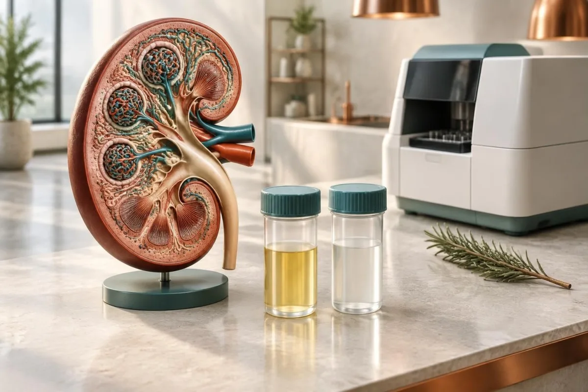
スポット尿検体から1日のタンパク質喪失量を推定できますが、数値は単位、採尿条件、腎機能、臨床的背景を併せて確認して初めて意味を持ちます。

要点
- 測定内容: スポット尿のタンパク/クレアチニン比は、時間を定めた24時間蓄尿を行わずにタンパク質の排泄量を推定します。1 g/gは、タンパク質約1 g/日の排泄量に相当します。
- 成人の一般的な結果: 150 mg/g未満、または0.15 mg/mgの比率は、通常、妊娠していない成人の一般的な範囲内とみなされますが、検査機関によって異なる場合があります。
- 高値: 尿タンパク/クレアチニン比が500 mg/gを超える場合は、一般に医療機関での評価が必要です。3,500 mg/gを超える値は、ネフローゼ域に該当します。
- 単位が重要です: 0.3 g/gは300 mg/g、約34 mg/mmolに相当します。これらは同じ比率を異なる形式で表示したものであり、タンパク質喪失量の異なる段階を示すものではありません。
- 検体採取が重要です: 発熱、激しい運動、尿路感染、脱水、月経、検体の汚染により、随時尿の測定値が一時的に高くなることがあります。
- 尿蛋白とアルブミン: 総蛋白にはアルブミンとその他の蛋白が含まれるため、尿蛋白比とアルブミン・クレアチニン比は、関連はあるものの異なる問いに答える指標です。
- フォローアップ: 蛋白の持続的な高値、血尿、高血圧、eGFRの低下、または浮腫がある場合、通常は再検査と、より広範な腎機能評価が行われます。
スポット尿タンパク比が実際に測定するもの
A 尿蛋白・クレアチニン比 同一検体中の尿蛋白と尿クレアチニンを比較し、尿中に失われる蛋白量を推定します。1 g/gという結果は、1日あたり約1 gの蛋白が排泄されると解釈されることがよくありますが、これは推定値であり、24時間蓄尿による直接測定ではありません。

尿の濃度は一日を通じて変化しますが、クレアチニンは希釈の程度を大まかに補正するため、この比が機能します。たとえば、濃縮尿中の蛋白が120 mgで、希釈尿中の蛋白が40 mgでも、それぞれのクレアチニン濃度がそれに比例して異なれば、比は同程度になることがあります。
随時尿の蛋白・クレアチニン比は、腎臓の濾過機能を直接測定するものではありません。この比は尿中への蛋白の移行を反映します。一方、推算糸球体濾過量、血清クレアチニン、シスタチンCは、腎臓が小分子の老廃物をどれだけ効率的に排泄するかを評価します。
Kantestiは、血清クレアチニン、eGFR、アルブミン、グルコースを報告された尿比と並べて整理できるAI血液検査アナライザーですが、尿検査や臨床医による診察の代わりにはなりません。複数の検査マーカーを比較する読者には、当社の バイオマーカーリファレンスガイド が、どの結果が血液由来で、どの結果が尿由来なのかを理解するのに役立ちます。
尿タンパク/クレアチニン比の単位によって意味が変わる理由
同じ尿蛋白クレアチニン比でも、mg/g、mg/mg、g/g、mg/mmolのいずれかで示されることがあるため、結果を評価する前に単位を確認する必要があります。数値上、300 mg/gは0.3 mg/mgおよび0.3 g/gに等しく、約34 mg/mmolです。

一般的な表記では、1 mg/mgは1 g/gに等しく、1 g/gはクレアチニン約113 mg/mmolに相当します。実用的な換算方法は、g/gに約113を掛けてmg/mmolを推定することで、0.15 g/gは約17 mg/mmolです。
0.8 g/gという結果を0.8 mg/gと取り違えてはいけません。前者は800 mg/gに相当し、臨床的にかなり高値です。その差は1,000倍であり、接頭辞が抜けるだけで解釈が完全に逆になり得る理由です。
尿中クレアチニン濃度自体が、特に尿が非常に希釈されている場合、筋肉量が少ない人、筋肉質の成人、または採尿のタイミングが通常と異なる場合に、異常な低値または高値として示されることがあります。当ガイドでは、 尿中クレアチニンと希釈 分母によって比が不安定になりやすい理由を説明しています。
成人の尿タンパク/クレアチニン比の一般的な範囲
妊娠していない成人では、尿蛋白クレアチニン比150 mg/g未満、または0.15 g/gが、実用的な基準上限として一般的に用いられます。500 mg/gを超える結果は境界域の上昇より懸念されますが、持続性や裏付けとなる所見がなければ、単一のカットオフ値だけで慢性腎臓病と診断することはできません。
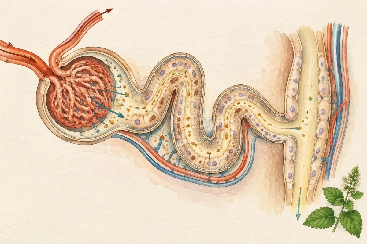
これらの区分は実用的な解釈ゾーンであり、普遍的な疾患ステージではありません。2024年のKDIGO慢性腎臓病ガイドラインは、単一の比を診断として扱うのではなく、異常な結果を確認し、アルブミン尿、eGFR、尿沈渣、臨床歴を総合的に評価することを重視しています(KDIGO、2024年)。
220 mg/gの比は、ある人では運動に伴う一時的な上昇を示し、別の人では初期腎疾患を示す可能性があります。この違いは、早朝第一尿での再検査、血圧測定、そして比の異常が少なくとも3か月間続くかどうかによって明らかになることがよくあります。
関連資料の 尿検査ガイド 尿濃縮の影響を受け、主にアルブミンを検出する試験紙所見とタンパク定量を区別するのに役立ちます。検査機関によって判定基準が若干異なる場合があるため、印字された基準範囲も解釈に含める必要があります。
| レベル | 範囲 | 意味 |
|---|---|---|
| 通常は低値 | <150 mg/g(<0.15 g/g;<17 mg/mmol) | Commonly within the usual adult range, though symptoms and other urine findings still matter. |
| 軽度に上昇 | 150-500 mg/g (0.15-0.50 g/g) | May be transient or clinically meaningful; repeat testing and context are usually needed. |
| 明らかに上昇 | 500-3,500 mg/g (0.50-3.50 g/g) | Often warrants kidney-focused assessment, especially with blood in urine, high blood pressure or reduced eGFR. |
| Nephrotic-range zone | >3,500 mg/g (>3.50 g/g; >396 mg/mmol) | Requires prompt clinical evaluation for substantial protein loss and possible glomerular disease. |
タンパク/クレアチニン比とアルブミン/クレアチニン比の違い
A protein-to-creatinine ratio measures total urine protein, while an albumin-to-creatinine ratio measures albumin specifically. The difference matters because a urine sample can contain albumin, low-molecular-weight proteins, immunoglobulin fragments or proteins from the urinary tract.

Albumin-to-creatinine ratio is usually preferred for detecting small increases in glomerular albumin leakage, especially in diabetes and hypertension. A PCR of 400 mg/g and an ACR of 30 mg/g do not describe the same protein pattern because most of the total protein may be something other than albumin.
A disproportionately high PCR with a relatively low ACR can suggest tubular protein loss, overflow proteins or urinary tract contributions, although laboratory methods and timing can also explain the mismatch. For example, a PCR of 1,000 mg/g with an ACR of 80 mg/g deserves a different workup from a PCR and ACR that are both near 1,000 mg/g.
Urine sediment can add another layer: red cells, casts and tubular cells may indicate an active process even when the ratio is only 250 mg/g. Our explanation of 尿沈渣所見に関する当社のガイド covers why microscopy can change the significance of a quantitative result.
採尿条件がスポット尿の結果に与える影響
A spot urine protein creatinine ratio is most interpretable when collected as a clean, preferably first-morning sample after avoiding unusual exertion. Fever, dehydration, urinary infection, menstruation, semen contamination and heavy exercise can produce temporary protein elevations that may fall within days.
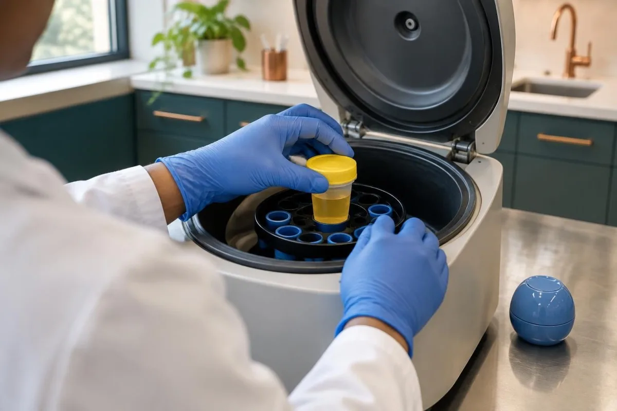
First-morning urine is often useful because it has been in the bladder for several hours and is less affected by daytime activity. A sample taken immediately after a long run may show a higher ratio than one collected after 24 to 48 hours of normal activity.
A dilute sample can create a ratio that is mathematically valid but less dependable when urine creatinine is very low. A practical collection plan includes washing hands, using the laboratory container, avoiding the first few seconds of flow when instructed and recording whether exercise or illness occurred within the previous 24 hours.
Persistent foamy urine is not proof of proteinuria, because rapid flow and concentrated urine can create bubbles, but foam that continues across several days deserves testing. Our article on 持続的な泡状の尿 explains when appearance should be replaced by measurement.
尿タンパク/クレアチニン比が高くなる原因
A high urine protein creatinine ratio can arise from glomerular leakage, tubular reabsorption problems, excess circulating proteins or inflammation below the kidneys. The reason clinicians combine the ratio with urine microscopy, eGFR and blood pressure is that the same numerical result can come from very different mechanisms.

Glomerular disorders often allow albumin and larger proteins to cross the filtration barrier, sometimes producing PCR values above 1,000 mg/g. Tubular disorders may produce smaller protein increases, while overflow states such as monoclonal light-chain production may require tests that a routine dipstick does not detect well.
A urinary infection can increase protein through inflammatory material and blood cells, particularly when leukocytes or nitrites are present. Treating an infection and repeating the ratio after approximately 1 to 2 weeks may clarify whether the elevation was temporary, although the exact interval depends on symptoms and clinical advice.
Protein without infection can also accompany high blood pressure, diabetes, autoimmune disease, heart failure or medication-related kidney stress. The presence of bacteria alone does not prove a symptomatic infection; our guide to bacteria in urine without symptoms explains why culture and symptoms must be considered together.
医療従事者が高値をどう解釈するか
A high urine protein creatinine ratio becomes more concerning when it is persistent, rising or accompanied by reduced eGFR, blood in urine, cellular casts, swelling or high blood pressure. A single value of 600 mg/g is a signal for assessment, not a diagnosis of irreversible kidney disease.
Magnitude changes the urgency: 180 mg/g is borderline in many adult laboratories, 900 mg/g is clearly abnormal, and 4,000 mg/g enters the nephrotic-range zone. The reason nephrotic-range values receive faster attention is that substantial protein loss can lower blood albumin and contribute to edema, altered lipids and clotting complications.
The pattern matters more than a solitary peak. For example, a hypothetical 52-year-old marathon runner with a PCR of 700 mg/g after a race and 90 mg/g on a rested first-morning repeat has a different risk profile from someone with 700 mg/g on three samples over 3 months.
The 2021 KDIGO glomerular disease guideline recommends disease-specific evaluation when proteinuria is substantial or accompanied by active urine sediment; it does not treat every elevated ratio as the same condition (KDIGO, 2021). A temporary eGFR reduction after dehydration can also distort the picture, as described in our 脱水と eGFR に関するガイド.
尿タンパク比を再検査すべき時期
Clinicians commonly repeat an unexpected urine protein creatinine ratio when the patient is well, the sample was not first-morning, or a temporary trigger could explain the result. Confirmation is particularly useful for borderline values between 150 and 500 mg/g, while very high results or red-flag symptoms should not wait for routine monitoring.

A repeat may be arranged within days to weeks after strenuous exercise, fever or urinary symptoms have resolved. Chronic kidney disease generally requires an abnormal kidney marker lasting at least 3 months, so a result on day 1 alone cannot establish chronicity.
Repeating the same test is not always enough. If PCR remains high, clinicians may add ACR, urine microscopy, serum creatinine and eGFR, electrolytes, albumin, glucose and blood pressure; if a monoclonal protein is suspected, serum or urine electrophoresis and free-light-chain testing may be considered.
Trends should use comparable conditions whenever possible, such as first-morning samples from the same laboratory. Our 採血タイムラインのガイド explains why illness, exercise, medication changes and timing can make two nearby results look more different than they truly are.
比率を解釈する際に参考となる血液検査
Serum creatinine, eGFR, albumin, electrolytes, glucose and blood pressure are the main companion data for a urine protein ratio. A high PCR with normal eGFR still deserves attention, while a high PCR plus falling eGFR or low serum albumin usually raises the urgency.
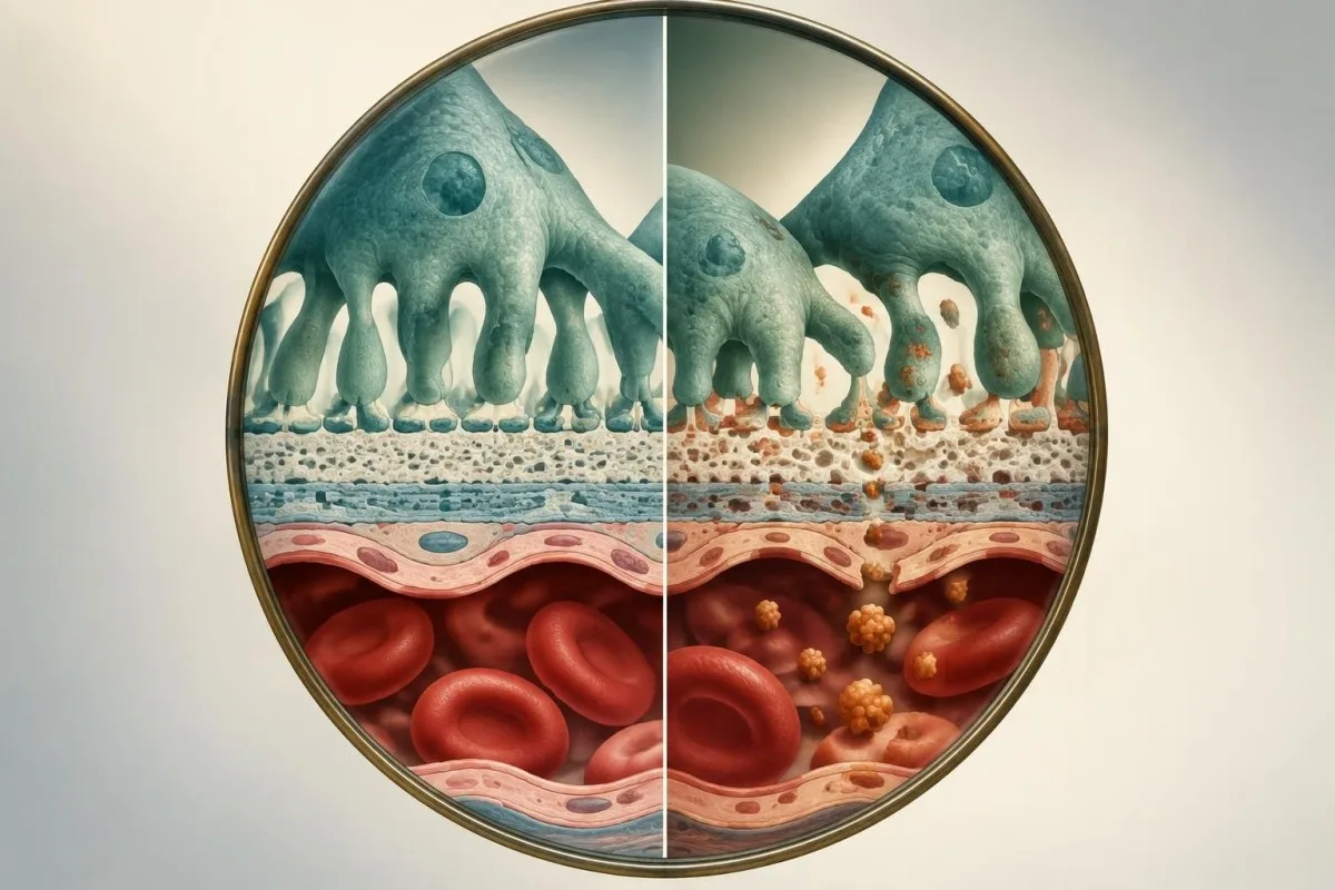
Serum creatinine can appear reassuring in a person with low muscle mass because creatinine production is lower, while a muscular adult may have a higher baseline without kidney disease. An eGFR of 58 mL/min/1.73 m² and PCR of 800 mg/g create a more concerning combination than either result viewed alone.
Low serum albumin below approximately 3.0 g/dL alongside PCR above 3,500 mg/g suggests that protein loss may be physiologically significant, although liver disease, nutrition and inflammation can also lower albumin. The reason clinicians ask about swelling is that reduced plasma oncotic pressure can shift fluid into tissues.
BUN and creatinine are influenced by hydration, diet and muscle turnover, so they do not quantify urinary protein. Our BUN-to-クレアチニン比の指標 explains why those blood markers should not be substituted for a urine protein measurement.
妊娠中の比率が意味すること
During pregnancy, a urine protein creatinine ratio of 0.3 g/g, or 300 mg/g, is commonly used as a threshold for significant proteinuria when assessing hypertensive disorders. The ratio must be interpreted with blood pressure, symptoms, platelet count, liver enzymes, creatinine and gestational age rather than used alone.
A pregnant person with blood pressure of 145/95 mmHg and PCR of 0.34 g/g needs prompt clinical assessment, even if the urine appearance is normal. Severe headache, visual change, upper abdominal pain, sudden swelling or shortness of breath require urgent maternity advice because they can indicate maternal organ involvement.
The evidence on the exact spot-ratio threshold is not perfectly uniform, especially near 0.3 g/g and in different populations. A systematic review by Côté and colleagues in the BMJ found that spot protein-to-creatinine testing can reduce the need for timed collection, but clinical context still determines whether additional testing is needed (Côté et al., 2008).
Pregnancy changes kidney filtration and can alter the meaning of creatinine-based estimates, so non-pregnant reference assumptions may mislead. The related 妊娠GFRガイド describes why pregnancy-specific review is preferable to applying adult cutoffs mechanically.
年齢や筋肉量が比率に与える影響
The creatinine denominator makes a spot urine protein ratio less straightforward in children, frail older adults, highly muscular people and those with low muscle mass. Two people with the same protein excretion can receive different ratios if their daily creatinine production differs substantially.

In small children, clinicians often adjust interpretation for age and body surface area rather than applying adult limits such as 150 mg/g. A toddler result of 0.2 mg/mg cannot be interpreted in the same way as a healthy adult result of 0.2 mg/mg without pediatric reference data.
An older adult with low muscle mass may have low urine creatinine and an apparently high ratio from a modest protein concentration. Conversely, a heavily muscled adult may have a larger creatinine denominator that makes the ratio look lower than the absolute protein concentration might suggest.
A family history of kidney disease, especially with ancestry-associated risk variants, can alter the threshold for investigation. Our overview of APOL1遺伝子検査 explains one example of inherited risk without implying that a ratio alone identifies a genetic cause.
高値がより緊急性を要する症状
A high urine protein creatinine ratio needs faster medical review when it occurs with swelling, breathlessness, markedly reduced urine output, visible blood in urine, severe headache or very high blood pressure. Many kidney conditions cause no symptoms at ratios below 1,000 mg/g, so feeling well does not rule out persistent protein loss.
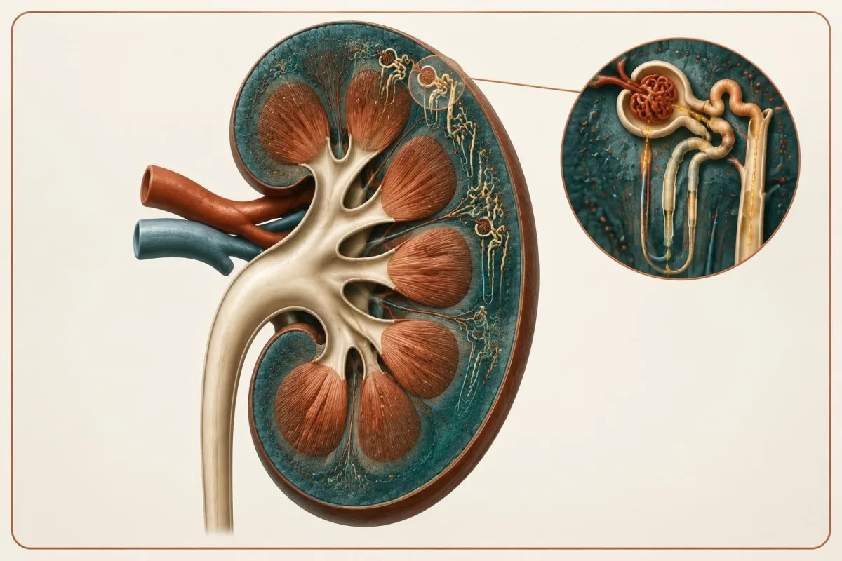
Sudden facial or ankle swelling with PCR above 3,500 mg/g may indicate heavy protein loss, especially when serum albumin is below 3.0 g/dL. New breathlessness can reflect fluid accumulation and should not be attributed to the urine result without prompt assessment.
Visible tea-colored or pink urine, reduced urine output over 12 hours, confusion or severe flank pain needs urgent care regardless of whether PCR is 100 mg/g or 1,000 mg/g. The ratio can be modest when blood or inflammation is the dominant clue.
Persistent protein with an eGFR below 30 mL/min/1.73 m² generally warrants specialist involvement under many referral pathways, though local criteria differ. Our low-eGFR warning guide explains why symptoms and filtration level can change the speed of evaluation.
その後に行われる可能性のある追加検査
Persistent or substantial proteinuria may lead to repeat ACR and PCR, urine microscopy, kidney ultrasound, immune testing, protein electrophoresis or specialist review. The specific sequence depends on whether the pattern is albumin-dominant, blood-associated, nephrotic-range, tubular or linked to another illness.
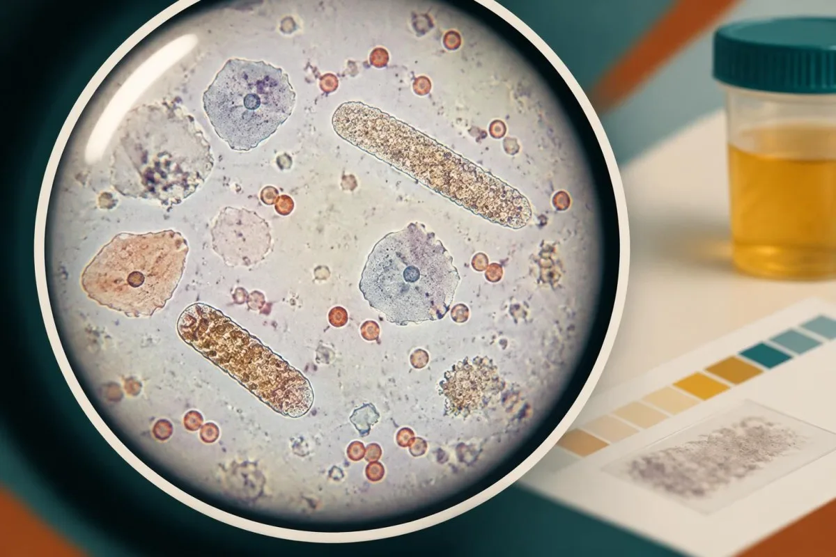
Albumin-dominant protein with red-cell casts may prompt assessment for glomerular inflammation, including complement levels and autoimmune antibodies. Marked protein with little blood may point clinicians toward diabetes, membranous disease or another filtration-barrier disorder, but these patterns are not diagnostic by themselves.
A kidney ultrasound can assess size, obstruction and structural features, while a biopsy is reserved for selected cases where tissue information would change treatment. A PCR of 2.0 g/g does not automatically mean biopsy; age, eGFR, ACR, sediment and treatment options all influence that decision.
Anti-PLA2R antibodies can support evaluation of some membranous nephropathy patterns, but a positive result still requires clinical interpretation. Our article on PLA2R antibody results explains why one specialized test cannot replace the wider kidney assessment.
薬剤や健康状態が経過観察に与える影響
Management of an elevated urine protein creatinine ratio targets the underlying cause and often includes blood-pressure control, diabetes care, medication review and avoidance of preventable kidney stress. Treatment decisions should follow confirmation and clinical assessment rather than a self-calculated ratio alone.

Clinicians may review non-steroidal anti-inflammatory drugs, contrast exposure, supplements and medicines that alter kidney filtration or potassium balance. An adult with PCR of 900 mg/g, eGFR of 52 and potassium of 5.5 mmol/L needs a different plan from someone with PCR of 900 mg/g and stable potassium of 4.2 mmol/L.
Angiotensin-converting enzyme inhibitors, angiotensin receptor blockers and selected newer kidney-protective medicines can reduce albuminuria in appropriate patients, but they require individualized monitoring. A change in serum creatinine or potassium after starting treatment may be clinically expected in some situations yet unsafe in others.
Kantesti is an AI blood test interpretation platform that can place repeated creatinine, eGFR, potassium, albumin and glucose results into a trend view; it does not prescribe treatment or decide whether a medicine is safe. Medication changes should be discussed with the prescriber, particularly when eGFR is below 60 mL/min/1.73 m².
尿検査と血液検査の結果を整理する方法
A urine protein ratio is easiest to interpret when the report includes its unit, collection date, urine creatinine concentration, laboratory reference interval and related blood results. A structured timeline can reveal whether a value of 250 mg/g is isolated, recurring or rising over 6 months.
Before interpreting a report, record whether the sample was first-morning, whether you had fever or strenuous exercise, and whether urinary symptoms or menstruation were present. Also record serum creatinine, eGFR, ACR, albumin, potassium and blood pressure from the same period when available.
Kantesti is an AI-powered blood test analysis tool that helps explain related blood biomarkers in context, but a urine ratio may not be available in every blood-test report. Our AIテクノロジーガイド describes why automated interpretation can organize data while still leaving clinical decisions to qualified professionals.
Do not compare 0.25 g/g from one laboratory directly with 25 mg/mmol from another without conversion; those values are approximately 250 mg/g and 25 mg/mmol, respectively. If the report lacks a unit or appears internally inconsistent, ask the laboratory or clinician to verify the original result before drawing conclusions.
結果について医療従事者に連絡すべき時期
Contact a clinician about a urine protein creatinine ratio above the laboratory range, especially if it exceeds 500 mg/g or persists on repeat testing. Seek urgent advice for heavy protein-range results with swelling, breathlessness, very high blood pressure, visible urine discoloration or markedly reduced urine output.
A result of 160 mg/g without symptoms may be handled differently from 1,600 mg/g with blood in urine, but both should be interpreted rather than ignored. The first may need a clean repeat sample; the second may need blood tests, microscopy and timely kidney review.
Bring the complete report rather than only the flagged number, including units, reference interval and urine creatinine. A clinician also needs medication doses, recent illness, exercise history and pregnancy status because those details can alter the probability of a transient or clinically significant result.
Kantesti can help users organize blood-test trends before an appointment, while our 医学的検証に関する情報 explains the role of clinical standards and oversight in automated interpretation. AI-generated context should support, not delay, care when warning symptoms are present.
よくある質問
尿タンパク/クレアチニン比の正常値はどのくらいですか?
尿蛋白/クレアチニン比が150 mg/g(または0.15 g/g)未満であれば、一般に妊娠していない成人の通常範囲内とみなされます。検査機関によって基準範囲が異なる場合があり、血尿、円柱、eGFRの低下、高血圧が認められる場合は、結果が正常に見えても腎疾患を除外できません。3か月以上にわたって異常が持続する場合は、境界域の結果が一度だけ認められる場合よりも重要です。
尿蛋白/クレアチニン比が0.3というのは高いですか?
0.3 g/gという比率は300 mg/gに相当し、成人の通常の基準値である150 mg/gを上回ります。妊娠中は、高血圧の評価において0.3 g/gが有意な蛋白尿の一般的な基準値として用いられるため、速やかに臨床状況を踏まえて評価する必要があります。妊娠していない場合、臨床医は通常、結果を再検査し、意味を判断する前にeGFR、尿沈渣、血圧、アルブミンを確認します。
尿タンパク/クレアチニン比が高いとはどういう意味ですか?
尿蛋白クレアチニン比が高い場合、尿中クレアチニンに対する蛋白量が通常より多いことを示しますが、この値だけで原因を特定することはできません。500 mg/gを超える値では腎臓に重点を置いた評価が行われることが多く、3,500 mg/gを超える値はネフローゼ域に該当します。運動、発熱、感染症、脱水、糸球体疾患、尿細管疾患、オーバーフロー蛋白はいずれも高値の原因となり得ます。
尿蛋白クレアチニン比は24時間蓄尿の代わりになりますか?
随時尿の尿蛋白クレアチニン比は、24時間蓄尿を行わずに1日の蛋白喪失量を推定できることが多く、1 g/gはおよそ1 g/日に相当します。尿中クレアチニン値が異常に低いまたは高い場合、採尿条件が通常と異なる場合、あるいは蛋白排泄量が急速に変化している場合は、信頼性が低くなります。随時尿の結果が症状、アルブミン値、その他の検査結果と一致しない場合、医療従事者は時間蓄尿を依頼することがあります。
尿蛋白比が高値の場合、どのくらいの期間を空けて再検査すべきですか?
150~500 mg/gの予期しない境界域の結果は、発熱、尿路症状、激しい運動などの一時的な要因が解消した後、臨床上の助言に応じて通常は数日から数週間以内に再検査されることがよくあります。慢性腎臓病と診断するには、腎臓の異常が少なくとも3か月持続している必要があるため、1回の検査結果だけでは慢性であると判断できません。非常に高い比率、むくみ、目に見える尿の変色、または尿量の減少がある場合は、定期的な再検査を待たずに医療機関で評価を受けてください。
尿検査におけるPCRとACRの違いは何ですか?
PCRは尿中総蛋白を測定するのに対し、ACRはアルブミンを特異的に測定します。PCRが1,000 mg/gでACRが80 mg/gの場合、PCRとACRがどちらも約1,000 mg/gの場合とは異なる蛋白パターンが示唆されます。臨床医はこの比較を用いて、糸球体からのアルブミン漏出、尿細管蛋白、オーバーフロー蛋白、または尿路由来の寄与を検討します。
血液検査結果の見方を確認しましょう
検査報告書をアップロードすると、KantestiのAIが数秒で各バイオマーカーを平易な言葉で説明します。世界中で200万人以上が利用しています。.
参考文献
- Klein, T., Mitchell, S., & Weber, H. (2026). 尿中ウロビリノーゲン検査:完全尿検査ガイド2026。. Kantesti AI医学研究。. 掲載日
- Klein, T., Mitchell, S., & Weber, H. (2026). 鉄検査ガイド:TIBC、鉄飽和度、鉄結合能。. Kantesti AI医学研究。. 掲載日
- KDIGO (2024). 慢性腎臓病の評価と管理に関するKDIGO 2024年臨床診療ガイドライン。. Kidney International. 掲載日
- KDIGO (2021). KDIGO 2021 Clinical Practice Guideline for the Management of Glomerular Diseases. Kidney International. 掲載日
- Côté AM et al. (2008). Diagnostic accuracy of urinary spot protein:creatinine ratio for proteinuria in hypertensive pregnant women: systematic review. BMJ。. 掲載日 パブメッド
- 200万以上分析対象の検査
- 127+国
- 100+言語
医療チームについて


E-E-A-T 信頼性シグナル
経験
医師主導による、検査結果解釈ワークフローの臨床レビュー。.
専門知識
臨床的な文脈においてバイオマーカーがどのように振る舞うかに焦点を当てた検査医学。.
権威
Thomas Klein医師による医学的レビュー。Sarah Mitchell医師およびHans Weber教授の協力。.
信頼性
アラームを減らすための明確なフォローアップ経路を備えた、エビデンスに基づく解釈。.


医療免責事項
この記事は教育目的のみを対象としており、医療助言を構成するものではありません。診断や治療の判断を行う際は、必ず有資格の医療提供者にご相談ください。.
公開 · 編集チームにお問い合わせください
Kantesti Ltdが発行。イングランドおよびウェールズで登録、会社番号. 17090423, 、英国ロンドン。.