
תוצאה חיובית של בדיקת נוגדנים אינה מחייבת טיפול באופן אוטומטי. תסמינים, היסטוריית חשיפה, ספירת אאוזינופילים ובדיקת עיניים יכולים להבחין בין ממצא מקרי לבין מחלה הדורשת טיפול.

נקודות עיקריות
- נוגדנים חיוביים מעידים על תגובה חיסונית לאנטיגנים של טוקסוקרה; תוצאה חיובית אחת אינה יכולה לקבוע אם זחלים גורמים כעת למחלה.
- חשיפה בעבר יכולים להותיר IgG שניתן לזהותו במשך שנים, ולכן חיוביות מתמשכת אינה בדיקה מהימנה לכישלון הטיפול.
- אאוזינופיליה פירושה בדרך כלל ספירה מוחלטת של אאוזינופילים מעל 0.5 × 10⁹/L; היא מצדיקה בירור אך אינה מזהה את טוקסוקרה כגורם.
- ספירות אאוזינופילים גבוהות יותר של לפחות 1.5 × 10⁹/L מצריכות הערכה אם הן מתמשכות או מלוות בתסמינים של איברים, ולא טיפול אוטומטי נגד טפילים.
- תסמיני עיניים כגון טשטוש ראייה חדש, גופים צפים או ירידה בראייה בעין אחת מחייבים הערכה מהירה; אובדן ראייה פתאומי מצריך טיפול חירום.
- טיפול לטוקסוקריאזיס ויסצרלית עשוי לכלול אלבנדזול 400 מ״ג פעמיים ביום למשך 5 ימים, תחת השגחת רופא, בהתאם להנחיות CDC.
- מחלת עיניים דורש טיפול בהנחיית מומחה, משום ששליטה בתגובה של הרקמה עשויה להיות חשובה לא פחות מטיפול בטפיל.
- מעקב צריך לעקוב אחר התסמינים והמגמות הרלוונטיות בבדיקות המעבדה; ירידה במדד הנוגדנים מ־2.0 ל־1.5 אינה מוכיחה, כשלעצמה, החלמה.
האם תוצאה חיובית של נוגדנים לטוקסוקרה מצריכה טיפול?
נוגדנים חיוביים לטוקסוקרה אינם מצריכים טיפול באופן אוטומטי. נוגדנים שניתן לזהות עשויים להעיד על חשיפה בעבר ולא על טוקסוקריאזיס פעילה; החלטות הטיפול תלויות בתסמינים תואמים, בהיסטוריית החשיפה ובממצאים תומכים כגון אאוזינופיליה. תסמיני עיניים חדשים מחייבים הערכה מהירה, גם כאשר תוצאת הנוגדנים חיובית במידה חלשה או בדיקות אחרות תקינות.
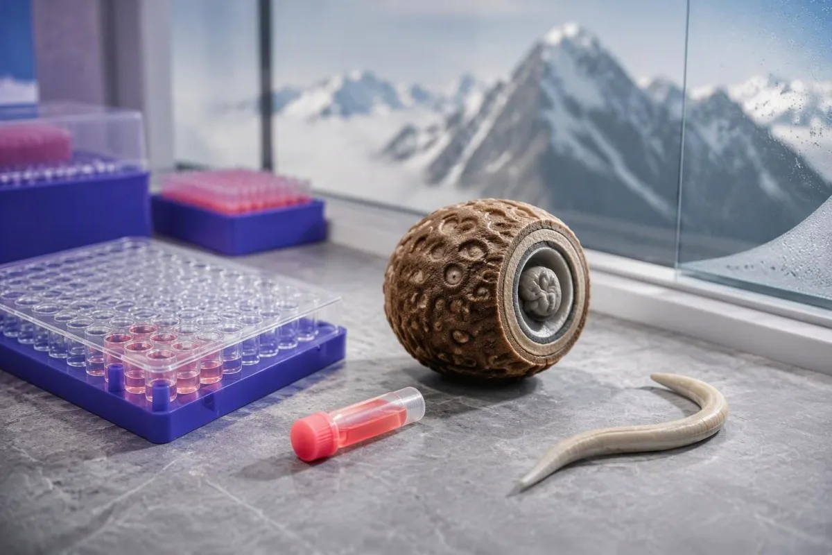
בדיקת נוגדנים חיובית מזהה תגובה חיסונית, ולא סופרת טפילים חיים. לכן, רופאים מבחינים בין 3 שאלות: האם הייתה חשיפה, האם קיימת מחלה, והאם סביר שהטיפול יועיל; תוצאת המעבדה עונה רק על חלק מהשאלה הראשונה.
לדוגמה, נבחן אדם היפותטי בן 38 עם תוצאת סקר חיובית, ללא תסמינים ועם ספירת אאוזינופילים מוחלטת של 0.18 × 10⁹/L. שילוב זה אינו מבסס טוקסוקריאזיס פעילה, ואילו אותה תוצאה לצד שיעול מתמשך, אאוזינופיליה וחשיפה רלוונטית תצדיק הערכה ממוקדת יותר.
הטיפול צריך להיות מכוון לתסמונת קלינית, ולא להפוך תוצאת נוגדנים לשלילית. Magnaval et al. (2001) מתארים את הקושי להבחין, באמצעות סרולוגיה בלבד, בין חשיפה בעבר לבין טוקסוקריאזיס בעלת משמעות קלינית; המדריך שלנו בנושא פרשנות לתוצאות נוגדנים חיוביות מסביר מדוע בדיקות נוגדנים שונות מובילות להחלטות שונות.
קנטסטי הוא מנתח בדיקות דם בינה מלאכותית שיכול לסייע בארגון תוצאת נוגדנים לצד CBC ופאנל כבד, אך אינו יכול לקבוע את חיוניות הזחלים על סמך דוח אחד. קראו על הארגון שלנו להבחנה בין שירות הפרשנות שלנו לבין ההערכה בהובלת רופא הנדרשת כדי לרשום טיפול.
מדוע נוגדנים לטוקסוקרה יכולים להישאר לאחר חשיפה בעבר
ניתן לזהות IgG לטוקסוקרה במשך שנים לאחר החשיפה, והוא עשוי להישאר לאחר הטיפול. נוגדנים הם חלק מהזיכרון החיסוני, ולא חותמת זמן המעידה מתי התרחשה החשיפה. דגימה חיובית אחת אינה יכולה להבחין באופן מהימן בין חשיפה בעבר הרחוק, זחלים שנותרו בגוף ללא תסמינים נוכחיים, וזחלים הגורמים למחלה פעילה ברקמות.
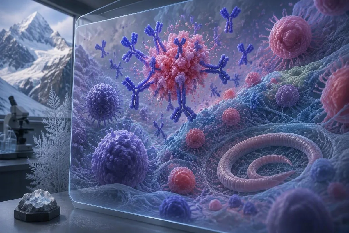
בני אדם נושאים בדרך כלל זחלים של טוקסוקרה ולא תולעים בוגרות במעי. שני המינים העיקריים הקשורים למחלה בבני אדם הם Toxocara canis ו־Toxocara cati; הזחלים עשויים לנדוד לרקמות, שבהן התגובות החיסוניות והתסמינים שונים מאלה של זיהום רגיל בתולעים עגולות במעי.
זחלים שנותרים בגוף ונוגדנים שנותרים בגוף הם מושגים קשורים, אך אינם בני החלפה. לאדם עשוי להיות IgG שניתן לזהות 3 שנים לאחר חשיפה חשודה, ללא עדות לפגיעה נוכחית באיברים; תוצאת הנוגדנים לבדה אינה יכולה לקבוע אם זחלים שנותרו בגוף עדיין חיים.
תוצאה חיובית מאוד אינה מאפשרת לקבוע באופן מהימן מתי התרחשה החשיפה. חשיפה חוזרת יכולה להעלות את רמות הנוגדנים, ובדיקות שונות מזהות הכנות אנטיגן שונות; לכן, אי אפשר לתרגם מדד מספרי של 4.0 לכמות זחלים גדולה פי 4 או לחומרת מחלה גדולה פי 4.
נוגדנים ארוכי־טווח מבדילים בין טוקסוקריאזיס לבין זיהומים שבהם ניתן לזהות בקלות את האורגניזם ישירות. הדיון שלנו בנושא נוגדנים לבבזיה לעומת זיהום ממחיש את אותה הבחנה כללית, אף שבדיקות האישור וכללי הטיפול בבבזיה אינם חלים על טוקסוקרה.
כיצד לפרש מדדי נוגדנים, טיטרים ותוצאות גבוליות
יש לפרש סרולוגיה לטוקסוקרה בהתאם לשיטת המעבדה המדווחת ולערך הסף שלה. There is no universal antibody concentration in IU/mL that separates past exposure from active disease. ELISA indexes, optical-density ratios and dilution titres are different measurements, so 2 laboratories can report the same specimen differently.
An antibody index is usually an assay-specific signal rather than an absolute concentration. In a hypothetical laboratory using positivity above 1.1, an index of 1.3 would be just above the cutoff; that invented example is not a reference range to apply to your own report.
Borderline results deserve particular attention when symptoms and exposure are absent. A laboratory may recommend another specimen or a more specific confirmatory method, such as an immunoblot; our explanation of תוצאות איכותיות וכמותיות helps distinguish a positive flag from a genuinely comparable numerical measurement.
The probability of disease before testing changes what a positive result means. In an explicitly simulated group of 1,000 people, with 10% disease prevalence, 80% sensitivity and 90% specificity, testing would produce 80 true positives and 90 false positives—only about 47% of positive results would represent disease.
Those simulated figures are not performance estimates for a particular Toxocara assay. Kantesti AI can help identify the method and reference information on a report, but cannot replace missing validation data; antibody index confirmation also requires assay-specific reasoning, not borrowing cutoffs from another infection.
אילו היסטוריות חשיפה הופכות טוקסוקריאזיס לסבירה יותר?
Toxocariasis is most often acquired by swallowing infective eggs from contaminated soil, hands or food. Dog and cat exposure matters because these animals can shed eggs, but simply owning a pet does not establish infection. Eggs generally require approximately 2–4 weeks in the environment to become infective.

Fresh animal faeces and established soil contamination are not identical exposure situations. Because eggs require environmental development, a garden or play area contaminated over several weeks can be more relevant than a single recent contact; daily pet-waste removal helps prevent eggs from reaching the infective stage.
Ask about 3 practical routes: soil or sand exposure, unwashed produce or hands, and consumption of undercooked tissues from infected animals. Foodborne transmission through larvae in animal tissues is recognised, but its contribution varies by setting; Trichinella antibody assessment involves a different parasite and different exposure clues.
A cat scratch is not the usual mechanism of Toxocara acquisition. Scratches instead raise other diagnostic possibilities, including Bartonella; our guide to cat scratch antibody testing explains why 1 animal-related event should not be used to label every positive infection test.
Geography changes the background likelihood of exposure but does not establish active disease. A hypothetical adult who gardened barefoot for 10 years may have a plausible exposure history, yet current symptoms, eosinophils and examination findings still determine whether that history explains the positive result.
אילו תסמינים וספירות אאוזינופילים תומכים במחלה פעילה?
Active visceral toxocariasis becomes more plausible when positive antibodies accompany compatible organ symptoms and eosinophilia. Fever, persistent cough, wheeze, abdominal discomfort or liver enlargement can support investigation, but none is specific. An absolute eosinophil count above approximately 0.5 × 10⁹/L is generally considered elevated in adults.
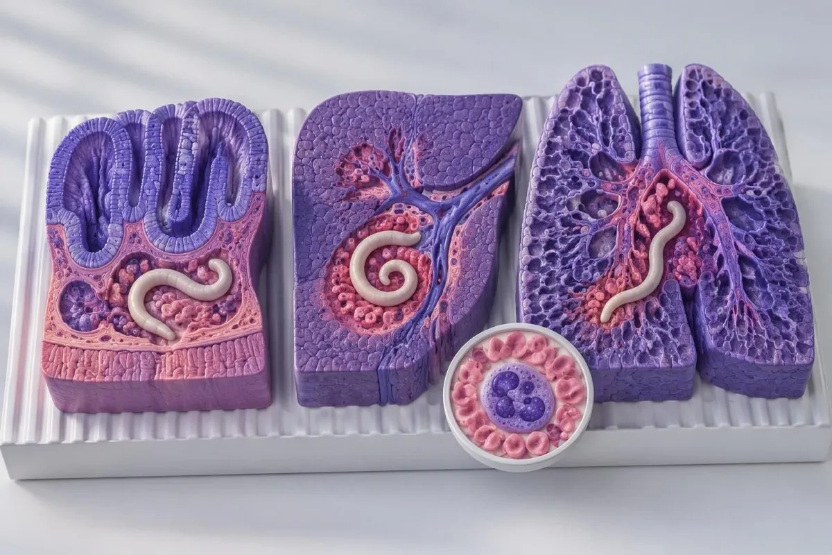
Eosinophilia is a clue to tissue responses, not a parasite identification test. Allergic disease, medicines, other helminths and some haematological disorders can raise eosinophils; a count of 0.8 × 10⁹/L therefore requires context rather than an automatic albendazole prescription.
Persistent counts of at least 1.5 × 10⁹/L warrant evaluation, especially when accompanied by respiratory, cardiac, neurological or other organ symptoms. That threshold reflects concern about hypereosinophilia and potential organ injury—not a validated Toxocara treatment cutoff; high eosinophil assessment מסביר את הדיפרנציאל הרחב יותר.
Normal eosinophils do not exclude ocular toxocariasis. Disease confined to 1 eye may produce little systemic laboratory response, so a normal CBC cannot overrule new visual symptoms; this distinction is why eye complaints are assessed separately from the visceral disease pattern.
Mild or so-called covert toxocariasis is harder to establish because fatigue, itch and abdominal symptoms have many competing explanations. Magnaval et al. (2001) discuss these less specific presentations; when only 1 vague symptom is present, clinicians must avoid treating an incidental antibody result as proof of its cause.
אילו תוצאות מעבדה אחרות מסייעות לפרש נוגדנים לטוקסוקרה?
A CBC with an absolute eosinophil count usually adds more diagnostic context than the antibody index alone. Liver tests, total IgE and inflammatory markers may help assess a suspected clinical syndrome, but none confirms toxocariasis. Read the actual values and units, not just whether 1 result is flagged red.

The absolute eosinophil count equals the white-cell count multiplied by the eosinophil fraction. A hypothetical WBC of 8.0 × 10⁹/L with 12% eosinophils gives 0.96 × 10⁹/L; 12% alone is less informative because the same percentage produces a different absolute count when total WBC changes.
Inflammatory markers cannot reliably separate Toxocara from allergy or another infection. CRP of 3 mg/L does not exclude localised eye disease, and CRP of 35 mg/L does not prove visceral toxocariasis; our guide to אי־התאמות בין WBC ל־CRP מסביר מדוע הסמנים האלה עונים על שאלות שונות.
Liver enzyme abnormalities describe tissue stress rather than its cause. A hypothetical ALT of 72 IU/L with a laboratory upper limit of 40 IU/L is 1.8 times that limit, but medicines, metabolic liver disease and other infections remain possibilities; פרשנות של פאנל כבד עוזר להפריד בין הדפוסים האלה.
קנטסטי הוא פלטפורמת פרשנות ביומרקרים של AI that can place eosinophils, ALT and antibody results in 1 organised summary for clinician review. Total IgE is particularly nonspecific: an elevated value may fit an allergic or helminth-related response, but no IgE threshold reliably establishes active toxocariasis.
מדוע תסמיני עיניים מחייבים הערכה מהירה?
New blurred vision, floaters or reduced vision—especially in 1 eye—need prompt assessment for ocular disease, regardless of the Toxocara antibody strength. Sudden visual loss, a curtain-like shadow or severe eye pain requires emergency care. Ocular toxocariasis can occur with low or negative serum antibodies and normal eosinophils.
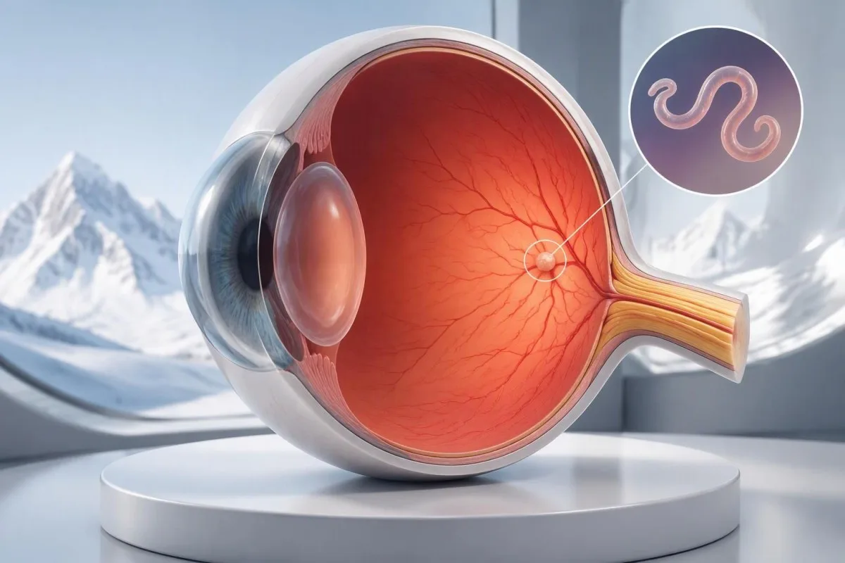
Ocular toxocariasis often affects only 1 eye. Ahn et al. (2014) describe retinal granulomas, vitreous tissue responses and other complications; the small local parasite burden helps explain why serum testing may be less informative than in systemic disease.
A positive antibody result does not identify the cause of every visual complaint. Uveitis, retinal tears, glaucoma and other conditions require different management, and HLA-B27-associated eye symptoms illustrate another reason that 1 positive laboratory marker cannot replace an eye examination.
Ocular toxocariasis diagnosis rests heavily on specialist examination. A dilated retinal examination and, when indicated, OCT or ocular ultrasound assess structures the antibody test cannot show; a weakly positive index of 1.2 should neither confirm the diagnosis nor delay referral in a symptomatic patient.
Do not wait several weeks for a repeat antibody result if vision has changed. Contact an urgent eye service the same day for new visual symptoms; in a child, a newly observed squint or white pupil also warrants urgent assessment because several sight-threatening conditions—not only Toxocara—can present that way.
כיצד רופאים מאבחנים טוקסוקריאזיס?
Toxocariasis diagnosis usually combines exposure history, compatible clinical findings, serology and exclusion of better explanations. No single routine test reliably confirms every form of the disease. Clinicians consider at least 3 evidence domains—exposure, organ findings and laboratory support—rather than treating a positive antibody flag as a diagnosis.
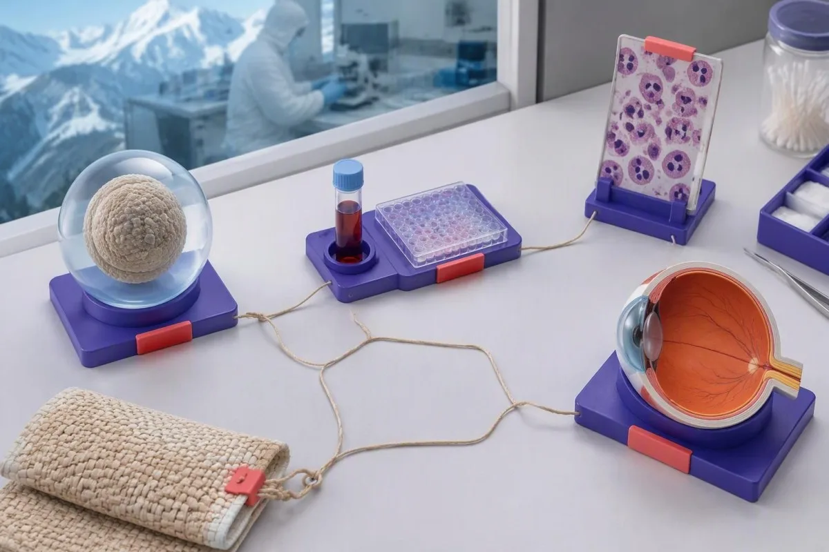
Human stool testing does not normally detect Toxocara eggs. Humans are accidental hosts, and larvae generally do not mature into egg-producing adult worms; 3 negative stool samples therefore do not rule out toxocariasis, although stool testing may help identify other parasites.
Cross-reactivity with other helminths can reduce serological specificity, particularly where multiple parasites circulate. Before prescribing systemic corticosteroids, clinicians may assess Strongyloides risk because steroids can precipitate severe strongyloidiasis; Strongyloides IgG follow-up follows different treatment principles from incidental Toxocara positivity.
Imaging should answer a specific organ-related question. Liver ultrasound or other imaging may be useful when symptoms, examination or laboratory findings suggest organ involvement, but scanning an asymptomatic person because of 1 positive antibody test can discover unrelated abnormalities without clarifying the diagnosis.
Kantesti can help organise these findings, but cannot resolve a discordant clinical picture from laboratory data alone. If antibodies are positive, eosinophils are 0.1 × 10⁹/L and symptoms point strongly toward another condition, the appropriate next step is clinician-led reassessment—not labelling every complaint as a parasite effect.
מתי סביר להסתפק במעקב לאחר תוצאה חיובית?
Observation can be reasonable when a person has positive Toxocara antibodies but no compatible symptoms or evidence of organ involvement. This is a clinician-led decision, not a rule that all asymptomatic positives can be ignored. One unexplained laboratory abnormality, such as persistent eosinophilia, may still require investigation.
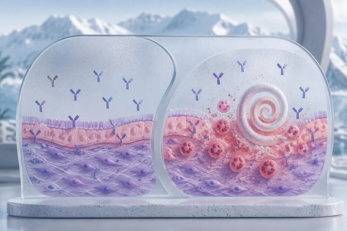
Evidence for treating every asymptomatic seropositive person is limited. Antibody persistence makes eradication difficult to measure, and the balance between benefit and medicine-related harm differs from symptomatic visceral disease; there is no universal antibody index, such as 2.0, that mandates treatment by itself.
An observation plan should specify what will be reviewed and what should trigger earlier contact. For example, a hypothetical clinician might arrange symptom and CBC review in 4–6 weeks when mild unexplained eosinophilia is present; that interval is an illustrative plan, not a universal toxocariasis follow-up guideline.
Routine parasite antibody screening is not usually useful solely because someone feels well and owns a pet. Broad screening increases the chance of finding evidence of past exposure without actionable disease; our guide to blood work without symptoms explains how risk-based testing reduces unnecessary follow-up.
Safety-net instructions make observation an active decision rather than neglect. New visual symptoms, persistent fever, worsening breathing or neurological symptoms should prompt earlier assessment, and a rising eosinophil count from 0.7 to 1.8 × 10⁹/L deserves review even if the antibody result is unchanged.
באיזה טיפול משתמשים לטוקסוקריאזיס תסמינית?
Clinicians commonly use albendazole for symptomatic visceral toxocariasis, while ocular disease needs specialist-directed treatment. CDC Clinical Care guidance lists albendazole 400 mg orally twice daily for 5 days for adults and children, but also states that the optimal treatment duration is undefined. This is prescribing information, not a self-treatment instruction.
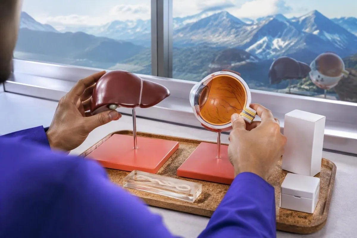
Treatment duration can differ across specialist practice because evidence is limited. CDC also lists mebendazole 100–200 mg twice daily for 5 days, while longer albendazole courses appear in published clinical practice; different schedules should not be interpreted as proof that a short course always fails.
Ocular management aims to protect sight and control the tissue response. Ahn et al. (2014) discuss antiparasitic therapy, corticosteroids and procedural treatment in selected cases; 1 standard visceral regimen should not be copied into an eye-disease treatment plan without ophthalmology input.
Longer albendazole courses may require liver-enzyme and blood-count monitoring. Baseline liver disease, interacting medicines and treatment duration affect the monitoring plan; our guide to ניתוח מגמות בטיחות תרופות explains why changes from an individual baseline can matter more than a single flag.
Kantesti does not prescribe antiparasitic medicines or corticosteroids. A hypothetical ALT rise from 30 to 110 IU/L during treatment needs clinician review rather than an AI-directed dose change; pregnancy, age under 2 years, liver disease and other medicines also require individual prescribing assessment.
כיצד ילדים, היריון ומצב חיסוני משנים את ההחלטות?
Children, pregnant people and patients needing immunosuppression require individual assessment rather than automatic treatment after positive serology. Exposure behaviours, eye findings and medicine safety differ across these groups. CDC notes limited albendazole safety evidence in children under 6 years, despite experience with younger children in public-health programmes.
Young children may have greater hand-to-mouth soil exposure, particularly with pica. A 3-year-old with repeated soil ingestion and eosinophilia has a different exposure profile from an asymptomatic adult, but neither age nor behaviour proves that a positive test represents active disease.
Paediatric laboratory interpretation needs age-appropriate reference information and an adult who can provide a reliable symptom history. Our discussion of פירוש AI לילדים explains why 1 automated summary cannot replace assessment of growth, visual behaviour and medicine suitability.
Pregnancy does not convert Toxocara IgG into proof of a new infection. Unlike toxoplasmosis, toxocariasis does not have an established routine prenatal serological screening pathway; an isolated positive result at 10 weeks of pregnancy requires contextual review, not assumptions about fetal infection or automatic antiparasitic treatment.
Before prolonged corticosteroid treatment, parasite assessment should match travel and exposure history. The immediate safety concern may be Strongyloides rather than Toxocara, and a patient taking prednisone 40 mg daily should not independently add or stop medicines because of an antibody report.
האם יש לחזור על בדיקת נוגדנים לטוקסוקרה לאחר הטיפול?
Repeat Toxocara antibodies are not a reliable stand-alone test of cure because IgG can remain positive for years. Follow-up should prioritise symptom improvement, relevant organ findings and eosinophil trends. One antibody measurement after treatment cannot establish either eradication or failure.
Use the same laboratory method when numerical trends are clinically needed. A result of 2.4 on one assay and 1.7 on another may reflect different calibrators or antigen preparations, rather than biological improvement; switching assays can create a trend that does not exist.
Symptoms and eosinophils often provide a more practical follow-up signal than persistent IgG. In a hypothetical case, cough improving alongside an eosinophil decline from 1.2 to 0.4 × 10⁹/L supports clinical improvement, but does not prove that every larva has been eliminated.
A small numerical change may fall within assay variation. An index shifting from 1.4 to 1.3 should not be treated as meaningful without laboratory information about reproducibility; our guide to שינויים משמעותיים בבדיקות דם explains the distinction between analytical variation and a real trend.
Ocular follow-up requires eye examination rather than antibody tracking alone. Stable serum IgG cannot show whether a retinal affected area is improving, and persistent or recurrent visual symptoms after a 5-day medicine course need specialist reassessment rather than repeated unsupervised treatment.
כיצד בינה מלאכותית יכולה לסייע בלי לאבחן ביתר טוקסוקריאזיס פעילה?
AI can help organise Toxocara antibodies, eosinophils and liver results, but cannot diagnose active disease from serology alone. A useful interpretation preserves uncertainty and highlights missing clinical information. At minimum, the report should be considered with 3 details: symptoms, exposure history and the laboratory’s assay method.
קנטסטי הוא פלטפורמת פענוח בדיקות דם של AI that can summarise related results and help prepare questions for a clinician. Our הסבר טכנולוגיית AI describes the interpretation workflow; a response generated in approximately 60 seconds is not an examination, a parasite-viability test or permission to delay urgent eye care.
Clinical reasoning should remain traceable to the original report. Check that an eosinophil value of 0.8 × 10⁹/L has not been confused with 0.8%, and that borderline and confirmed-positive findings are distinguished; our מידע על תיקוף ופיקוח קליני information describes how we discuss methodology without claiming every output establishes a diagnosis.
Privacy also matters when sharing laboratory documents. Remove unnecessary identifiers and obtain consent before submitting another person’s report; our מידע על פרטיות ונתונים explains data handling, but privacy safeguards do not change the medical limitations of interpreting 1 antibody result.
Bring 4 items to the appointment: the complete report, symptom dates, exposure details and a medicine list. Ask whether the result explains a defined clinical syndrome, whether another diagnosis fits better, what would justify treatment, and which symptoms require urgent reassessment.
פרסומי מחקר: אילו ראיות תומכות בהחלטות הטיפול האלה?
Treatment decisions in this article rely on Toxocara-specific clinical literature and CDC guidance, not unrelated laboratory publications. The 2 peer-reviewed reviews listed below explain diagnostic limitations and ocular management. The separate Zenodo records cover iron and coagulation testing; they do not establish how positive Toxocara antibodies should be treated.
A review article is not the same as a randomised treatment trial. Magnaval et al. (2001) and Ahn et al. (2014) bring together clinical observations and published evidence, but important uncertainties remain about optimal treatment duration and the benefit of treating asymptomatic seropositivity.
Kantesti’s 2 listed Zenodo publications address different laboratory topics. The מדריך רקע לבדיקות ברזל may help with an independently identified iron abnormality, while the coagulation testing background guide addresses clotting tests; neither is a confirmatory test for toxocariasis.
A useful medical article must separate evidence, inference and illustration. The hypothetical antibody indexes and worked examples here are not patient records or validated treatment thresholds; our תקני תוכן רפואי explain that approach, including why 1 positive antibody finding should never be presented as a complete diagnosis.
שאלות נפוצות
האם תוצאה חיובית בבדיקת נוגדנים לטוקסוקרה פירושה שעליי ליטול אלבנדזול?
0. תוצאה חיובית בבדיקת נוגדנים לטוקסוקרה אינה אומרת בהכרח שעליכם ליטול אלבנדזול. ניתן לזהות IgG במשך שנים, ולכן רופאים מחפשים תסמינים תואמים, היסטוריה של חשיפה וממצאים תומכים, במקום לטפל על סמך תוצאה חיובית אחת בלבד. ה-CDC מציין אלבנדזול במינון של 400 מ״ג פעמיים ביום למשך 5 ימים לטוקסוקריאזיס ויסצרלית, אך ההחלטה ומשך הטיפול מחייבים השגחה של רופא. תסמינים חדשים בעיניים מחייבים הערכה מהירה של רופא עיניים במקום טיפול עצמי.
האם נוגדנים לטוקסוקרה יכולים להישאר חיוביים לאחר הטיפול?
נוגדנים לטוקסוקרה עשויים להישאר חיוביים במשך שנים לאחר החשיפה, וייתכן שיישארו ניתנים לזיהוי גם לאחר הטיפול. תוצאה שעדיין חיובית כעבור 6 חודשים אינה מוכיחה כשלעצמה שהטיפול נכשל. מעקב אחר תסמינים, מגמות במספר האאוזינופילים ובדיקות של האיברים הרלוונטיים מועיל בדרך כלל יותר להערכת המעקב. יש להשוות מגמות מספריות של רמות הנוגדנים רק כאשר שיטות המעבדה עקביות במידה מספקת.
איזו ספירת אאוזינופילים מצביעה על טוקסוקריאזיס?
ספירת אאוזינופילים מוחלטת מעל כ־0.5 × 10⁹/L מצדיקה בירור, אך אינה מזהה טוקסוקריאזיס באופן ספציפי. אלרגיה, תרופות, טפילים אחרים ומחלות המטולוגיות עלולים לגרום לאותו ממצא. ספירות מתמשכות של לפחות 1.5 × 10⁹/L מצריכות הערכה, במיוחד בנוכחות תסמינים באיברים. רמות אאוזינופילים תקינות אינן שוללות מחלה המוגבלת לעין.
האם טוקסוקריאזיס עינית יכולה להופיע עם בדיקת דם שלילית?
טוקסוקריאזיס עינית יכולה להופיע עם רמות נמוכות או שליליות של נוגדנים בנסיוב, משום שהמחלה עשויה להיות מוגבלת לעין אחת. גם ספירת אאוזינופילים תקינה אינה שוללת מעורבות עינית. רופא עיניים מעריך ממצאים ברשתית ובזגוגית, ולעיתים נעזר ב-OCT או באולטרסאונד כדי להבהיר את האבחנה. טשטוש ראייה חדש או הופעת נקודות צפות מחייבים בדיקה בהקדם, ואילו אובדן ראייה פתאומי מצריך טיפול חירום.
האם בדיקת צואה שלילית שוללת טוקסוקרה?
בדיקת צואה שלילית אינה שוללת טוקסוקריאזיס בבני אדם. בני אדם מארחים בדרך כלל זחלים ברקמות ולא תולעי טוקסוקרה בוגרות המטילות ביצים, ולכן אפילו 3 דגימות צואה שליליות אינן יכולות לשלול את המחלה. בדיקת צואה עדיין עשויה לזהות טפילי מעיים אחרים. הערכת טוקסוקריאזיס משלבת במקום זאת ממצאים קליניים, היסטוריית חשיפה, בדיקות סרולוגיות והערכה ספציפית לאיבר בעת הצורך.
מה המשמעות של מדד נוגדנים גבולי לטוקסוקרה?
אינדקס גבולי של נוגדנים לטוקסוקרה פירושו שאות הבדיקה קרוב לערך הסף של המעבדה לצורך קבלת החלטה, ולא שאושרה מחלה פעילה. לדוגמה, אינדקס של 1.3 עשוי להיות מעט מעל ערך סף היפותטי של 1.1, אך המספרים האלה אינם ערכי ייחוס אוניברסליים. ייתכן שהמעבדה המדווחת תמליץ על בדיקה חוזרת או על שיטת אימות ספציפית יותר. התסמינים וההיסטוריה של חשיפה קובעים אם יש תועלת קלינית בהבהרה.
קבלו הסבר על תוצאות בדיקות הדם שלכם
העלו את דוח המעבדה שלכם, וה-AI של Kantesti יסביר כל סמן ביולוגי בשפה פשוטה בתוך שניות. יותר מ-2 מיליון אנשים ברחבי העולם משתמשים בו.
הפניות
- Klein, T., Mitchell, S., & Weber, H. (2026). מדריך לבדיקות ברזל: TIBC, ריווי ברזל ויכולת קשירת ברזל. מחקר רפואי של Kantesti AI. DOI
- Klein, T., Mitchell, S., & Weber, H. (2026). טווח תקין של aPTT: מדריך לקרישת דם בנושא D-Dimer ו-Protein C. מחקר רפואי של Kantesti AI. DOI
- Magnaval JF et al. (2001). Highlights of human toxocariasis. The Korean Journal of Parasitology. DOI
- Ahn SJ et al. (2014). Ocular toxocariasis: clinical features, diagnosis, treatment, and prevention. Asia Pacific Allergy. DOI
- Centers for Disease Control and Prevention (n.d.). Clinical Care of Toxocariasis. CDC Parasites.
- 2 מיליון+בדיקות שנותחו
- 127+מדינות
- 100+שפות
על הצוות הרפואי


אותות אמון E-E-A-T
הִתנַסוּת
סקירה קלינית בהובלת רופא של תהליכי עבודה לפענוח בדיקות מעבדה.
מוּמחִיוּת
רפואה מעבדתית מתמקדת באופן שבו סמנים ביולוגיים מתנהגים בהקשר קליני.
סמכותיות
נבדק רפואית על ידי ד״ר Thomas Klein, בתרומתם של ד״ר Sarah Mitchell ופרופ׳ ד״ר Hans Weber.
אֲמִינוּת
פרשנות מבוססת-ראיות עם מסלולי המשך ברורים כדי להפחית בהלה.
הצהרה רפואית
מאמר זה מיועד למטרות חינוכיות בלבד ואינו מהווה ייעוץ רפואי. תמיד יש להתייעץ עם גורם מוסמך בתחום הבריאות לצורך החלטות אבחון וטיפול.
פורסם · צרו קשר עם צוות העריכה
פורסם על ידי Kantesti Ltd, הרשומה באנגליה ובוויילס, מספר חברה. 17090423, לונדון, בריטניה.
