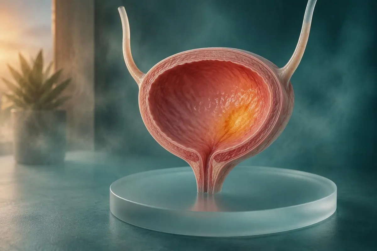
Painless visible blood in urine is the bladder cancer warning sign people should never explain away. Most urinary symptoms have benign causes, but the pattern, timing, and your risk factors determine how quickly to act.


- Visible blood in urine should be assessed promptly even if it happens once, causes no pain, and disappears within hours.
- Painless blood in urine is a classic bladder cancer warning sign; urinary infection and stones can also cause it, so testing identifies the cause.
- Hématurie microscopique means 3 or more red cells per high-power field on a properly collected urine microscopy test.
- Symptômes d’urgence include inability to pass urine, large clots, fever with flank pain, fainting, or rapidly worsening weakness.
- Smoking history remains the strongest modifiable bladder cancer risk factor and matters even after quitting.
- Urine dipsticks detect heme but cannot diagnose bladder cancer or reliably distinguish red cells from myoglobin.
- Cystoscopy directly examines the bladder lining and remains the key test when visible hematuria or higher-risk microscopic hematuria is present.
- analyses sanguines can identify anemia or kidney impairment but do not rule out bladder cancer when results are normal.
Which bladder cancer symptoms should prompt action now?
Painless visible blood in urine needs prompt medical assessment, even when the urine clears after one trip to the toilet and you otherwise feel well. Bladder cancer can cause urgency, frequency, burning, pelvic discomfort, or no symptom beyond red, pink, or cola-coloured urine; most people with these symptoms do not have cancer, but the visible-blood pattern is not safe to self-diagnose.
As of October 2, 2026, I advise patients to contact a clinician the same day or next working day for new visible hematuria. A clot, inability to pass urine, dizziness, fever above 38.0°C, or severe side or back pain changes that advice to urgent or emergency care. The useful starting point is our detailed guide to signes d’alerte du sang dans les urines.
In my clinical experience, the misleading reassurance is, “It only happened once.” Intermittent bleeding is compatible with a stone, infection, an enlarged prostate, kidney disease, medication-related bleeding, and bladder cancer; the stop-start nature does not tell us which cause is present.
Kantesti est un Analyseur de test sanguin AI that helps people understand accompanying results such as hemoglobin, creatinine, and inflammatory markers, but it cannot inspect the bladder or exclude cancer. Dr. Thomas Klein’s practical rule is simple: a normal full blood count never cancels the need to investigate visible urinary blood.
A note on colour
Pink, red, rusty-brown, and tea-coloured urine can all represent urinary bleeding, although food pigments, medicines, bilirubin, and muscle pigment can mimic it. Photographing the colour before flushing can help the clinician, but do not delay evaluation to collect a perfect sample.
Why painless blood in urine carries special weight
Painless visible blood in urine is the urinary symptom most associated with bladder cancer evaluation. It does not prove cancer, and infection remains common, yet painlessness removes the reassuring clue that many people expect from a stone or acute cystitis.
Visible hematuria occurs in roughly 80% to 90% of people who present with bladder cancer, although the reverse is not true: many visible-hematuria episodes arise from benign causes. The clinical question is therefore not “Is cancer likely from colour alone?” but “Has the urinary tract been examined enough to safely exclude an important cause?”
The EAU non-muscle-invasive bladder cancer guideline identifies hematuria as the most common presenting sign and supports cystoscopy as part of diagnostic assessment when bladder cancer is suspected (Babjuk et al., 2022). Red urine after beetroot, blackberries, rifampicin, phenazopyridine, or dehydration may be harmless, but a dipstick and microscopy settle that uncertainty far better than guesswork.
Do not assume anticoagulants explain the finding. Apixaban, rivaroxaban, warfarin, and aspirin can make bleeding more noticeable, but they do not create a bladder source from nothing; clinicians generally still evaluate unexplained hematuria. If you take warfarin, an unexpectedly high INR deserves separate attention in our guide des résultats INR.
What microscopic blood in urine means
Microscopic hematuria means at least 3 red blood cells per high-power field on urine microscopy from one properly collected specimen. A positive dipstick alone is not microscopic hematuria because exercise, menstrual contamination, and muscle pigment can make the strip positive without urinary red cells.
The American Urological Association guideline defines microhematuria at the threshold of 3 red cells per high-power field, then uses age, smoking exposure, persistence, and prior gross hematuria to guide risk-based testing (Barocas et al., 2020). The threshold is deliberately low because microscopy is a triage signal, not a cancer diagnosis.
A clean-catch midstream sample collected away from menstruation and at least 48 hours after strenuous endurance exercise reduces avoidable false alarms. Red cells with substantial protein, casts, or dysmorphic shapes can point toward a kidney-filter problem rather than a bladder source; our urine sediment explainer shows why morphology matters.
Persistence matters more than a single borderline count. In a low-risk younger adult with 3 to 10 red cells per field after a recent viral illness, a clinician may repeat testing; in a 67-year-old former smoker, the same count usually warrants more direct urologic discussion.
Can urgency, burning, or frequency be early signs?
Urgency, frequent urination, burning, and new nighttime urination can occur with bladder cancer, but infection, menopause-related tissue change, diabetes, stones, and overactive bladder are much more common. The concern rises when symptoms persist after treatment, recur without a positive culture, or appear alongside blood.
I see this pattern most often when a patient receives two or three antibiotic courses for “UTIs” but cultures repeatedly show no bacterial growth. A urine culture taken before antibiotics is especially valuable: symptoms plus pyuria can occur without conventional bacterial infection, while a negative culture should prompt clinicians to widen the differential.
Burning that lasts more than 2 weeks after a treated infection, or returns within 6 weeks, deserves review rather than endless empirical antibiotics. Frequency without burning can also reflect high glucose, diuretic medication, excess fluid intake, sleep apnea, or diabetes; compare these pathways in our frequent urination blood test guide.
Carcinoma in situ, a flat high-grade bladder change, may cause marked irritative symptoms while imaging appears unrevealing. That is one reason a clinician may recommend cystoscopy even when a urine scan looks reassuring.
When urinary bleeding becomes an emergency
Go to urgent care or an emergency department for urinary bleeding with clots, inability to urinate, fever, severe flank pain, fainting, or new confusion. These combinations can signal blockage, a serious kidney infection, significant blood loss, or another condition requiring immediate treatment.
Clot retention is more than an inconvenience: clots can obstruct urine flow and stretch the bladder painfully over hours. If you have not passed urine for 6 to 8 hours despite a strong urge, especially with visible clots, do not wait for a routine appointment.
Fever above 38.0°C with chills, flank pain, vomiting, or rapid heart rate points more toward upper urinary infection than bladder cancer, but both can coexist. A clinician may check urine culture, kidney function, complete blood count, and imaging; our overview of les résultats de la leucocyte estérase explains why one strip result is not enough.
Heavy bleeding can lower hemoglobin, yet a normal hemoglobin level does not mean an episode is trivial because acute changes may lag by several hours. New shortness of breath, chest pressure, collapse, or pale clammy skin needs emergency assessment regardless of the urine colour.
Common causes that mimic bladder cancer warning signs
Urinary infection, stones, vigorous exercise, menstrual contamination, enlarged prostate, kidney disease, and certain medicines commonly mimic bladder cancer symptoms. A plausible benign explanation is useful only when the symptom resolves and objective testing supports that explanation.
A stone often causes sudden one-sided colicky pain that comes in waves, nausea, and blood in urine, whereas bladder cancer bleeding is often painless. That distinction is imperfect: small stones may be quiet, and cancer can coexist with infection or stones, so symptoms should guide testing rather than replace it.
Distance runners can develop transient hematuria after prolonged impact and dehydration, usually clearing within 24 to 72 hours. I still recommend a urine check if it is visible, repeats, lasts beyond 72 hours, or occurs without a hard workout; see hématurie du coureur for a careful return-to-training approach.
Cloudiness is not the same as blood. Crystals, vaginal discharge, phosphate sediment, and concentrated urine can all make urine look cloudy, while true bleeding can leave it clear-looking; our cloudy urine guide separates those visual clues.
Who has a higher pre-test risk of bladder cancer?
Age over 50, current or previous smoking, occupational aromatic-amine exposure, pelvic radiation, cyclophosphamide, and chronic bladder irritation increase bladder cancer risk. Risk factors do not diagnose cancer, but they lower the threshold for complete evaluation when hematuria appears.
Tobacco exposure is the dominant modifiable factor; smoking is linked to roughly one-half of bladder cancer cases in many high-income populations. The risk falls after quitting but does not instantly return to baseline, so a remote 20-pack-year history remains clinically relevant decades later.
Ask specifically about work with dyes, rubber, leather, paint, printing, combustion products, and some chemical manufacturing, especially before modern protective standards. The question is not meant to assign blame—exposure histories are often incomplete—but it helps a urologist select the appropriate work-up.
Long-term cyclophosphamide exposure and previous pelvic radiotherapy deserve explicit mention at referral. Dr. Thomas Klein also asks about family patterns of urothelial cancers and Lynch syndrome, though inherited causes account for a small minority of cases; our family cancer testing overview explains what routine blood panels can and cannot screen for.
Why bladder symptoms in women can be missed
Women with bladder cancer are more likely to have early symptoms attributed to urinary infection, particularly when burning and frequency occur without dramatic bleeding. Any visible blood, recurrent culture-negative symptoms, or symptoms that persist after treatment should reset the clinical assessment.
A practical safeguard is to ask for the actual culture result, not merely whether an antibiotic was prescribed. A culture with no growth after symptoms recur twice in 6 months should make clinicians consider stone disease, atrophic change, interstitial cystitis, tuberculosis in relevant settings, and bladder pathology.
Menstrual blood can contaminate a urine specimen, but it should lead to repeat collection after bleeding ends—not dismissal of persistent findings. Midstream collection is helpful, and catheter-collected specimens are occasionally used when contamination repeatedly obscures the result.
Kantesti AI est une Outil d’analyse de prise de sang alimenté par l’IA that can organize kidney function, glucose, and anemia trends for a clinician conversation, but urinary symptoms still require urine-based and anatomical testing. For urinary bacteria found without symptoms, read our separate guide on when bacteriuria needs treatment.
What tests are used after blood is found in urine?
Initial hematuria assessment usually includes urinalysis with microscopy, urine culture when infection is plausible, kidney function testing, and risk-based imaging plus cystoscopy. The exact sequence varies by age, kidney function, pregnancy status, contrast allergy, and whether blood is visible.
Urinalysis checks red cells, white cells, protein, nitrite, glucose, and specific gravity; microscopy confirms whether red cells are actually present. A culture is most informative before antibiotics, while proteinuria or casts may redirect referral toward nephrology as well as urology.
Serum creatinine and estimated GFR help clinicians choose imaging safely, particularly when CT contrast is considered. An eGFR below 30 mL/min/1,73 m² often triggers a more individualized contrast discussion, rather than an automatic cancellation of all imaging; see our guide de la fonction rénale.
Kantesti’s service d’interprétation des tests de laboratoire par l’IA can place creatinine, eGFR, hemoglobin, and platelet results into longitudinal context after a clinician orders them. We describe safeguards and clinical limits in our approche de validation médicale.
Which blood test results matter—and what they cannot tell you
A complete blood count and kidney panel can show consequences or alternative explanations for urinary symptoms, but no routine blood test screens reliably for bladder cancer. Normal hemoglobin, creatinine, CRP, and white-cell counts cannot rule out a small bladder growth.
Hemoglobin below approximately 120 g/L in many non-pregnant women ou 130 g/L in many adult men meets common anemia definitions, although laboratory ranges vary. Persistent urinary bleeding can contribute to iron deficiency, but gastrointestinal and gynecologic sources are often more common and must be considered in parallel.
Creatinine can rise with dehydration, urinary obstruction, medicines, or chronic kidney disease; one number without a baseline is less informative than a trend. A sudden fall in eGFR together with reduced urine output is more urgent than an isolated mildly low eGFR on a stable outpatient result.
Urine cytology looks for abnormal shed cells and is more sensitive for some high-grade cancers than low-grade growths, but a negative cytology does not clear a person with visible hematuria. Our urine dipstick explanation details the difference between a strip result and a diagnosis.
What cystoscopy and imaging can find
Cystoscopy examines the bladder lining directly and is the definitive first-line visual test for many people with visible hematuria or higher-risk microscopic hematuria. Ultrasound, CT urography, and sometimes MRI evaluate the kidneys, ureters, and surrounding structures that a cystoscope cannot fully assess.
Flexible outpatient cystoscopy usually takes only several minutes and commonly causes brief burning or urinary frequency afterward. Patients often fear it more than the procedure warrants; discomfort is real but most tolerate it with local anesthetic gel and clear explanation.
CT urography provides detailed upper-tract imaging and can detect stones, kidney masses, and urothelial abnormalities, but it involves ionizing radiation and contrast considerations. Ultrasound avoids radiation and is useful in many lower-risk situations, though it can miss small bladder or ureter abnormalities.
The final choice should reflect risk rather than a one-size-fits-all scan. The AUA risk-stratified approach was designed partly to avoid exposing low-risk patients to unnecessary CT radiation while ensuring visible hematuria is not minimized (Barocas et al., 2020).
What if symptoms continue after normal tests?
Persistent or recurrent visible blood in urine needs reassessment even after an initially normal work-up, particularly when the bleeding pattern changes or new urinary symptoms develop. A normal evaluation is reassuring, but it describes what was detectable at that point in time rather than offering a lifetime guarantee.
Keep a short symptom record: date, colour, clots, pain score from 0 to 10, fever, exercise, medication changes, and culture results. This is more clinically useful than a vague report of “on and off for months,” and it can reveal whether bleeding follows anticoagulant dosing, exercise, or infection episodes.
Recurrent microscopic hematuria after a negative evaluation is handled differently across health systems because evidence on repeat timing is not perfect. In my experience, shared decision-making works best when the clinician explains what was checked, what could still be missed, and what specific change should trigger earlier return.
Kantesti can help preserve a chronological record of blood results while you prepare for review, and our guide chronologique des analyses sanguines shows how illness and hydration alter values. Do not use lab tracking as a substitute for repeat urine testing or urology follow-up.
What not to do while waiting for assessment
Do not self-treat unexplained urinary bleeding with leftover antibiotics, stop prescribed anticoagulants without advice, or assume hydration will solve persistent red urine. Hydration can improve concentration-related dark urine, but it does not diagnose the source of confirmed red cells.
Antibiotics can sterilize a culture and obscure whether infection was actually present, especially if started before sample collection. If you are feverish or acutely unwell, seek care urgently rather than delaying treatment to obtain an ideal specimen.
Non-steroidal anti-inflammatory drugs such as ibuprofen may worsen bleeding risk in some people and can affect kidney function, particularly with dehydration or ACE inhibitors. Bring every medication, supplement, and dose to the appointment; even herbal products can influence bleeding or urine colour.
Avoid high-dose vitamin C immediately before a urine dipstick if possible, because it can occasionally interfere with some strip reactions. That is a technical detail, but it has prevented more than one confusing repeat test in my own clinic.
How to prepare for a useful bladder symptom appointment
Bring a concise timeline, medication list, smoking and exposure history, prior urine cultures, and any photographs of visible urine changes to a hematuria appointment. These details often determine whether a clinician repeats a urine test, orders imaging, or refers directly for cystoscopy.
Write down whether blood appeared at the beginning, throughout, or only at the end of urination, because the timing can occasionally offer anatomical clues. Also record whether there was pain, fever, recent exercise, travel, a new medicine, or vaginal bleeding; none is definitive alone, but together they sharpen the assessment.
Ask three direct questions: Was microscopy performed? Was a culture positive before antibiotics? Do my age, smoking, and visible-blood history mean I need cystoscopy or upper-tract imaging? Most patients find these questions cut through rushed consultations without sounding confrontational.
Kantesti est un Plateforme d’interprétation des biomarqueurs par IA built to make laboratory reports more understandable across 75+ languages, while medical decisions remain with qualified clinicians. Our medical content is reviewed with oversight from the Conseil consultatif médical, et notre guide des biomarqueurs can help you decode the blood tests ordered alongside urinary evaluation.
A simple safety-net plan for bladder warning signs
Treat painless visible blood in urine as a prompt-evaluation symptom, treat clots or inability to urinate as urgent, and treat repeated unexplained urinary symptoms as a reason to revisit the diagnosis. This approach catches serious disease without pretending that every urinary symptom is cancer.
For a single new red or pink episode without emergency symptoms, arrange medical contact within 24 to 48 hours where local services allow. For persistent microscopic blood, follow the repeat-testing or referral plan your clinician gives you; the right interval can range from weeks to months depending on individual risk.
For fever, vomiting, severe back pain, clots, faintness, or urinary retention, seek same-day urgent assessment. These red flags are about immediate complications, not simply cancer probability, and waiting for a scheduled scan is the wrong trade-off.
If anxiety is high, say so plainly. A clear plan with named tests, expected result dates, and return precautions is better medicine than casual reassurance; our guide de technologie d'IA clinique explains how we communicate uncertainty around lab interpretation.
Questions fréquemment posées
Des saignements indolores dans les urines sont-ils toujours un cancer de la vessie ?
La présence de sang indolore dans les urines n'est pas toujours un cancer de la vessie, mais elle mérite toujours une évaluation médicale rapide. Les infections urinaires, les calculs, les maladies rénales, l'hypertrophie de la prostate, les médicaments et l'exercice physique peuvent également provoquer des saignements visibles. Le cancer de la vessie se manifeste souvent par une hématurie indolore intermittente, de sorte que même un seul épisode qui disparaît en 24 heures ne doit pas être négligé. Consultez les urgences plus rapidement en cas de caillots, de rétention urinaire, de fièvre supérieure à 38,0°C, de douleurs intenses, de malaise ou de faiblesse.
Quels sont les premiers signes de cancer de la vessie ?
Le premier signe de cancer de la vessie est souvent la présence visible de sang dans les urines sans douleur, bien que certaines personnes développent une envie pressante persistante, une sensation de brûlure, une fréquence ou une miction nocturne. Le cancer de la vessie à un stade précoce peut ne pas entraîner de perte de poids, de bilan sanguin anormal ou de douleur intense. Les symptômes qui persistent après un traitement pour une infection urinaire présumée, en particulier lorsque les cultures d'urine sont négatives, justifient une nouvelle évaluation clinique. La cystoscopie et l'imagerie appropriée, et non les symptômes seuls, déterminent si un cancer de la vessie est présent.
Une analyse d'urine peut-elle détecter le cancer de la vessie ?
Un test d'urine peut détecter du sang, des indices d'infection, des protéines et des cellules anormales, mais il ne peut pas diagnostiquer ou exclure de manière fiable le cancer de la vessie à lui seul. L'hématurie microscopique est définie comme 3 globules rouges ou plus par champ de haute puissance au microscope, tandis qu'une bandelette urinaire nécessite une confirmation car elle peut détecter de l'hème provenant de sources autres que les globules rouges. La cytologie urinaire peut soutenir l'évaluation d'une maladie de haut grade mais peut manquer les tumeurs de la vessie de bas grade. L'hématurie macroscopique ou l'hématurie microscopique à risque plus élevé peuvent toujours nécessiter une cystoscopie et une imagerie.
Quand dois-je m'inquiéter de la présence de sang dans les urines après l'exercice ?
Le sang dans les urines après un exercice d'endurance intense disparaît souvent en 24 à 72 heures, mais le sang visible doit tout de même être vérifié au moins une fois. Consultez rapidement si cela persiste au-delà de 72 heures, récidive, apparaît sans exercice intense, inclut des caillots, ou survient chez une personne de plus de 50 ans ou ayant des antécédents de tabagisme. L'exercice peut expliquer une hématurie transitoire mais n'exclut pas sans risque les calculs, les maladies rénales ou le cancer de la vessie. Une microscopie urinaire après repos et bonne hydratation est généralement plus informative qu'une bandelette réactive seule.
Can bladder cancer cause UTI-like symptoms?
Bladder cancer can cause urgency, frequency, burning, and nighttime urination that resemble a urinary tract infection. Infection is much more common, but repeated symptoms with negative cultures or poor response to antibiotics should trigger reassessment. A urine culture obtained before antibiotics is particularly helpful because treatment can make later cultures falsely unrevealing. Visible blood in urine alongside UTI-like symptoms needs timely evaluation even if burning is present.
Do normal blood tests rule out bladder cancer?
Normal blood tests do not rule out bladder cancer because many early bladder growths do not cause anemia, kidney impairment, raised CRP, or abnormal white-cell counts. A full blood count may show anemia when bleeding is prolonged, and creatinine helps clinicians select safe imaging, but neither test visualizes the bladder. Hematuria assessment relies on urine microscopy, risk history, cystoscopy when indicated, and upper urinary tract imaging. A normal hemoglobin result should never be used to dismiss painless visible blood in urine.
Obtenez dès aujourd’hui une analyse de sang par IA
Rejoignez plus de 2 millions d’utilisateurs dans le monde qui font confiance à Kantesti pour une analyse instantanée et précise des analyses de laboratoire. Téléversez vos résultats prise de sang et recevez une interprétation complète des biomarqueurs de 15,000+ en quelques secondes.
📚 Publications de recherche citées
Klein, T., Mitchell, S., & Weber, H. (2026). Diarrhée après le jeûne, présence de points noirs dans les selles et guide gastro-intestinal 2026. Recherche médicale par IA Kantesti.
Klein, T., Mitchell, S., & Weber, H. (2026). Guide de santé pour les femmes : Ovulation, ménopause et symptômes hormonaux. Recherche médicale par IA Kantesti.
📖 Références médicales externes
⚕️ Avertissement médical
Cet article est fourni à des fins éducatives uniquement et ne constitue pas un avis médical. Consultez toujours un professionnel de santé qualifié pour les décisions de diagnostic et de traitement.
Signaux de confiance E-E-A-T
Expérience
Revue clinique guidée par un médecin des flux d’interprétation des analyses.
Compétence
Orientation en médecine de laboratoire sur la façon dont les biomarqueurs se comportent dans un contexte clinique.
autorité
Rédigé par le Dr Thomas Klein, avec relecture par le Dr Sarah Mitchell et le Prof. Dr Hans Weber.
Fiabilité
Interprétation fondée sur des preuves, avec des parcours de suivi clairs pour réduire l’alarme.