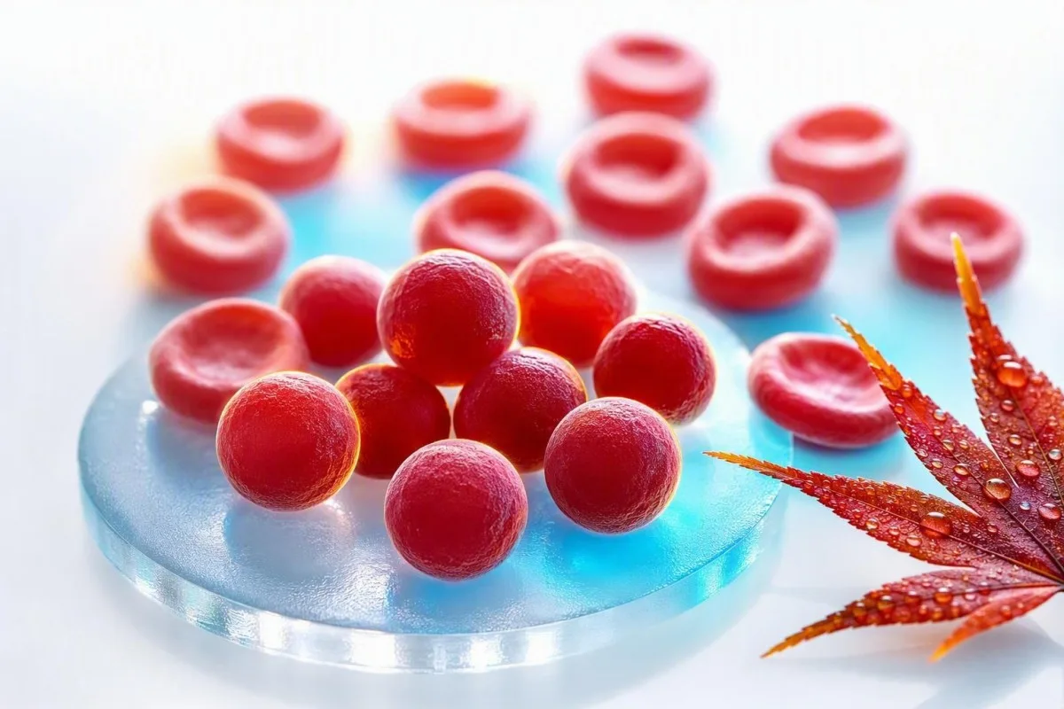
Alta MCHC estas nekomuna kaj ofte pli utilas kiel sugesto ol kiel diagnozo. La praktika demando estas ĉu la ruĝaj ĉeloj estas vere densaj kaj sferaj, aŭ ĉu la specimeno misformis la kalkulon.


- MCHC-gamo kutime estas 32-36 g/dL (320-360 g/L) ĉe plenkreskuloj, kvankam la propra gamo de la laboratorio prioritatas.
- Alta MCHC super 36 g/dL estas nekutima kaj devus instigi kontrolon por sferocitoj, hemolizo, brulvundoj, aŭ analiza interfero.
- Sferocitoj estas malgrandaj, densaj ruĝaj ĉeloj sen centra paleco kaj povas okazi en hereda sferocitozo aŭ aŭtoimuna hemoliza anemio.
- Malvarmaj aglutininoj povas false altigi MCHC per buliĝo de ruĝaj ĉeloj, malaltigante la mezuritan RBC-kalkulon, kaj misformante kalkulitajn indicojn.
- Lipemio povas supertaksi hemoglobinon sur kelkaj analiziloj, kreante altigitan MCHC eĉ kiam la formo de la ruĝaj ĉeloj estas normala.
- Brula vundo povas produkti veran altan MCHC per perdo de ruĝ-ĉela membrano, kutime kune kun anemio kaj laboratoria pruvo de hemolizo.
- Hemolizaj sugestoj inkluzivas plialtigitajn retikulocitojn, indirektan bilirubinon kaj LDH, kun malalta haptoglobino kiam ruĝaj ĉeloj rompiĝas en la korpo.
- Ripeta testado kun freŝe kolektita, konvene traktita EDTA-specimeno ofte estas la unua prudenta paŝo kiam MCHC estas izolita kaj nur modere alta.
Alta MCHC en CBC: la rekta respondo
Alta MCHC kutime signifas, ke ruĝaj ĉeloj estas nekutime densaj, sed ĝi ankaŭ povas signifi, ke la analizilo estis trompita de la specimeno. Ĉe plenkreskuloj, MCHC super ĉirkaŭ 36 g/dL meritas kuntekston: veraj altecoj plej ofte indikas sferocitojn aŭ signifan termikan vundon, dum malvarmaj aglutinoj, lipemio kaj specimena hemolizo estas oftaj laboratoriaj klarigoj. Ekde la 23-a de aŭgusto 2026, mi ne diagnozus sang-malsanon nur per ĉi tiu indekso.
MCHC estas kalkulita kiel hemoglobino dividita per hematokrito, multobligita per 100, do ĝi estas koncentriĝo prefere ol rekta ĉela mezurado. Hemoglobino de 15.0 g/dL kaj hematokrito de 40% produktas MCHC de 37.5 g/dL; la nombro povas pliiĝi ĉar hemoglobino estas trolegita, hematokrito estas sublegita, aŭ ambaŭ. Nia CBC biomerkila gvidilo estas utila kiam pluraj indeksoj estas flagigitaj samtempe.
Kiam mi revizias panelon kun MCHC 36.5-37.5 g/dL sed normala hemoglobino, bilirubino, retikulocitoj kaj RDW, artefakto ofte estas pli verŝajna ol malsano. MCHC de 39-42 g/dL estas fiziologie malfacile konservi en netaj cirkulantaj ruĝaj ĉeloj, do la laboratorio devas aktive konsideri interferon antaŭ ol iu ajn panikas.
Kantesti estas AI-sangotesta analizilo kiu legas MCHC kune kun hemoglobino, hematokrito, MCV, RDW, bilirubino, LDH, kaj antaŭaj rezultoj prefere ol trakti unu ruĝan flagon kiel diagnozon. La klinika regulo de D-ro Thomas Klein estas simpla: kontrolu la aritmetikon kaj la specimenon antaŭ ol ordoni ampleksan serĉadon; nia detala RDW kaj ruĝĉela indeksa gvidilo klarigas kial ĉi tiu padron-bazita aliro gravas.
MCHC ne estas MCH aŭ MCV
MCHC priskribas hemoglobinan koncentriĝon interne de la pakita ruĝ-ĉela volumo, dum MCH priskribas hemoglobinan kvanton por ĉelo kaj MCV priskribas ĉelan grandecon. La konfuzo de ĉi tiuj tri indeksoj estas unu kialo, pro kiu rutina CBC-signalo povas soni pli alarmante ol ĝi estas.
MCH estas kutime 27-33 pg por ĉelo, dum MCV estas kutime 80-100 fL en plenkreskuloj; neniu el la gamoj difinas MCHC. Grandaj ruĝaj ĉeloj povas porti pli da hemoglobino kaj tial pliigi MCH, tamen ilia MCHC povas resti normala ĉar ilia volumo kreskis proporcie. Vidu nian praktikan komparon de MCV kaj MCH se ambaŭ valoroj estas signitaj.
Vera alta MCHC ofte pariĝas kun normala aŭ malalt-normala MCV ĉar sferocitoj perdis membranan surfacareon kaj iĝis pli rondaj, ne ĉar ili enhavas senfinan hemoglobinon. Tiu geometrio gravas: sfero tenas malpli da surfaco por sia volumo, do ĝi ne havas palan centron sur bone preparita ĉela specimenoglifo.
La afero estas, ke levita MCHC-sanga testo povas esti matematike reala dum same biologiige misgvida. Iomete subtaksita hematokrito post ruĝ-ĉela aglutinado plenigos la rilaton eĉ se la ruĝaj ĉeloj de la paciento ne estas densaj entute; jen kial kompleta sangokalkula revizio kompleta sangokalkula revizio devus antaŭi konkludojn.
Sferocitoj: la klasika vera MCHC-sugesto
Sferocitoj estas la plej rekonebla kaŭzo de vera alta MCHC, ĉar la perdo de membrano faras ruĝajn ĉelojn kompaktaj kaj dense hemoglobinigitaj. Ili estas viditaj en hereda sferocitozo kaj aŭtoimuna hemoliza anemio, sed trouverez trovo devas esti interpretita kun simptomoj kaj hemolizaj markiloj.
Hereda sferocitozo ofte produktas MCHC-valorojn super 36 g/dL, retikulocitozon, intermitan ictison, galŝtonojn, splenomegalion, aŭ familia historio de anemio. La 2011-datita brita socio por hematologia gvidlinio notas ke MCHC subtenas la diagnozon sed ne estas sufiĉe sentema aŭ specifa por konfirmi ĝin memstare (Bolton-Maggs et al., 2012).
En aŭtoimuna hemoliza anemio, antikorpoj markas ruĝajn ĉelojn por parta membrano forigo en la lieno, lasante sferocitojn malantaŭe. Rekta antiglobina testo, retikulocita kalkulo, bilirubino, LDH, kaj haptoglobino estas pli informaj kune ol iu ajn izolita indekso; nia gvidilo pri malalta haptoglobino preter hemolizo kovras facile maltroveblan limigon.
Unu klinika problemo: lastatempa transfuzo povas dilui aŭ kaŝi la denaskan sferocitan ŝablonon de paciento dum pluraj semajnoj. Christensen et al. priskribas eozin-5-maleimidan ligon per fluocitometrio kiel utilan teston por suspektata hereda sferocitozo, kvankam hematologo devus elekti kaj temigi provojn zorge (Christensen et al., 2015).
Kiel scii ĉu alta MCHC venas kun hemolizo
Alta MCHC fariĝas pli klinike signifa kiam ĝi akompanas anemion, levitan retikulocitan kalkulon, nerektan bilirubinan altigon, LDH-altigon, kaj malaltan haptoglobinon. Tiu grupo indikas pliigitan ruĝ-ĉelan turnoveron, kvankam neniu unu markilo estas perfekta.
Procento de retikulocitoj super 2% ankoraŭ povas esti neadekvata en signifa anemio, do klinikistoj ofte kalkulas absolutajn retikulocitajn nombrojn aŭ retikulocitan produktan indicon anstataŭe. Ekzemple, 4% retikulocita nombro kun hematokrito 24% povas reprezenti nur modestan ostan medulan respondon post korekto; nia artikolo pri altaj retikulocitaj indicoj trairas la distingon.
Nekonjugita bilirubino altiĝas, kiam hemo estas prilaborata pli rapide ol la hepato povas konjugi ĝin, dum LDH altiĝas ĉar ruĝaj ĉeloj enhavas abundan LDH. Haptoglobino sub la laboratorio malsupera limo subtenas intravascularan hemolizon, sed ĝi povas ŝajni normala dum inflamo ĉar haptoglobino estas akutfaza proteino. Barcellini kaj Fattizzo diskutas tiun diagnozan kaptilon en sia revizio de aŭtoimuna hemoliza anemio (Barcellini & Fattizzo, 2015).
Malhela urino, rapide kreskanta laceco, manko de spiro ripoze, brusta doloro, sveno, aŭ flavaj okuloj kun febro meritas samtagan klinikan taksadon. Fragmentitaj ĉeloj estas malsama kaj pli urĝa ŝmirklarigo ol sferocitoj; nia klarigo pri kiam skistocitoj bezonas urĝan revizion helpas klarigi kial.
Kial brulvundoj povas kaŭzi vere altigitan MCHC
Ampleksa termika vundo povas kaŭzi veran altan MCHC ĉar varmo damaĝas ruĝ-ĉelajn membranojn, produktante membranen perdon, sferocitsimilajn ĉelojn, kaj hemolizon. Tio estas ĝenerale hospitala trovaĵo post klinike evidentaj brulvundoj, ne klarigo por hazarda ambulatoria CBC rezulto.
Termika damaĝo povas krei mikrosferocitojn ene de horoj post gravaj brulvundoj, kaj la MCHC povas altiĝi dum la membrano-al-volumena rilatumo falas. Hemoglobino ankaŭ povas fali dum la postaj tagoj pro fluidaj ŝanĝoj, procedoj, flebotomio kaj daŭra hemolizo, do ununura MCHC ne povas kvantigi vundan severecon.
Mi foje vidis pacientojn fokusiĝi pri MCHC de 37.1 g/dL post krizokaza alokasiĝo dum ignori multe pli ageblajn anomaliojn: falantan hemoglobinon, kreskantan kreatininon, kaliajn ŝanĝojn, aŭ laktaton. En tiu situacio, kuracistoj administras la personon kaj brulfiziologion, ne la indicon; nia posthospitala testhoraro klarigas kial valoroj povas moviĝi rapide.
Kantesti AI estas AI-biomarkila interpretada platformo kiu povas flamiĝi altan MCHC kune kun dinamikaj hemoglobinaj kaj renorezultoj, sed akraj brulvundoj postulas rektan hospitalan taksadon kaj ne povas esti sekure administritaj de raporta interpretado. Rezulto post severa brulvundo devus esti komparata kontraŭ la antaŭaj 24-72 horoj, ne kontraŭ ĝenerala populacia mezumo.
Malvarmaj aglutinoj: la alta MCHC-artefakto kiun kuracistoj serĉas
Malvarmaj aglutinoj povas produkti falsan altan MCHC kaŭzante ruĝajn ĉelojn kunbuliĝi ĉe ĉambra temperaturo antaŭ analizo. La analizilo povas kalkuli plurajn aliĝintajn ĉelojn kiel unu partiklon, kondukante al malfalse malalta RBC nombro, malfalse alta MCV, kaj ŝvelinta kalkulita MCHC.
La klasika malvarma aglutina padrono estas malalta RBC nombro kun neproporcie alta MCV, alta MCH, kaj alta MCHC, ofte sen egala klinika rakonto. Varmigi la EDTA tubon al 37°C kaj reaperi la CBC ofte korektas la valorojn, kio estas multe pli informa ol ripeti hazardan teston semajnojn poste.
Malvarmaj aglutinoj povas okazi transiente post iuj spiraj infektoj aŭ persisti en malvarma aglutina malsano kaj certaj limfoproliferaj malordoj. Simptomoj kiel koloroŝanĝo aŭ malkomforto en fingroj kun malvarma ekspozicio aldonas klinikan pezon, sed la laboratoria padrono mem povas esti la unua klarigo; rekta ripeta testo post hemolizo ofertas praktikajn redrawing demandojn.
Ne varmigu vin agreseme aŭ provu manipuli la rezulton hejme. Demandu ĉu la laboratorio vidis ruĝ-ĉelan aglutinadon, ĉu la specimeno estis varmigita kaj reanalizita, kaj ĉu mana hematokrito estis farita; tiuj tri respondoj ofte solvas la aferon.
Lipemio kaj specimena hemolizo povas misformi MCHC
Lipemio kaj en-vitra prova hemolizo povas malfalse altigi MCHC ĉar multaj analiziloj mezuras hemoglobinon fotometre post kiam ruĝaj ĉeloj estas lizie. Turbidaj lipidoj aŭ libera hemoglobino en la specimeno povas pliigi optikan absorbon kaj igi hemoglobinon ŝajni pli alta ol ĝi vere estas.
La lipemia es visible con mayor frecuencia después de triglicéridos muy altos, generalmente por encima de unos 400-500 mg/dL, aunque la interferencia depende del instrumento y de la longitud de onda del ensayo. Un laboratorio puede utilizar un blanco de plasma, una longitud de onda alternativa o un enfoque de reemplazo de plasma en lugar de simplemente aceptar el resultado automatizado de hemoglobina.
La hemólisis in vitro ocurre después de la recolección o manipulación, no dentro del cuerpo, y puede afectar a más que la MCHC. El potasio, la LDH, la AST y el fosfato pueden aumentar en una muestra dañada, por lo que una MCHC alta aislada junto con un potasio alto inesperado requiere una verificación de la calidad de la muestra; consulte nuestra guía de errores de potasio relacionados con la hemólisis.
Una capa de plasma visiblemente rosada o roja es una pista de laboratorio, no una prueba de anemia hemolítica en el paciente. El seguimiento práctico es una recolección fresca con manipulación cuidadosa y procesamiento rápido, especialmente si el informe original incluye un índice de hemólisis o un comentario de que los resultados pueden verse afectados.
Problemoj pri kolekto, stokado kaj tuboj, kiuj ŝanĝas CBC-indicojn
La mala mezcla de la muestra, el llenado insuficiente, la coagulación y el análisis retrasado pueden alterar los índices de CBC, aunque el almacenamiento retrasado con mayor frecuencia disminuye en lugar de aumentar la MCHC al permitir que las células se hinchen. Por lo tanto, una MCHC alta debe leerse con los comentarios de la muestra del laboratorio, no simplemente con el número en el portal del paciente.
Las muestras de CBC generalmente se recolectan en tubos de EDTA, y un tubo con llenado insuficiente puede tener una proporción anticoagulante/muestra excesiva que cambia las mediciones celulares. Los coágulos pequeños pueden eliminar selectivamente las células del análisis y hacer que el recuento automatizado sea internamente inconsistente; nuestro resumen de aditivos para tubos de sangre explica por qué el tipo de tubo no es un detalle trivial.
El almacenamiento a temperatura ambiente durante períodos prolongados hace que los glóbulos rojos absorban agua, aumentando el MCV y tendiendo a disminuir la MCHC. Esa dirección de cambio es útil: una MCHC muy alta después de una muestra retrasada no se explica automáticamente solo por la edad, por lo que la aglutinación en frío o la interferencia óptica aún pueden estar presentes.
Una muestra capilar puede ser apropiada en algunas situaciones, pero es más propensa a la variabilidad de la recolección que una muestra de sangre venosa con EDTA bien recolectada. Si una MCHC alta no tiene síntomas coincidentes o anomalías relacionadas, generalmente prefiero una nueva extracción venosa limpia antes de tratarla como un nuevo diagnóstico crónico.
Uzu antaŭajn CBC por decidi ĉu la ŝanĝo estas reala
Un aumento repentino de la MCHC desde una línea de base estable de 33-35 g/dL a 38 g/dL es más sugestivo de un problema de muestra o analizador que de esferocitosis hereditaria en lenta evolución. Las tendencias no reemplazan un frotis, pero son una poderosa pista de control de calidad.
La mayoría de las personas tienen un rango personal de MCHC relativamente estrecho a lo largo del tiempo, a menudo variando menos de 1 g/dL cuando las condiciones de prueba son comparables. Una verificación delta de laboratorio compara un resultado nuevo con uno anterior para identificar cambios implausibles antes de que se publiquen los resultados; nuestro artículo sobre deltokontrolojn muestra cómo funciona esa red de seguridad.
Kantesti estas AI-labora testa interpretservo que compara los valores de CBC actuales y anteriores, lo que ayuda a distinguir una señal persistente de un valor atípico único. En nuestro flujo de trabajo de revisión, un cambio en la MCHC importa más cuando viaja con hemoglobina, MCV, bilirrubina y reticulocitos en una dirección biológicamente coherente.
Hay excepciones. La nueva hemólisis autoinmune, una quemadura importante, una reacción transfusional o una aglutinación en frío marcada pueden cambiar genuinamente los resultados rápidamente, por lo que un valor histórico estable es tranquilizador pero nunca una razón para ignorar ictericia nueva, orina oscura o anemia.
Kondiĉoj ofte kulpigitaj pri MCHC sed malofte respondecaj
La deshidratación, la deficiencia de hierro, la deficiencia de vitamina B12 y el estrés común no suelen causar una MCHC verdaderamente alta. Pueden cambiar otros valores de CBC, pero no deben usarse como explicaciones convenientes para una MCHC de 38 g/dL sin confirmar la muestra.
Fer-manko tipe malaltigas MCH kaj MCHC kaj ofte malaltigas MCV kiam la manko estas establita. Frua fer-manko povas havi normalan MCV, sed ĝi ne klarigas hiperkromiajn indeksojn; nia klarigo de malalta transferrina saturiĝo helpas apartigi feran haveblecon de CBC-koncentriĝo.
Manko de vitamino B12 aŭ folato pli ofte kaŭzas makrocitozon, kun MCV super 100 fL, prefere ol alta MCHC. Se MCV kaj MCH estas ambaŭ altaj sed MCHC estas normala, tio estas aliaj diagnozaj direktoj; vidu altaj MCV-sekvaj konsiloj.
G6PD-manko kaŭzas epizodan hemolizon post specifaj oksidaj ellasiloj ĉe sentemaj homoj, sed MCHC ne estas fidinda ekzamena testo por ĝi. Dum akuta epizodo, la smiro kaj turnover-markiloj povas esti pli rivelantaj, dum enzimaj testoj eble bezonas ripeton post resaniĝo ĉar junaj ruĝaj ĉeloj enhavas pli da G6PD.
La plej utilaj postaj testoj post levita MCHC
La plej bona sekva paŝo por izolita alta MCHC estas kutime ripeto de CBC kun laboratorio-revizio, sekvata de periferia smiro kaj hemoliza panelo nur se la altigo daŭras aŭ aliaj konsiloj ĉeestas. Ordi ĉiun anemian teston tuj povas krei pli da bruo ol respondoj.
Fokusa unua-linia revizio inkluzivas ripetan CBC, ĉelan provaspektra glito-revizion, retikulocitan kalkulon, totalan kaj indirektan bilirubinon, LDH, haptoglobinon, kaj rektan antiglobulinan testadon kiam aŭtoimuna hemolizo estas kredinda. La smiro povas identigi sferocitojn, aglutinadon, fragmentojn, polikromazion, aŭ normalan morfologion, kiu faras artefakton pli verŝajna.
Se hereda sferocitozo restas kredinda, hematologo povas konsideri eozino-5-maleimido-ligon, osmanan fragilecan testadon, aŭ genetikan testadon. Tiuj testoj havas malsamajn falsajn negativajn riskojn post transfuzo kaj dum akuta hemolizo, pro kio la testordono devus sekvi klinikan historion prefere ol kontrollisto; nia G6PD testogvidilo ilustras la pli vastan tempoproblemon.
Kantesti AI interpretas altan MCHC per kontrolado de la kalkulo kontraŭ rilataj CBC kaj hemolizaj markiloj, poste identigante demandojn por preni al klinikisto. Ĝi ne anstataŭas ĉelan morfologian revizion, precipe kiam la laboratorio eldonis analizilan flagon aŭ kiam anemio plimalboniĝas.
Kiam alta MCHC bezonas urĝan medicinan taksadon
Alta MCHC mem ne estas kriz-sojlo, sed alta MCHC kun signoj de akuta hemolizo, severa anemio, aŭ grava brulvundo povas postuli samtagan zorgon. Simptomoj kaj la rapid-eco de ŝanĝo determinas urĝecon multe pli bone ol ĉu la flago estas markita H.
Serĉu urĝan taksadon por nova manko de spiro ripoze, brustodoloro, sinkopo, konfuzo, rapide plimalboniĝanta malforteco, te-kolora urino, aŭ flaviĝo de la okuloj kun febro. Tiuj simptomoj povas akompani signifan anemion aŭ hemolizon, precipe kiam hemoglobino falis je pli ol 2 g/dL de konataj bazlinio.
Kontaktu la klinikiston kiu ordonis la teston ene de tagoj se MCHC restas super 36 g/dL sur ripeto-provo, anemio ĉeestas, aŭ la raporto mencias aglutinadon aŭ sferocitojn. En stabila, sana persono kun MCHC 36.2 g/dL kaj alie normalaj rezultoj, ne-urĝa re-desegnado ofte estas racia; nia gvidilo pri bilirubina ŝablono povas helpi formi la sekvan diskuton.
Ne komencu feron, folaton, steroidojn, aŭ suplementojn simple por korekti MCHC. Neniu traktas la ĉefajn verajn kaŭzojn, kaj steroidoj precipe povas obskuri la taksadon de aŭtoimuna hemolizo se komencitaj sen klinika superrigardo.
Kiel Kantesti metas MCHC en klinika kunteksto
Kantesti legas altan MCHC kiel padrono-rekono-problemo: veraj dens-ĉelaj malordoj kaj provaĵaj artefaktoj produktas malsamajn kombinaĵojn de CBC, kemio, flagoj, kaj antaŭaj tendencoj. La celo estas fari la sekvan klinikan demandon pli klaran, ne por etikedi raran malsanon el unu kalkulita indekso.
Kantesti estas AI-movita ilo por analizo de sangotestoj uzata de pli ol 2 milionoj da homoj en 127 landoj por organizi laboratoriajn rezultojn en kunteksto. Por alta MCHC, nia AI kontrolas ĉu hemoglobino kaj hematokrito matematike subtenas la rezulton, ĉu MCV estas neatendite alta, kaj ĉu bilirubino, LDH, retikulocitoj, aŭ haptoglobino indikas ruĝ-ĉelan turniĝon.
Nia platformo ankaŭ serĉas internajn kontraŭdirojn: MCHC 40 g/dL kun normala ŝmiraj komentoj kaj sen anemio povas postuli artefakton prompton, dum MCHC 37 g/dL kun sferocitoj, retikulocitozo, kaj ne-rekta bilirubina altigo meritas medicinan sekvadon. La teknika aliro kaj limoj estas priskribitaj en nia Gvidilo de AI-teknologio.
Mi konsilas al pacientoj konservi la laboran PDF, dato de kolekto, fasta stato, lastatempa malsano, transfuz-historio, kaj ajnaj laboratoriaj komentoj. Tiu malgranda rekordo ofte klarigas ŝajnan diskrepancojn pli rapide ol ripeti larĝan testadon, precipe kiam rezultoj estas komparataj monatojn poste.
Demandoj, kiuj faras alt-MCHC-rendevuon pli produktiva
Demandu ĉu la MCHC estis ripetita, ĉu la laboratorio vidis sferocitojn aŭ aglutinadon, kaj ĉu anemio aŭ hemolizaj markiloj ĉeestas. Tiuj demandoj ŝanĝas la konversacion de “kial unu nombro estas alta?” al konkreta, testebla klinika vojo.
Utilaj vortumoj inkluzivas: “Ĉu la specimeno estis hemolizita aŭ lipema?”, “Ĉu la analizilo flagis ruĝ-ĉelan aglutinadon?”, kaj “Ĉu ni povas kompari ĉi tiun rezulton kun mia antaŭa MCHC?” Man-maniero de ŝmirado estas precipe valora kiam MCHC estas senĉese super 36 g/dL aŭ anemio estas neklarigita.
D-ro Thomas Klein rekomendas preni liston de medikamentoj, lastatempaj infektoj, malvarm-induktitaj simptomoj, familia historio de galŝtonoj aŭ anemio, kaj transfuzoj ene de la pasintaj 3 monatoj. Tiu historio povas indiki heredaĵan sferocitozon, aŭtoimunan hemolizon, aŭ transientan malvarman aglutininon multe pli rapide ol ĝenerala interreta serĉo.
La medicina enhavo de Kantesti estas reviziita kun kuracista kontrolado, sed via propra kuracisto povas ekzameni vin, peti varmigitan re-rulon, kaj paroli rekte kun la laboratorio. Legantoj, kiuj volas kompreni nian klinikan revizian kadron, povas renkonti la medicina konsila komitato, dum persistantaj aŭ simptomaj nenormalaĵoj ĉiam devas esti diskutitaj persone.
Esplorado, necerteco kaj la limoj de unuopa MCHC
MCHC estas utila ekzamena konduko, ne memstara diagnoza testo por heredaĵa sferocitozo, aŭtoimuna hemolizo, brulvundoj, aŭ malvarma aglutinina malsano. Ĝia valoro akre kreskas kiam ripetita specimeno, ĉela morfologio, turniĝaj markiloj, kaj klinika historio ĉiuj rakontas la saman rakonton.
Neniu universala MCHC-limo diagnozas heredaĵan sferocitozon ĉar analizilaj metodoj, aĝo, retikulocitozo, kaj kunekzistanta ferodeficienco povas ŝanĝi sentivecon. La BSH-gvidlinio subtenas kombini familian historion, ŝmirajn trovojn, retikulocitojn, bilirubinon, kaj konfirmantan testadon kie necese prefere ol fidi je ununura sojlo (Bolton-Maggs et al., 2012).
Kantesti subtenas informitan sekvadon identigante rezultajn ŝablonojn kaj necertecon, ne per eldonado de definitiva hematologia diagnozo. Nia aliro por klinika validigo klarigas kial nekutimaj valoroj, analizilaj flagoj, kaj krizaj simptomoj restas kialoj por homa laboratoria kaj kuracista revizio.
Por rilata hematologia kunteksto, vidu la esploran publikaĵon Gvidilo pri B-negativa sangotipo, LDH-sangotesto kaj retikulocita kalkulo kaj la pli vastan klinikan komunikresurson Diareo Post Fastado, Nigraj Makuloj en Feko & Gvidilo pri GI 2026. Ambaŭ estas suplementaj edukaj publikaĵoj prefere ol anstataŭaĵoj por periferia ŝmirado aŭ specialista takso.
Oftaj Demandoj
Kio signifas alta MCHC-nivelo en sangokontrolo?
Alta nivelo de MCHC kutime signifas, ke la hemoglobina koncentriĝo ene de la ruĝ-ĉela volumo superas la laboratorian intervalon, kutime super 36 g/dL ĉe plenkreskuloj. Veraj altigoj okazas kun sferocitoj pro hereda sferocitozo, aŭtoimuna hemoliza anemio, aŭ grava termika vundo. Malvarmaj aglutininioj, lipemio kaj prova hemolizo ankaŭ povas krei falsan altan rezulton. Ripeta CBC, revizio de la ĉela provaĵo kaj hemolizaj markiloj klarigas, kiu klarigo taŭgas.
Ĉu alta MCHC estas danĝera?
Alta MCHC ne estas danĝera mem; ĝi estas kalkulita CBC indekso prefere ol malsano. MCHC de 36,2 g/dL kun normala hemoglobino kaj sen simptomoj ofte estas traktata kun rutina ripettesto, dum alta MCHC plus falanta hemoglobino, iktero, malluma urino, aŭ sputmanĝo necesas prompta taksado. Valoroj super 39 g/dL precipe verŝajne reflektas specimenan aŭ analizilan enmiksiĝon. La klinika risko venas de la subesta kaŭzo, kiel signifa hemolizo, ne de la MCHC nombro sole.
Ĉu dehidratiĝo povas kaŭzi altan MCHC?
La senhidratiĝo kutime ne kaŭzas vere altan MCHC, ĉar ĝi koncentras plasman volumon pli ol ŝanĝi hemoglobinan koncentriĝon ene de individuaj ruĝaj globuloj. La senhidratiĝo povas pliigi hemoglobinon kaj hematokriton kune, lasante MCHC proksime de ĝia kutima 32-36 g/dL intervalo. Se MCHC estas 37-40 g/dL, klinikistoj devus konsideri sferocitojn, malvarman aglutinadon, lipemion, aŭ mezuran problemon anstataŭe. Ripetita bone kolektita CBC estas pli utila ol provi korekti la valoron per ekstraj fluidoj.
Ĉu sferocitoj ĉiam altigas la MCHC?
Sferocitoj ofte altigas MCHC super 36 g/dL, sed ili ne ĉiam faras tion kaj alta MCHC ne pruvas la ĉeeston de sferocitoj. Hereda sferocitozo povas esti kaŝita per feromanko, lastatempa transfuzo, aŭ milda malsano, dum aŭtoimuna hemolizo povas montri variajn trovojn depende de la tempo. Necesas ĉelspecimena glito reviziita de sperta laboratoria profesiulo por identigi sferocitojn. Retikulocita kalkulo, bilirubino, LDH, haptoglobino kaj rekta antiglobulina testo aldonas diagnozan kuntekston.
Ĉu malvarmaj aglutinoj povas kaŭzi altan MCHC?
Malvarmaj aglutinoj povas false altigi MCHC per buligi ruĝajn ĉelojn antaŭ ol la aŭtomata CBC estas mezurata. La padrono ofte inkluzivas malaltan RBC-kalkulon, altan MCV, altan MCH, kaj MCHC super 36 g/dL kiu ne konformas al la simptomoj de la paciento. Varmigi la EDTA specimenon al 37°C kaj refari ĝin povas normaligi la kalkulojn. Persistanta malvarma aglutinado aŭ hemolizaj simptomoj devas esti taksitaj de klinikisto aŭ hematologo.
Ki testojn mi devus peti post alta MCHC?
Post mallonga alta MCHC, la kutimaj sekvaj testoj estas ripeto de CBC, revizio de ĉela specimen-slideTo, retikulocita nombro, bilirubinaj frakcioj, LDH, haptoglobino, kaj foje rekta antiglobina testo. Kliniko povas peti eozino-5-maleimido-ligon aŭ alian specialecan testadon se hereda sferocitozo estas suspektata. MCHC super 36 g/dL kun normala ripetita testado kaj sen anemio eble ne bezonas ampleksan esploradon. La plej bona testsekvenco dependas de simptomoj, antaŭaj CBC-valoroj, familia historio, kaj laboratoriaj flagoj.
Akiru hodiaŭ AI-movitan analizon de sangoanalizo
Aliĝu al pli ol 2 milionoj da uzantoj tutmonde, kiuj fidas je Kantesti por tuja, preciza analizo de laboratoriaj testoj. Alŝutu viajn rezultojn de sangoanalizo kaj ricevu ampleksan interpretadon de 15,000+-biomarkiloj en sekundoj.
📚 Referencitaj esplorpublikaĵoj
Klein, T., Mitchell, S., & Weber, H. (2026). G Negativa sangotipo, gvidilo pri LDH-sangotesto kaj retikulocita kalkulo. Kantesti AI Medicina Esploro.
Klein, T., Mitchell, S., & Weber, H. (2026). Diareo Post Fastado, Nigraj Makuloj en Fekaĵo & Gista Sistemo Gvidilo 2026. Kantesti AI Medicina Esploro.
📖 Eksteraj medicinaj referencoj
⚕️ Medicina Deklaro
Ĉi tiu artikolo estas nur por edukaj celoj kaj ne konsistigas medicinan konsilon. Ĉiam konsultu kvalifikitan sanprovizanton por diagnozaj kaj traktadaj decidoj.
Signaloj de fido E-E-A-T
Sperto
Kuracista gvidata klinika revizio de laboratoriaj interpretaj laborfluoj.
Kompetenteco
Laboratoria medicino fokusiĝas pri kiel biomarkiloj kondutas en klinika kunteksto.
Aŭtoritateco
Skribita de d-ro Thomas Klein kun revizio de d-ro Sarah Mitchell kaj prof. d-ro Hans Weber.
Fidindeco
Evident-bazita interpretado kun klaraj sekvaj vojoj por redukti alarmon.