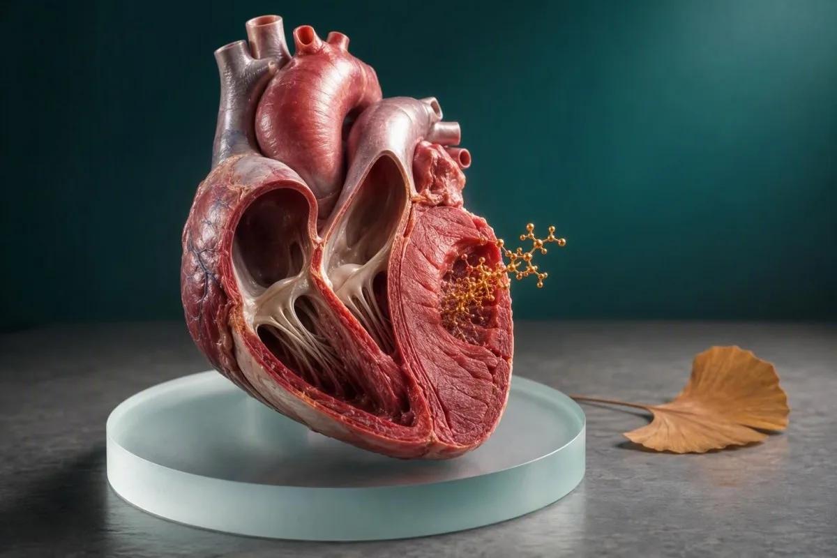
CK-MB is 'n ouer maar steeds nuttige merker van spierskade wat die hart kan betrek. Sy waarde hang af van tydsberekening, simptome, EKG-bevindinge, totale CK, en – gewoonlik – troponien.


- CK-MB betekenis: CK-MB staan vir kreatien kinase miokardiale band, 'n ensiemisoorm wat hoofsaaklik in hartspier voorkom, maar ook in skeletspier.
- Normale waardes: Baie CK-MB massa-toetse gebruik 'n boonste verwysingslimiet naby 3–5 ng/mL, maar die laboratorium-spesifieke reeks geniet altyd voorkeur.
- Tydsberekening: CK-MB styg algemeen 3–6 uur na miokardiale besering, bereik 'n piek op 12–24 uur, en keer dikwels terug na die basislyn binne 48–72 uur.
- Troponien eerste: Hoë-sensitiwiteit kardiale troponien is die verkieslike bloedmerker vir vermoedelike hartaanval in moderne noodsorg.
- Geen enkele toetsdiagnose: 'n Verhoogde CK-MB alleen kan nie 'n hartaanval diagnoseer nie, want strawwe oefening, spierbesering, chirurgie en spiersiekte kan dit verhoog.
- Relatiewe indeks: 'n CK-MB-indeks bo ongeveer 5% mag 'n kardiale bron ondersteun, maar dit word onbetroubaar wanneer totale CK aansienlik verhoog is.
- Dringende simptome: Nuwe borsdruk, kortasem, floute, sweet of pyn wat na die kakebeen of arm versprei, benodig noodassesserings ongeag 'n vorige CK-MB-resultaat.
- Tendens tel: 'n Styging en daling oor herhaalde monsters is meer insiggewend as 'n enkele geïsoleerde getal.
CK-MB is die miokardiale bandvorm van kreatien kinase
CK-MB staan vir kreatien kinase miokardiale band. Dit is 'n ensiemvorm wat in 'n CK-MB-bloedtoets gemeet word wanneer klinici moontlike besering aan hartspier ondersoek, hoewel 'n abnormale waarde op sigself nie 'n hartaanval bewys nie.
Kreatien kinase help selle om ATP vinnig te regenereer, die onmiddellike brandstof wat tydens spierwerk gebruik word. CK-MB is gekonsentreer in kardiale spier, terwyl CK-MM oorheers in skeletspier en CK-BB meer wydverspreid in brein- en gladde weefsels gevind word. Vir 'n grondslag oor die ouer ensiem, sien ons kreatienkinase-gids.
'n CK-MB-resultaat word gewoonlik gerapporteer as massakonsentrasie in ng/mL of, minder dikwels, ensiemaktiwiteit in U/L. Baie huidige massatoetse stel 'n boonste limiet rondom 3–5 ng/mL, maar geslagsspesifieke limiete en toetskalibrasie verskil genoeg dat 'n getal van een laboratorium nie na 'n ander oorgedra kan word nie.
Kantesti is 'n KI-bloedtoetsontleder wat CK-MB saam met die toetsverwysingsinterval, totale CK, troponien, en die versamelingstyd lees. Dit tel, want 'n waarde van 6 ng/mL op 2 uur na simptome dra 'n baie ander vraag oor as 6 ng/mL na 'n marathon.
Die agtervoegsel kan definitief klink, en pasiënte hoor verstaanbaar “miokardiale” as “hartaanval.” In werklike kliniese werk beteken dit dat die laboratorium een leidraad oor spierselbesering gemeet het—nie die oorsaak, ligging, of erns van daardie besering nie.
Waarom die naam steeds op verslae verskyn
Sommige hospitale behou CK-MB in nood- of binnepasiënt bestelstelle, veral wanneer hulle 'n kortstondige merker wil hê om moontlike herhalende besering te assesseer. In baie gemeenskapsinstellings is dit egter grootliks vervang deur hoë-sensitiwiteit troponien vir aanvanklike hartaanval-evaluering.
CK-MB en totale CK meet verwante maar verskillende dinge
Totale CK meet ensiem wat uit alle spierryke weefsels vrygestel word, terwyl CK-MB een CK-fraksie meet. Totale CK kan tot duisende U/L styg na strawwe aktiwiteit of spierbesering sonder om 'n hartprobleem aan te dui.
'n Totale CK van 1,500 U/L na ongewone oefening kan skeletspier-spanning, dehidrasie, medikasie-effekte, of rabdomiolise weerspieël; dit identifiseer nie die bron op sy eie nie. CK-MB kan ook in daardie omgewing toeneem omdat skeletspier klein hoeveelhede van die MB-fraksie bevat.
Die ou CK-MB relatiewe indeks word bereken as CK-MB gedeel deur totale CK, dan vermenigvuldig met 100. 'n Indeks onder 3% leun tradisioneel na skeletspier, terwyl 'n indeks bo 5% 'n kardiale bron kan ondersteun—maar hierdie afsnypunte is ondersteunende bewyse, nie 'n vonnis nie.
Wanneer totale CK uiters hoog is, kan die indeks mislei omdat beide die teller en die noemer vir verskillende biologiese redes verander. Dit is een rede waarom klinici nou groter gewig heg aan reeks hoë-sensitiwiteit troponien en die EKG eerder as om wiskundig 'n dubbelsinnige CK-MB resultaat te probeer red.
'n Praktiese voorbeeld: 'n sterkte-atleet met totale CK 4,000 U/L en CK-MB 12 ng/mL mag 'n lae relatiewe proporsie hê en geen isgemiese simptome nie. Dieselfde CK-MB waarde in 'n sittende persoon met nuwe vergruisende borsdruk verdien 'n heel ander reaksie.
Hoekom totale CK hoog mag wees
Oefening, val, langdurige onbeweeglikheid, aanvalle, binnespierse inspuitings, hipotiroïdisme, statiene en inflammatoriese spierafwykings kan totale CK verhoog. Ons gids tot gevaarlike hoë CK verduidelik wanneer donker urine, ernstige swakheid, of verminderde urine-uitset die dringendheid verander.
Waarom 'n geneesheer steeds 'n CK-MB bloedtoets mag aanvra
Klinici mag CK-MB bestel wanneer bors simptome geëvalueer word, moontlike herhalende miokardiale besering, of 'n onduidelike verhoging in ander spierensieme. Dit word gewoonlik saam met EKG toetsing en troponien bestel eerder as 'n selfstandige siftingsondersoek.
In die eerste 24–72 uur na 'n bevestigde kardiale gebeurtenis, kan CK-MB se relatief vinnige afname soms help om 'n moontlike tweede besering te identifiseer. Troponien bly dikwels langer verhoog, so 'n vars CK-MB styging mag konteks byvoeg, hoewel plaaslike protokolle verskil en bewyse nie perfek uniform is nie.
'n Klinikus mag ook CK-MB gebruik wanneer 'n ouer laboratorium informasie stelsel dit steeds met totale CK bundel. Na my ervaring, word pasiënte dikwels gealarmeer deur 'n outomaties gegenereerde CK-MB vlag wat hul behandelende span nooit as bewyse van infarksie beskou het nie omdat die troponien en EKG gerusstellend was.
CK-MB is nie 'n siftings toets vir stille koronêre plaak, toekomstige hartrisiko, of algemene fiksheid nie. Bloeddruk, diabetes status, LDL cholesterol, ApoB, rook, nierfunksie, en familiegeskiedenis beantwoord meer nuttige voorkomende vrae; ons ApoB en ApoA1 verhouding gids verduidelik een van daardie risiko patrone.
Indien jy simptoomvry is en CK-MB op 'n verslag van 'n beroeps-, sport-, of binnepasiënt paneel sien, vra hoekom dit bestel is en waarmee jou klinikus dit vergelyk het. Die bestel konteks vertel ons dikwels meer as 'n effens abnormale resultaat.
'n Toets is nie 'n diagnose nie
Hartaanvalle is kliniese diagnose gebaseer op simptome, ondersoek, EKG of beelding bewyse, en biomarker dinamika. Laboratorium merkers identifiseer miokardiale besering; hulle vestig nie onafhanklik of 'n geblokkeerde koronêre aar dit veroorsaak het nie.
Die CK-MB tydsverloop verduidelik waarom 'n enkele resultaat kan mislei
CK-MB begin gewoonlik styg ongeveer 3–6 uur na akute miokardiale besering, bereik 'n hoogtepunt rondom 12–24 uur, en val dikwels terug na basislyn teen 48–72 uur. 'n Normale vroeë monster kan dus te vroeg wees om gerus te stel.
Tydsberekening is een van die minste gewaardeerde dele van kardiale bloed toetsing. 'n Persoon wat 45 minute na skielike simptome opdaag, mag 'n CK-MB binne die reeks hê ten spyte van werklike besering, terwyl iemand wat 2 dae na 'n gebeurtenis getoets word, 'n dalende maar steeds abnormale waarde mag hê.
Dr. Thomas Klein het dit meestal na vertraagde aanbieding gesien: 'n pasiënt voel snags onwel, wag tot die volgende oggend, en wonder dan hoekom klinici 'n toets herhaal wat reeds hoog is. Die herhaal monster soek na 'n betekenisvolle verandering, nie bloot om te bevestig dat een nommer 'n lyn oorgesteek het nie.
Hoë-sensitiwiteit troponien kan vroeër as CK-MB styg, dikwels binne 1–3 uur, afhangende van die toets en die tydsberekening van die besering. Reeks toets protokolle gebruik algemeen monsters by aanbieding en 1–3 uur later; jou noodafdeling volg sy gevalideerde plaaslike pad.
Harde opleiding skep nog 'n tydsberekening strik. CK en CK-MB kan 24–72 uur na ekstensiewe oefening toeneem, veral afdraande hardloop of onbekende weerstand werk. Ons marathon-atleet laboratoriumgids verduidelik waarom die tydsberekening van die oefening in die kliniese geskiedenis hoort.
Die tersaaklike klok begin by die aanvang van simptome.
Laboratoriums teken die tyd van monsterversameling aan, maar klinici benodig ook die beste skatting van wanneer borsdruk, asemloosheid, ineenstorting, of 'n ander kommerwekkende simptoom begin het. Indien simptome kom en gaan, dokumenteer die eerste en mees ernstige episode eerder as om later te raai.
CK-MB verwysingsreekse en resultaatpatrone
Die meeste CK-MB massa toetse definieer normaal as ongeveer 0–3 of 0–5 ng/mL, maar jou laboratorium se boonste verwysingslimiet is die enigste toepaslike afsnypunt vir jou verslag. 'n Klein verhoging benodig kliniese korrelasie; daar is geen universele CK-MB getal wat 'n hartaanval aandui nie.
Laboratoriums stel reekse vas deur hul eie toets, kalibrasie, en verwysingspopulasie te gebruik. Sommige Europese laboratoriums rapporteer CK-MB aktiwiteit in U/L eerder as massa in ng/mL, wat direkte internetvergelykings veral nutteloos maak.
'n CK-MB waarde van 7 ng/mL wanneer die boonste limiet 5 ng/mL is, is 'n 1.4-voudige verhoging, nie 'n diagnostiese etiket nie. 'n Resultaat 10 keer die boonste limiet is meer kommerwekkend vir aansienlike spierbesering, maar selfs dan moet klinici identifiseer of die bron kardiaal of skeletagtig is.
AST kan styg na beide kardiale en skeletagtige spierspanning, wat soms 'n ou-styl ensiempaneel bemoeilik. Oorsig van hoë AST patrone kan help verklaar waarom AST, CK, en lewer toetse soms saam beweeg na oefening.
Die praktiese reël is eenvoudig: behou die presiese eenhede, verwysingsreeks, versamelingstyd, simptome, en onlangse aktiwiteit wanneer jy CK-MB bespreek. 'n Geknipte skermskoot wat net die rooi vlag wys, is selde genoeg om veilig te interpreteer.
Waarom 'n rooi vlag nie outomaties gevaarlik is nie
Verwysingsintervalle beskryf waar resultate in 'n geselekteerde populasie val; hulle sorteer nie mense in veilige en onveilige groepe nie. Vir 'n breër verduideliking van vlae en laboformaat, lees wat buite reeks beteken.
CK-MB teenoor troponien: hoekom troponien gewoonlik lei
Hoë-sensitiwiteit kardiale troponien is meer kardiaal-spesifiek en sensitiewer as CK-MB vir die diagnose van akute miokardiale besering. CK-MB mag geselekteerde inligting byvoeg, maar dit moet nie 'n gevalideerde troponienpad oorheers nie.
Troponien I en troponien T is proteïene wat integraal tot die hartspier se sametrekkingsapparaat is. CK-MB is 'n ensiemisoform wat oorwegend, maar nie uitsluitlik nie, in hartspier voorkom; hierdie biologiese verskil is waarom CK-MB vs troponien nie 'n gelyke kompetisie vir aanvanklike diagnose is nie.
Die 2021 ESC akute koronêre sindroom riglyn beveel gevalideerde hoogsensitiewe hartspiertroponien-algoritmes aan wat die toets-spesifieke 99ste-persentiel boonste verwysingslimiet en seriele verandering gebruik (Collet et al., 2021). 'n Troponien bo daardie limiet dui op miokardiale besering, maar dit beteken steeds nie outomaties dat 'n koronêre blokkasie dit veroorsaak het nie.
Troponien kan vir 7–14 dae verhoog bly na 'n groot infarksie, terwyl CK-MB dikwels binne 2–3 dae normaliseer. Daardie korter venster is die historiese rasioneel vir CK-MB in vermoedelike herinfarksie, hoewel baie sentrums nou kliniese, EKG-, beelding- en troponien-veranderingsbewyse gebruik in plaas daarvan.
Troponienresultate is ook toets-afhanklik: hs-cTnI en hs-cTnT waardes kan nie direk omgeskakel word nie, en “n ”normale" getal het 'n ander betekenis voor teenoor 8 uur na simptome. Vergelyk die toetse in ons troponien I teenoor T verduideliking.
Geen merker identifiseer die oorsaak op sy eie nie
Sepsis, vinnige aritmie, pulmonêre embolisme, miokarditis, ernstige anemie, nier siekte, en hartversaking kan troponien verhoog sonder 'n klassieke tipe 1 hartaanval. CK-MB het sy eie nie-hartspier oorsake, so die volle kliniese prent bly nie-onderhandelbaar.
Hoe geneeshere 'n hartaanval anders as CK-MB diagnoseer
'n Hartaanval vereis akute miokardiale besering plus bewyse van iskemie, nie bloot 'n verhoogde CK-MB of troponien nie. Bewyse kan iskemiese simptome, nuwe EKG-veranderinge, beelding-bevindinge, of 'n koronêre bloedklont insluit.
Die Vierde Universele Definisie van Miokardiale Infarksie definieer akute miokardiale besering as 'n hartspiertroponien-waarde bo die toets se 99ste persentiel met 'n styging en/of daling; miokardiale infarksie vereis ook kliniese bewyse van iskemie (Thygesen et al., 2019). CK-MB vervang nie hierdie raamwerk nie.
Kantesti is 'n AI bloedtoets interpretasie platform wat gerapporteerde waardes en tendense kan organiseer, maar dit kan nie 'n hartaanval uit 'n PDF of foto diagnoseer nie. Akute simptome vereis intydse evaluasie, EKG-interpretasie, vitale tekens, en klinikus se oordeel—nie 'n app-gebaseerde gerusstelling lus nie.
Oorweeg 'n 52-jarige hardloper met CK-MB 6 ng/mL na 'n 42-km wedloop: 'n normale EKG, lae stabiele troponien, en seer beenspiere dui in een rigting. 'n 52-jarige met dieselfde CK-MB plus nuwe sentrale druk, sweet, en 'n dinamiese troponienstyging behoort in noodsorg te wees.
Vir pasiënte met kortasem in plaas van duidelike pyn, kan risiko steeds beduidend wees, veral by ouer volwassenes, mense met diabetes, en vroue. Ons kortasem bloedtoets gids dek die breër laboratoriumkonteks.
Tipe 1 en tipe 2 miokardiale infarksie verskil
Tipe 1 miokardiale infarksie behels gewoonlik akute koronêre plaatversteuring en trombose. Tipe 2 miokardiale infarksie weerspieël suurstof voorsiening-aanvraag wanbalans, soos ernstige anemie of volgehoue vinnige aritmie, en bestuur teiken die sneller asook koronêre risiko.
Nie-kardiale redes waarom CK-MB verhoog kan wees
Skeletspierbesering, groot oefening, trauma, chirurgie, inflammatoriese miopatie, en nierdisfunksie kan 'n verhoogde CK-MB produseer. Hierdie oorsake is waarom klinici nooit CK-MB in isolasie interpreteer nie.
Skeletspier bevat 'n beskeie CK-MB fraksie, so beskadigde of regenereerende spiere kan genoeg vrystel om 'n abnormale toets te veroorsaak. Onlangse val, kneusbesering, langdurige immobilisasie, beslagleggings, spuitings in spiere, en ortopediese chirurgie moet almal gemeld word wanneer 'n resultaat bespreek word.
Chroniese spierafwykings kan aanhoudend abnormale totale CK en CK-MB patrone produseer wat niks met akute koronêre siekte te doen het nie. In seldsame gevalle skep makro-CK komplekse of toetsinterferensie verwarrende resultate, en die laboratorium mag ondersoek met isoensiem skeiding of 'n alternatiewe metode.
'n Ongepaardheid verdien aandag: merkbaar verhoogde CK-MB met normale reeks troponien en geen simptome moet lei tot 'n oorsig van spiergeskiedenis en analitiese kwessies eerder as reflektiewe kardiale beeldvorming. Indien die monster tydens insameling beskadig is, 'n hemolise hertekeningsgids mag relevant wees.
Nierversaking kan die interpretasie van verskeie biomerkers, veral troponien, kompliseer deur chroniese miokardiale besering en verminderde opruiming-verwante effekte. Dit gee nie toestemming om 'n stygende resultaat te ignoreer nie; dit maak tendense en simptome selfs waardevoller.
Die indeks het beperkings in spiersiekte
Die CK-MB-indeks is ontwikkel vir 'n nouer kliniese omgewing as wat baie pasiënte aanneem. In uitgebreide spierbesering of chroniese miopatie, kan 'n indeks bo 5% steeds misleidend wees, so kontemporêre klinici prioritiseer troponien-gebaseerde algoritmes.
Oefening kan CK-MB verhoog sonder 'n hartaanval
Langafstandhardloop, CrossFit-styl oefeninge, swaar eksentrieke optel, en uithouvermoë-byeenkomste kan totale CK en soms CK-MB vir 24–72 uur verhoog. Oefening-verwante verhoging is meer waarskynlik wanneer simptome afwesig is en troponien en EKG nie 'n iskemiese patroon toon nie.
Die ding is, spiere weet nie dat jou jaarlikse bloedtoets môre is nie. Afdraande hardloop en hoë-volume weerstand opleiding skep mikroskopiese spierontwrigting, en CK kan later as spierpyn piek—dikwels 24–48 uur na die sessie.
Ek adviseer pasiënte om nie te probeer om “n toets te ”slaag" deur anders te oefen sonder om hul klinikus in te lig nie. Teken die tipe oefening, duur, spierpyn, hidrasie, aanvullings, koors, en medikasie aan; hierdie inligting kan onnodige alarm voorkom en steeds beskerm teen die mis van ware siekte.
Donker cola-kleurige urine, ernstige swelling, diep swakheid, verwarring, of dalende urine-uitset na inspanning is anders as roetine vertraagde-spierpyn en benodig dringende assessering. Die risiko daar is rabdomiolise en nierbesering, nie bloot 'n ongerieflike hoë CK nie.
Indien borsstyfheid, ongewone kortasemigheid, ineenstorting, of hartkloppings tydens oefening voorgekom het, moenie 'n abnormale CK-MB aan fiksheid toeskryf nie. Raadpleeg ons hartkloppings bloedtoets gids terwyl jy dringende kliniese assessering reël.
Hoe lank om te rus voor 'n roetine bloedtrekking
Vir “n nie-noodgevalle basale CK-meting, is dit ”n redelike praktiese benadering om onbekende hoë-intensiteit oefening vir ongeveer 48–72 uur te vermy, tensy jou klinikus anders aanbeveel. Moenie noodtoetse vir borsimptome uitstel om eers te "rus" nie.
Hoe die laboratorium CK-MB meet en waar foute ontstaan
Die meeste moderne CK-MB toetse gebruik immuniese toetse wat CK-MB massa in ng/mL kwantifiseer, terwyl ouer metodes ensiemaktiwiteit gemeet het of iso-ensieme geskei het. Verskillende metodes kan verskillende getalle en verwysingsintervalle lewer.
Massa-analises identifiseer CK-MB proteïen met teenliggame en is oor die algemeen meer spesifiek as ouer aktiwiteits-analises. Tog kan heterofiele teenliggame, makro-CK, monsterkwaliteit probleme, en baie hoë totale CK af en toe 'n resultaat skep wat nie by die kliniese prentjie pas nie.
'n Laboratorium kan delta-kontrole gebruik—vergelyking met vorige resultate—om 'n biologies onplausibele sprong te merk. Skielike veranderinge is nie altyd foute nie, maar die vergelyking van monsteromstandighede, medikasieveranderinge, en onlangse aktiwiteit is sinvol; sien ons laboratorium-delta-kontrole verduideliker.
Vas is gewoonlik nie nodig vir CK-MB nie, en om water te drink is sinvol tensy jy vloeistofbeperkings het. Biotien beïnvloed hoofsaaklik sekere immuniese toetse eerder as CK-MB universeel, so maak hoë-dosis aanvullings bekend, tipies 5–10 mg/dag of meer, eerder as om voorgeskrewe behandeling op eie te stop.
Vanaf 25 Augustus 2026 behoort 'n goeie laboratoriumverslag die analiet, eenhede, plaaslike interval, versamelingstyd, en vlagstatus te identifiseer. Die ontbreking van enige van daardie elemente maak veilige afstandinterpretasie aansienlik swakker.
Vra watter analise gebruik is
Indien “n CK-MB-waarde skerp in stryd is met jou simptome of troponienresultate, vra of die laboratorium CK-MB massa of aktiwiteit gemeet het en of inmenging oorweeg is. Daardie gefokusde vraag is nuttiger as om te soek na ”n universele "normale CK-MB" waarde.
Hoe om 'n CK-MB verslag te lees sonder om te oorreageer
Lees CK-MB met sy eenhede, die laboratorium se boonste limiet, totale CK, troponienreeks, EKG-resultaat, en tydlyn van simptome. 'n Enkel hoë vlag, veral na oefening of besering, het lae diagnostiese spesifisiteit.
Begin met die presiese resultaat en sy plaaslike boonste verwysingslimiet. Vra dan of die monster geneem is voor simptome, binne 3 uur, 6–12 uur, of dae later; dieselfde konsentrasie het 'n verskillende betekenis op elke punt van daardie kurwe.
Kyk vervolgens vir 'n patroon. 'n Stygende troponien met isgemiese simptome en veranderende EKG-veranderinge weeg baie meer as CK-MB alleen, terwyl verhoogde CK na been-swaar opleiding met stabiele troponien dikwels na skeletspiere wys.
Kantesti se tendensanalise help gebruikers om vorige resultate en versamelingskonteks te bewaar eerder as om elke rooi vlag as 'n nuwe krisis te behandel. 'n Vorige CK, onlangse oefensessie, en herhaalde waarde beantwoord dikwels vrae wat 'n geïsoleerde verslag nie kan nie.
Moenie CK-MB van verskillende laboratoria vergelyk asof hulle identies is nie, veral as een U/L gebruik en 'n ander ng/mL. Hou 'n bloedtoets-tydlyn maak opvolgafsprake meer produktief.
Vrae wat die moeite werd is om na ’n afspraak te bring
Vra: Was dit CK-MB massa of aktiwiteit? Wat was my troponien-tendens? Het my EKG iskemie getoon? Kan onlangse spierbesering dit verklaar? Benodig ek 'n herhaalde monster, ekokardiogram, of geen verdere kardiale toetsing nie?
Simptome wat dringende sorg benodig, ongeag CK-MB
Bel dadelik nooddienste vir nuwe borsdruk wat langer as 5–10 minute duur, ernstige kortasemigheid, floute, koue sweet, of pyn wat na die kaak, arm, rug, of boonste buik versprei. Moenie wag vir 'n CK-MB resultaat of probeer om een tuis te interpreteer nie.
Hartaanval simptome is nie altyd dramaties nie. Vroue, ouer volwassenes, en mense met diabetes kan met naarheid, ongewone moegheid, kortasemigheid, rugongemak, of 'n gevoel dat iets verkeerd is, aanbied in plaas van klassieke borspyn.
Dr. Thomas Klein se reël in kliniese praktyk is direk: as simptome aktueel en kommerwekkend is, is die syfers wat jy gister gesien het sekondêr. Noodspanne kan vinnig 'n EKG verkry, hoë-sensitiwiteit troponien met die regte interval herhaal, en bloeddruk, ritme, suurstofversadiging, en alternatiewe diagnoses assesseer.
'n Hoë troponien is self nie sinoniem met 'n koronêre blokkasie nie, maar dit moet nooit self-getriëer word vanuit 'n portaalresultaat nie. Lees hoë troponien dringende tekens vir die situasies waar onmiddellike sorg die veiliger keuse is.
Indien simptome opgelos het, maar nuut, oefen-geïnduseerd, of herhalend was, soek dieselfde-dag mediese advies. Stabiele simptome verdien steeds 'n geneesheer se assessering, veral met bekende koronêre siekte, rook, diabetes, chroniese niersiekte, of 'n sterk familiegeskiedenis.
Moenie self bestuur as simptome ernstig is nie
Noodvervoer maak voorsiening vir monitering en vroeë behandeling indien 'n gevaarlike ritme ontwikkel. Kou of inname van medikasie moet volg op nood-afskakeling of geneesheer se advies, veral as jy bloedverdunners gebruik, allergieë het, of is aangesê om nie aspirien te neem nie.
Wie benodig opvolg na 'n abnormale CK-MB resultaat
'n Abnormale CK-MB resultaat benodig opvolg wanneer dit nuut, stygend, onverklaarbaar, gepaardgaande met simptome, of vergesel deur abnormale troponien- of EKG-bevindings is. Die volgende stap mag herhaalde toetsing wees, nie outomaties 'n skandering of prosedure nie.
Vir 'n lae-risiko, simptoomvrye persoon na duidelike strawwe oefening, kan 'n geneesheer CK en CK-MB herhaal na rus en hidrasie. Vir iemand met aktiewe bors simptome of 'n abnormale troponien, is nood assessering meer gepas as buitepasiënt her-toetsing.
Opvolg hang ook af van basislyn kardiovaskulêre risiko. Diabetes, vorige koronêre siekte, rook, hoë LDL, hoë lipoproteïen(a), hipertensie, en niersiekte kan die drumpel vir vinnige ondersoek verlaag; ons Gids vir harttoetse vir vroue dek algemene gemiste risikomerkers.
Kantesti is 'n KI-aangedrewe bloedtoets-analisehulpmiddel wat kombinasies kan onthul vir bespreking, insluitend CK-MB, CK, kreatinien, AST, en opeenvolgende datums. Dit moet die klinikus ondersteun—nie vervang nie—wat u simptome, ondersoek, EKG en medikasie ken.
Herhaalde toetse moet ideaal dieselfde laboratorium gebruik en op 'n klinies betekenisvolle interval versamel word. Om 'n CK-MB-monster 15 minute later te herhaal, kan min byvoeg, terwyl 'n troponienprotokol van 1–3 uur hoogs insiggewend kan wees wanneer dit deur daardie hospitaal gevalideer word.
Wat om mee te bring vir opvolg
Bring die volledige verslag, vorige waardes, medikasie- en aanvullingslys, 'n verslag van onlangse oefening of besering, en 'n duidelike simptoomtydlyn. Hierdie besonderhede kan u spaar van beide vals gerusstelling en onnodige toetsing.
Veilige gebruik van KI-interpretasie vir CK-MB resultate
KI kan CK-MB-terminologie verduidelik en tendense organiseer, maar dit kan nie bepaal of borsimptome 'n noodgeval is nie. Veiligheid hang af van die bewaring van die oorspronklike verslag, die erkenning van onsekerheid, en die eskaleer van dringende simptome na persoonlike sorg.
Kantesti KI interpreteer CK-MB-resultate deur eenhede, verwysingsintervalle, verwante biomerkers en vorige resultate in ag te neem, eerder as om 'n diagnose uit een vlag te verklaar. Ons biomerker verwysingsgids verduidelik waarom laboratoriumreekse en metingmetodes belangrik is.
Vir privaatheid, verwyder of beskerm onnodige identifiserende inligting voordat enige verslag gedeel word en gebruik dienste met duidelike databestuurstandaarde. Kantesti bedien gebruikers in meer as 127 lande en ondersteun veeltalige verslaginterpretasie, maar plaaslike nooddienste en u kliniese span bly die regte roete vir akute simptome.
Ons klinici hersien mediese inhoud teen kliniese standaarde, en ons tegnologiegids beskryf die beperkings van KI-ondersteunde laboratoriuminterpretasie. 'n Verklaring is die nuttigste wanneer dit u vertel watter inligting ontbreek en watter vraag om vervolgens te vra.
Gebruik nooit 'n KI-interpretasie om sorg vir nuwe drukagtige borsongemak, ineenstorting of ernstige asemloosheid te vertraag nie. 'n Veilige hulpmiddel behoort daardie grens baie duidelik te maak.
Wat 'n nuttige KI-verklaring insluit
'n Klinies verantwoordelike verklaring identifiseer die eenheid, die laboratoriumreeks, moontlike nie-kardiale oorsake, metgesel toetse, tydsbeperkings, en dringende simptome. Dit belowe nie dat 'n abnormale of normale CK-MB alleen 'n hartaanval insluit of uitsluit nie.
Die praktiese kernpunt vir CK-MB resultate
CK-MB is 'n hart-geassosieerde kreatienkinasiefraksie, maar hoogsensitiewe troponien, EKG-bevindinge, simptome en herhaalde toetse dryf nou die meeste hartaanvalbesluite. 'n CK-MB-resultaat word die beste behandel as een stuk bewys, nooit 'n diagnose op sigself nie.
As u CK-MB effens hoog is na strawwe oefening, besering, chirurgie, of spierpyn, moenie paniekerig raak nie—maar gee wel u klinikus die konteks. As dit hoog is met borsimptome, asemloosheid, sweet, floute, of 'n abnormale troponien, behandel dit as 'n tydgevoelige kliniese vraag.
Kantesti kan u help om 'n bondige vraelys op te stel en vorige verslae te vergelyk, terwyl ons kliniese valideringstandaarde verduidelik hoe ons veilige resultaatinterpretasie benader. Dit kan nie akute koronêre sindroom van die huis af bevestig of uitsluit nie.
Dr. Thomas Klein beveel aan om die oorspronklike verslag te hou eerder as om slegs die abnormale lyn te kopieer. Die presiese monster tyd, eenhede, plaaslike reeks, totale CK, troponienwaardes, EKG-verklaring, medikasie, en onlangse oefening is die data wat CK-MB interpreteerbaar maak.
Vir geneesheer-hersiene gesondheidsopvoeding en die mense agter ons kliniese standaarde, ontmoet die Kantesti mediese adviesraad. Die beste volgende stap is dié wat ooreenstem met u simptome en risiko—nie die nommer op die bladsy wat die skrikwekkendste lyk nie.
'n Laaste veiligheidsondersoek
Nuwe of voortdurende moontlike kardiale simptome geniet altyd voorkeur bo 'n vorige resultaat wat gerusstellend lyk. Soek eers noodhulp; bespreek die laboratoriumbesonderhede later, wanneer u veilig is.
Gereelde vrae
Waarvoor staan CK-MB op 'n bloedtoets?
CK-MB staan vir kreatienkinase miokardiale band, 'n vorm van die kreatienkinase-ensiem wat oorwegend in hartspier voorkom, maar ook in kleiner hoeveelhede in skeletspiere. Baie laboratoriums rapporteer CK-MB-massa in ng/mL, met boonste verwysingsgrense algemeen naby 3–5 ng/mL. 'n Hoë resultaat kan spierselbesering aandui, maar CK-MB alleen kan nie 'n hartaanval diagnoseer nie. Klinici interpreteer dit met simptome, EKG-bevindinge, reeks troponien, totale CK, en toets tydsberekening.
Wat is 'n normale CK-MB-vlak?
'n Normale CK-MB-massa is dikwels onder ongeveer 3–5 ng/mL, hoewel die korrekte normale reeks die interval is wat deur die laboratorium wat die toets uitgevoer het, gedruk word. Sommige laboratoriums rapporteer CK-MB-aktiwiteit in U/L in plaas daarvan, so waardes kan nie direk tussen metodes vergelyk word nie. 'n Waarde net bokant die boonste limiet kan voortspruit uit onlangse oefening of skeletspierbesering. Daar is geen enkele CK-MB-afsnypunt wat miokardiale infarksie bevestig of uitsluit nie.
Is CK-MB of troponin beter vir 'n hartaanval?
Hoogsensitiewe kardiale troponien is oor die algemeen beter as CK-MB vir die opsporing van akute miokardiale besering omdat dit sensitiewer en spesifieker vir hartspier is. Die 2021 ESC-riglyn gebruik toets-spesifieke seriële hoogsensitiewe troponien-algoritmes, dikwels met monsters by aanbieding en 1–3 uur later. CK-MB kan soms aanvullende inligting verskaf omdat dit dikwels binne 48–72 uur na die basislyn terugkeer. Geen biomerker alleen stel 'n hartaanval vas sonder bewyse van iskemië soos simptome, EKG-veranderinge, beeldingbevindinge, of 'n koronêre bloedklont nie.
Kan oefening CK-MB verhoog?
Ja, strawwe oefening kan CK-MB verhoog, want skeletspiere bevat 'n klein CK-MB fraksie. Uithouvermoë-gebeurtenisse, onbekende swaar optel, afdraande hardloop en hoë-intensiteit interval opleiding kan totale CK en soms CK-MB vir ongeveer 24–72 uur verhoog. Oefening maak nie borspyn, floute, ernstige asemloosheid of sweet veilig om te ignoreer nie. Daardie simptome benodig steeds dringende kliniese assessering met EKG en seriële troponien toetsing.
Wat beteken 'n CK-MB-indeks bo 5%?
'n CK-MB-relatiewe indeks van bo ongeveer 5% het tradisioneel voorgestel dat 'n groter deel van die gemete kreatienkinase uit hartspier eerder as skeletspier mag kom. Die indeks word bereken as CK-MB gedeel deur totale CK, vermenigvuldig met 100, en waardes onder 3% het histories skeletspier bevoordeel. Hierdie berekening word onbetroubaar na ernstige spierbesering, met baie hoë totale CK, of in chroniese spiersiekte. Moderne sorg steun dus meer op reeks hoogsensitiewe troponien en EKG-bevindinge.
Hoe vinnig styg CK-MB na 'n hartaanval?
CK-MB begin gewoonlik om 3–6 uur na akute miokardiale letsel te styg, bereik 'n piek rondom 12–24 uur, en keer dikwels binne 48–72 uur na die basislyn terug. 'n Normale CK-MB-uitslag wat binne die eerste 1–2 uur van simptome verkry word, kan te vroeg wees om letsel uit te sluit. Nooddepartemente gebruik herhaalde monsters en gevalideerde hoogsensitiewe troponienprotokolle eerder as om op een CK-MB-meting te vertrou. Die tyd van simptoomaanvang moet so akkuraat moontlik gedeel word.
Kry vandag KI-aangedrewe bloedtoets-analise
Sluit aan by meer as 2 miljoen gebruikers wêreldwyd wat Kantesti vertrou vir onmiddellike, akkurate laboratoriumtoetsanalise. Laai jou bloedtoetsresultate op en ontvang omvattende interpretasie van 15,000+-biomerkers binne sekondes.
📚 Verwysde navorsingspublikasies
Klein, T., Mitchell, S., & Weber, H. (2026). Nipah-virusbloedtoets: Vroeë opsporing en diagnosegids 2026. Kantesti KI Mediese Navorsing.
Klein, T., Mitchell, S., & Weber, H. (2026). B Negatiewe Bloedgroep, LDH-bloedtoets en retikulosiettelling-gids. Kantesti KI Mediese Navorsing.
📖 Eksterne mediese verwysings
⚕️ Mediese Vrywaring
Hierdie artikel is slegs vir opvoedkundige doeleindes en vorm nie mediese advies nie. Raadpleeg altyd ’n gekwalifiseerde gesondheidsorgverskaffer vir besluite oor diagnose en behandeling.
E-E-A-T Vertrouenseine
Ervaring
Kliniese oorsig gelei deur ’n geneesheer van laboratorium-interpretasie-werksvloei.
Kundigheid
Laboratoriumgeneeskunde fokus op hoe biomerkers in ’n kliniese konteks optree.
Gesagsvermoë
Geskryf deur dr. Thomas Klein met hersiening deur dr. Sarah Mitchell en prof. dr. Hans Weber.
Betroubaarheid
Bewysgebaseerde interpretasie met duidelike opvolgpaaie om alarm te verminder.