A negative tularemia antibody test during the first 2–3 weeks of symptoms does not exclude infection. Paired samples collected about 2–3 weeks apart can reveal seroconversion or a fourfold titer rise, but suspected tularemia needs clinical assessment—and sometimes treatment—before antibodies become detectable.

اہم نکات
- Early negatives are expected in some infections because Francisella tularensis antibodies often remain undetectable until 2–3 weeks after symptoms begin.
- جوڑی کے نمونے usually involve an acute specimen collected early and a second specimen approximately 2–3 weeks later.
- چار گنا اضافہ means two dilution steps: a hypothetical change from 1:40 to 1:160 supports recent infection when symptoms and exposure fit.
- واحد مثبت results cannot reliably distinguish current infection from previous exposure; antibodies can persist for years.
- IgM and IgG may appear around the same time in tularemia, so an IgM-positive result alone does not establish a new infection.
- Exposure history should include ticks, handling rabbits, and mowing or brush cutting near animal habitats; symptoms commonly begin 3–5 days after exposure.
- Clinical assessment should happen promptly when fever accompanies a skin sore, enlarged lymph nodes, or respiratory symptoms after a plausible exposure.
- لیبارٹری حفاظت requires clinicians to alert the laboratory before submitting specimens when tularemia is suspected; routine specimen handling may need modification.
What does a tularemia test actually measure?
A tularemia test may measure antibodies against Francisella tularensis, detect bacterial genetic material, or attempt to grow the organism. These methods answer different questions: an antibody result reflects your immune response, which often takes 2–3 weeks to become detectable, rather than directly measuring bacteria in your body.
Francisella tularensis antibodies are usually tested in serum using agglutination assays or other laboratory-specific methods. A report reading 1:160 describes a dilution endpoint, not 160 bacteria, and a qualitative positive report may contain no titer at all; our guide to مثبت اینٹی باڈی کے نتائج یہ فرق واضح کرتی ہے۔.
دی test name and method are two details worth checking before interpreting any result. An enzyme immunoassay index, an agglutination titer, and a PCR detected result cannot be placed on the same numerical scale—even when all three reports mention the same organism.
کنٹیسٹی ایک ہے۔ اے آئی بلڈ ٹیسٹ تجزیہ کار that helps explain laboratory terminology, but a tularemia result still requires the specimen date, symptom history, and clinical assessment. Our تنظیم اور مقصد describe this interpretation role; an explanation generated in approximately 60 seconds cannot remove a biological antibody window.
Why can tularemia antibodies be negative early?
Tularemia antibodies may be negative early because the immune response has not yet crossed the assay’s detection threshold. CDC diagnostic guidance states that antibodies are often not detectable until 2–3 weeks after symptom onset, so a negative first-week sample cannot safely exclude an otherwise plausible infection.
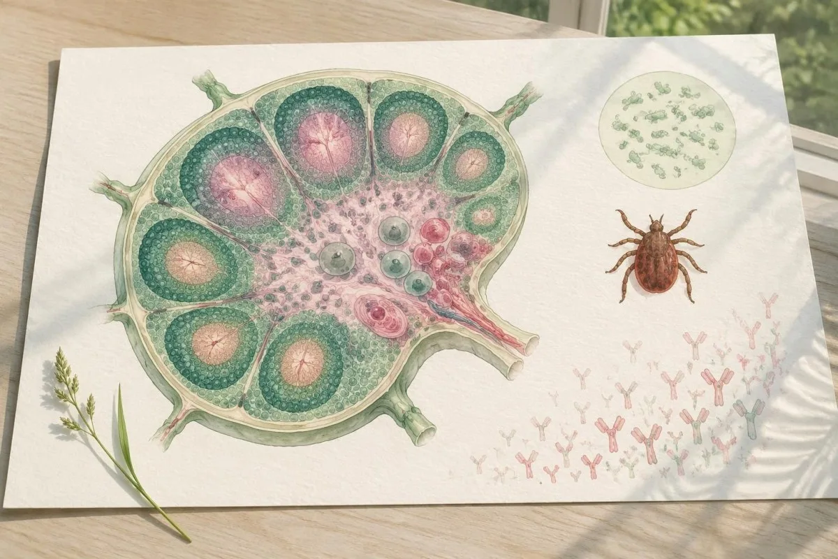
An early negative result is a timing problem, not proof that symptoms are unrelated. The CDC’s Clinical Testing and Diagnosis for Tularemia guidance recommends interpreting negative serology against illness duration; a sample taken on symptom day 4 is less informative than one taken after week 3.
A hypothetical 36-year-old gardener with fever, a small finger sore, and an enlarged elbow-area lymph node could have negative antibodies on day 6. The combination still warrants assessment because a localized entry site plus its draining lymph node suggests a specific route of exposure, whereas either finding alone has many explanations.
Different infections have different testing windows, so one negative tick-borne panel does not settle the entire differential diagnosis. Our explanation of early spotted fever negatives illustrates the shared principle without implying that tularemia and spotted fever have identical assays or 2–3-week antibody kinetics.
When should testing be timed from symptom onset?
Time tularemia serology from the first day of symptoms, not just the exposure date. Symptoms commonly start 3–5 days after exposure, although the reported incubation range is 1–21 days; the separate antibody-development interval then makes a calendar based only on a tick bite misleading.
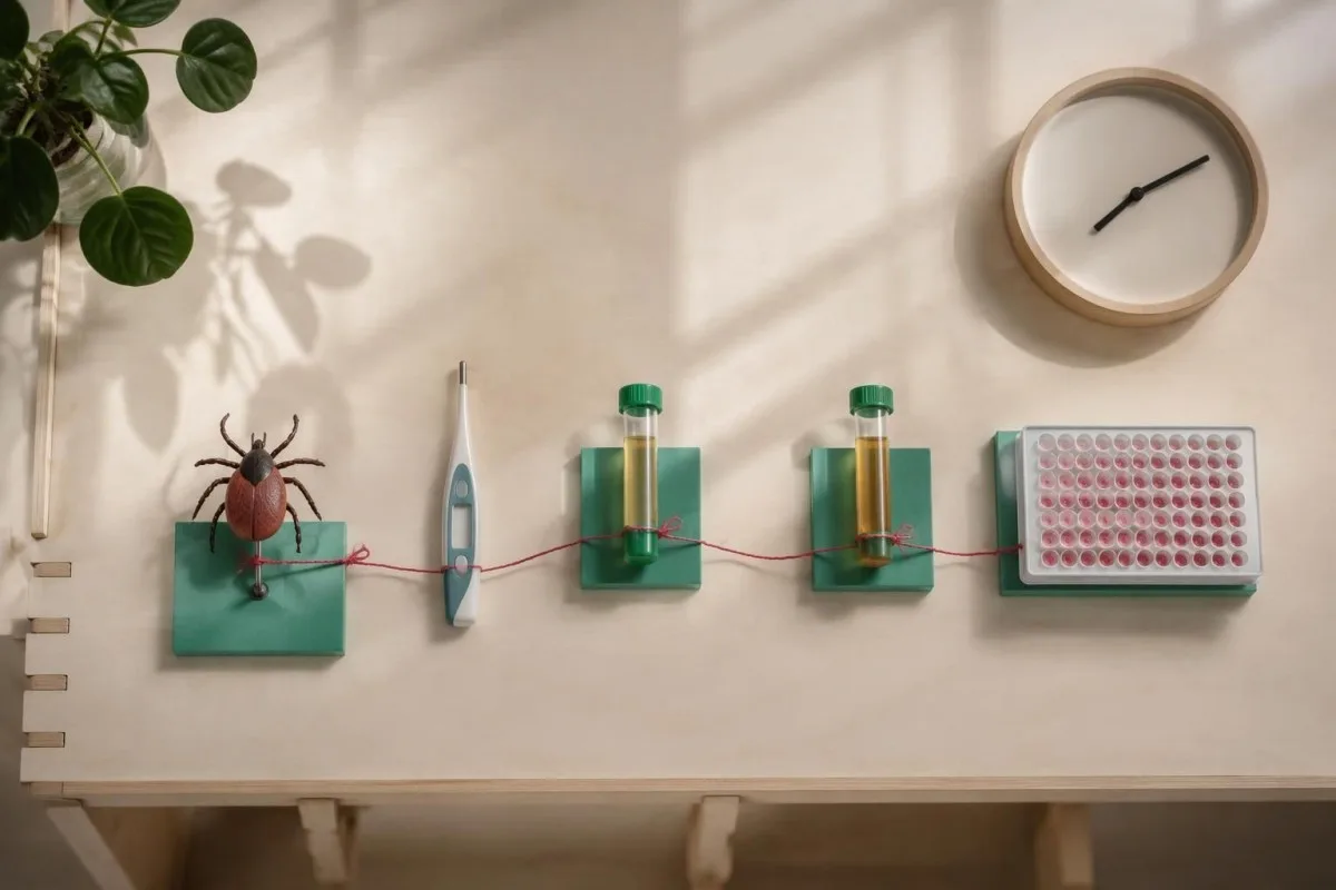
Three dates improve interpretation: the likely exposure date, the first symptom date, and each specimen collection date. For example, a tick bite on November 1 followed by fever on November 5 and testing on November 8 produces a symptom-day-3 sample—not a specimen collected seven days into illness.
A repeat sample approximately 2–3 weeks after the acute specimen is often more useful than repeating the same assay the next morning. The CDC recommends an acute specimen, ideally in the first week, followed by a convalescent specimen; our anaplasmosis sample timing guide explains why symptom dates also matter for other tick-associated tests.
Kantesti AI can help organize these three dates alongside the wording of a report, but the collection date takes priority over the date a PDF was downloaded. Record antibiotic start dates separately because treatment can affect direct-detection yield and complicate interpretation without creating a reliable formula for predicting seroconversion.
How do paired antibody samples confirm recent infection?
Paired tularemia antibody samples can demonstrate seroconversion or a fourfold rise in titer. A hypothetical change from 1:40 to 1:160 represents two dilution steps and supports recent infection when the clinical picture fits; both specimens should preferably be assessed with the same method and laboratory.
A fourfold rise is not the same as any increase. Moving from 1:80 to 1:160 is a twofold rise, whereas 1:80 to 1:320 is fourfold; the larger change helps distinguish a developing immune response from small differences caused by endpoint reading or assay variability.
Seroconversion means a change from negative to positive, but the exact interpretation depends on the initial detection limit and the laboratory’s confirmation criteria. If the first report says less than 1:20 and the second says 1:160, the initial value is censored below a threshold—there is no exact starting number from which to calculate a precise fold change.
Ask whether the laboratory can compare the two specimens together, rather than comparing independently produced reports months apart. Our guide to discordant ehrlichiosis results discusses paired-serology reasoning in another tick-borne condition, but its organism-specific cutoffs should not be transferred to a tularemia assay.
What does a positive antibody titer mean?
A single positive tularemia antibody titer supports exposure but does not independently prove an active infection. Some agglutination assays use a threshold such as 1:160, but there is no universal cutoff across methods; the reporting laboratory’s interpretation and the patient’s illness timing must accompany the number.
Higher titers do not provide a severity score. A hypothetical 1:640 result is not evidence of four times the illness severity of a 1:160 result; dilution endpoints measure antibody reactivity, while severity depends on breathing, circulation, organ involvement, and other clinical findings.
Cross-reactivity can produce misleading positive results, particularly with Brucella antibodies. A 2020 review in Frontiers in Cellular and Infection Microbiology discusses assay performance, persistent antibodies, and cross-reactions (Maurin, 2020); our brucellosis testing explanation shows why animal exposure alone does not identify one organism.
A stable positive titer on two samples can reflect prior exposure or a later-stage response that has already plateaued. For example, unchanged 1:320 titers collected three weeks apart do not demonstrate a fourfold rise, but they also cannot automatically exclude current illness if both specimens were obtained late.
Can IgM and IgG identify when tularemia started?
Tularemia IgM and IgG cannot reliably date an infection from one specimen. Both antibody classes may become detectable around 2–3 weeks after symptoms begin, and persistence complicates interpretation; the familiar shortcut that IgM always means new infection and IgG always means old infection is unreliable here.

Persistent IgM is a recognized interpretation problem in tularemia serology. The Frontiers review describes long-lasting antibody responses, including IgM in some people (Maurin, 2020), so an isolated IgM-positive result months or years after exposure should not automatically trigger treatment for a newly acquired infection.
ایک فرضی IgM-positive, IgG-negative result on symptom day 10 needs method-specific review, not an automatic diagnosis. Some assays detect different classes at different thresholds, and nonspecific reactivity remains possible; our guide to Mycoplasma IgM limitations explains the broader misconception without treating these infections as interchangeable.
کنٹیسٹی ایک ہے۔ AI خون کے ٹیسٹ کی رپورٹ کی تشریح that can explain the distinction between antibody class and dilution titer, but neither field alone establishes an infection date. Our کلینیکل ویلیڈیشن فریم ورک شامل ہے جو یہ بتاتے ہیں کہ لیب کی تشریحات کیسے ٹیسٹ کی جاتی ہیں اور طبی طور پر کیسے ریویو کی جاتی ہیں۔ Kantesti کا میڈیکل ریویو ماڈل ہمارے should not be read as proof of tularemia-specific diagnostic performance; no 1:160 result can substitute for an exposure history and examination.
When does a tick exposure raise suspicion?
Tick exposure raises suspicion for tularemia when fever develops with a skin entry-site sore and nearby enlarged lymph nodes. Symptoms commonly begin 3–5 days after exposure, but a tick may go unnoticed and the skin sore may be absent; neither a missing bite history nor a negative early antibody result excludes tularemia.
ایک فرضی صورتحال پر غور کریں 42-year-old hiker with fever four days after a tick bite and a tender groin node on the same side as an ankle sore. That drainage pattern makes an entry-site process plausible, but clinicians still consider bacterial skin infections and other tick-borne illnesses rather than diagnosing tularemia from the bite alone.
Tick-associated tests are organism-specific. A Lyme antibody or Western blot result does not measure Francisella tularensis antibodies, and a tularemia result does not exclude Lyme disease; our Lyme band interpretation guide explains why the name of each ordered assay matters more than calling the collection a tick panel.
Geography and blood-count patterns help prioritize additional testing, but they do not create a universal three-infection checklist. If anemia or evidence of red-cell breakdown accompanies fever, clinicians may investigate other causes; our Babesia antibody interpretation guide explains why exposure antibodies and active bloodstream infection require different evidence.
Why does handling rabbits matter for diagnosis?
Handling infected rabbits or hares can transmit tularemia through skin contact, including small unnoticed breaks in the skin. A finger or hand entry site may be followed by elbow-area or armpit lymph-node enlargement; an antibody specimen collected during the first 7 days of symptoms may still be negative.

ایک فرضی 29-year-old who handled a sick rabbit five days before fever should tell the clinician exactly what happened: direct handling, a scratch, contact with animal material, or possible dust exposure. These routes can lead to different clinical forms, and remembering protective gloves does not rule out every possible exposure.
The location of enlarged lymph nodes helps distinguish plausible entry routes. A hand sore and an armpit node form one coherent pattern, while generalized nodes suggest a broader differential; our Bartonella ٹائٹر کی وضاحت covers another animal-associated cause of regional node enlargement with different tests.
Do not collect or transport an animal yourself to prove the exposure. One additional handling episode can create avoidable risk; clinical services or public-health authorities can advise whether animal investigation is appropriate, while clinicians document the original contact and decide which human specimens can be collected safely.
Can mowing or landscaping cause pulmonary tularemia?
Mowing, brush cutting, and similar work can generate aerosols associated with pulmonary tularemia, particularly when contaminated animal material is disturbed. Fever, cough, chest discomfort, or breathlessness after such work requires assessment even without a skin sore; symptoms may precede detectable antibodies by 2–3 weeks.
ایک فرضی 54-year-old groundskeeper develops fever and cough four days after brush cutting where rabbit remains were present. That exposure history changes the diagnostic conversation because a routine respiratory virus panel does not test for tularemia, and a negative antibody result on symptom day 5 would be expected in some infections.
Animal-associated pneumonia has several possible causes, not just tularemia. The type of animal, travel location, and tasks performed help distinguish them; our کیو فیور اینٹی باڈی گائیڈ explains another exposure-linked illness whose phase-specific antibodies follow a different interpretation system.
A positive test for one pneumonia cause does not measure another. For example, a Legionella urine antigen result concerns a different organism and has its own detection limits; our Legionella testing explanation clarifies that distinction, while worsening breathlessness should prompt urgent care rather than waiting another two weeks for serology.
Can PCR or culture help before antibodies appear?
PCR or culture may provide direct evidence before tularemia antibodies become detectable, but the right specimen and advance laboratory notification are essential. A negative blood PCR does not exclude an infection concentrated in a skin site, lymph node, or lung; there is no single sensitivity percentage that applies to every specimen.
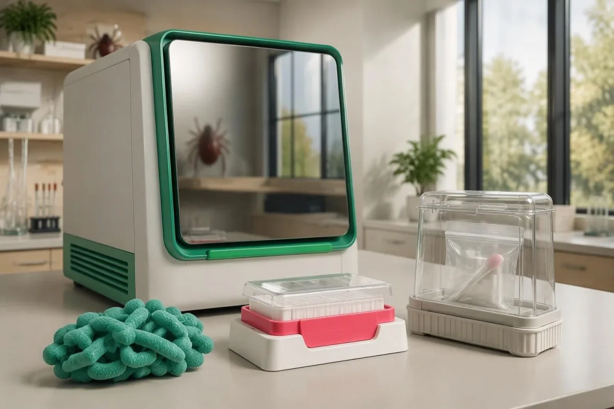
Clinicians should alert the laboratory when tularemia is suspected before specimens are processed. CDC diagnostic guidance emphasizes special safety procedures because Francisella tularensis poses an occupational risk; this instruction applies to routine cultures too, not only to a separately labeled tularemia test ordered after day 3.
The affected body site helps determine the most useful specimen. Depending on the clinical form, clinicians may consider material from an entry site, a lymph-node aspirate, respiratory specimens, or blood; the choice is a clinical and laboratory decision, not a recommendation for patients to obtain any of these four specimen types themselves.
Antibiotics and specimen timing can reduce culture yield, so a negative culture after several treatment days has a different meaning from a pretreatment specimen. Our guide to negative cultures in endocarditis discusses that general principle, but tularemia requires its own organism-specific processing and biosafety pathway.
Do WBC, CRP, or kidney results support diagnosis?
WBC, CRP, and kidney-function tests help assess illness severity and treatment safety, but they do not identify tularemia. A hypothetical WBC of 12.5 × 10⁹/L and CRP of 90 mg/L indicate an inflammatory response in context; neither value distinguishes Francisella tularensis from other infectious or noninfectious causes.
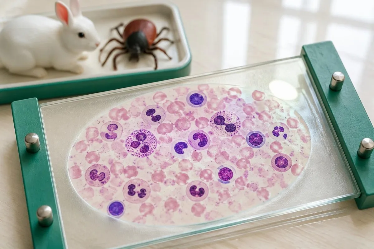
Normal inflammatory markers cannot independently rule out tularemia, especially when obtained early or in a localized illness. A hypothetical CRP of 8 mg/L on symptom day 2 should not outweigh a convincing exposure and examination; our WBC اور CRP کا موازنہ explains why these two markers may disagree.
Creatinine matters when dehydration or certain antibiotics are concerns. For example, a change from 70 to 130 µmol/L requires clinical interpretation against baseline kidney function, fluid status, and timing; our گردے کے تناسب کی تشریح گائیڈ is supporting education, not evidence that a particular kidney pattern diagnoses tularemia.
Kantesti AI can help explain changes across two laboratory reports, but a trend explanation is not a sepsis assessment or an antibiotic dosing service. The practical distinction is whether a result answers the identity question—what caused the illness—or the safety question—whether kidney function, hydration, or other organ findings need clinical attention.
Why should assessment not wait for antibody confirmation?
Suspected tularemia should prompt clinical assessment without waiting for positive antibodies because the diagnostic window can outlast the safe treatment window. Fever with compatible tick, rabbit, or mowing exposure merits prompt review; breathing difficulty, confusion, fainting, or rapidly worsening illness requires emergency evaluation even on symptom day 1.

Treatment may begin on clinical suspicion while confirmation is pursued. The CDC’s 2025 antimicrobial recommendations include ciprofloxacin, levofloxacin, gentamicin, and doxycycline, with selection influenced by severity, illness duration, pregnancy, age, and other factors; an early negative serology result is not a reason to delay a clinically indicated treatment decision.
Treatment duration depends on the selected medicine. The 2025 CDC recommendations generally use 10 days for ciprofloxacin, levofloxacin, or gentamicin and 14–21 days for doxycycline; severe illness or delayed treatment affects drug selection, and these durations are not instructions to self-start or modify a prescription.
Routine antibiotics do not all provide reliable tularemia treatment, so tell the clinician if fever persists despite an existing prescription rather than assuming antibiotics exclude the diagnosis. When gentamicin is selected, kidney function and drug exposure may need monitoring; our جینٹامائسن کی نگرانی کی رہنما explains why sample timing matters alongside the approximately 10-day treatment course.
What should follow-up after treatment include?
Tularemia follow-up should assess recovery, complications, and any planned convalescent specimen—not demand immediate antibody negativity. Francisella tularensis antibodies can persist for years, so a positive result after a 10-day treatment course does not by itself show treatment failure or establish a need for more antibiotics.
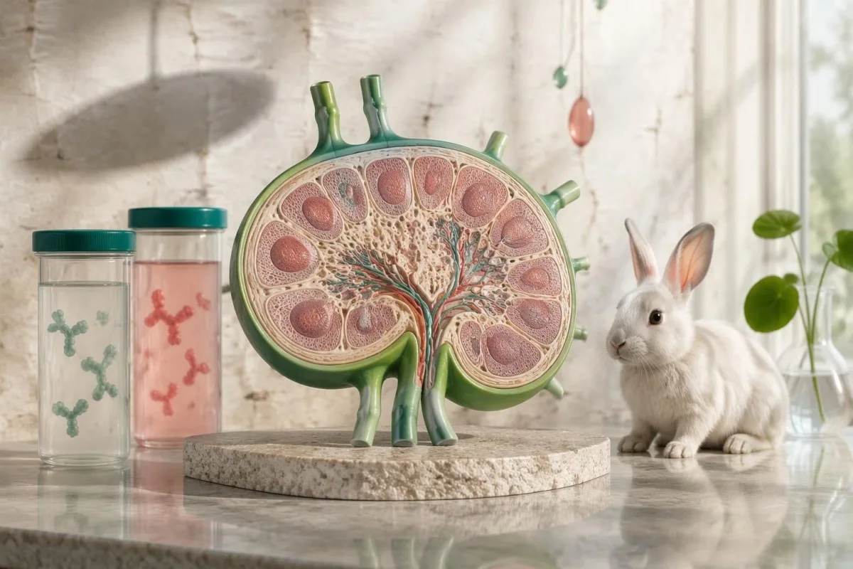
Persistent lymph-node enlargement can lag behind improvement in fever, but enlarging nodes, new fluid accumulation, or recurrent fever need reassessment. If a node remains prominent at a two-week review, the clinician considers its trajectory and examination rather than using the antibody titer as a stand-alone decision about drainage or further treatment.
ایک مفید follow-up record contains four items: symptom onset, exposure details, specimen collection dates, and antibiotic names with start dates. Keep the laboratory’s method and reference interpretation attached to each result; copying only positive into a personal tracker removes information needed to distinguish a screening finding from paired-sample confirmation.
کنٹیسٹی ایک ہے۔ اے آئی لیب ٹیسٹ کی تشریح کی خدمت that helps organize report terminology and trends, not a microbiology laboratory or a substitute for examination. Our اے آئی ٹیکنالوجی گائیڈ explains the interpretation workflow; comparing two uploaded reports cannot establish bacterial clearance when persistent antibodies are the only available measurement.
What evidence supports these testing recommendations?
Tularemia testing recommendations draw on clinical guidance and studies of antibody kinetics, assay performance, and specimen handling. CDC diagnostic guidance and a 2020 serology review support the central distinction: an early negative result cannot exclude disease, while a positive result requires timing and clinical context rather than an isolated cutoff.
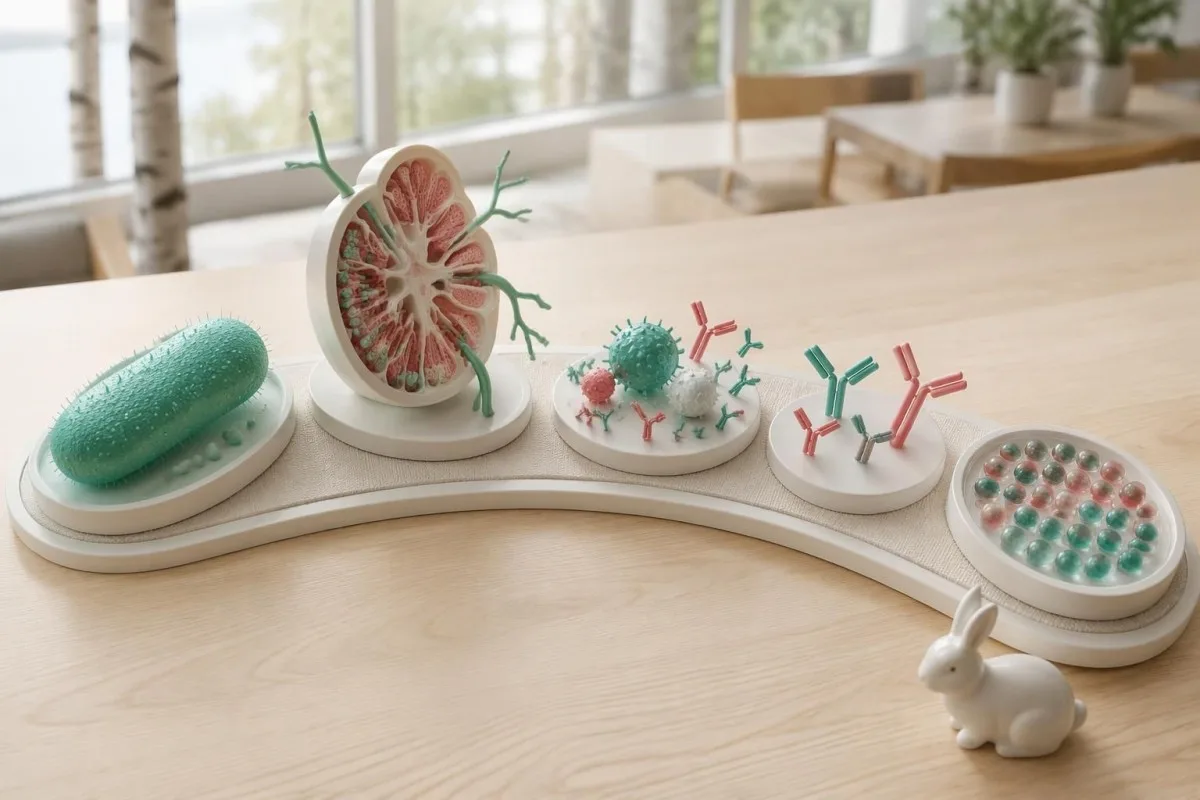
Published assay accuracy is not automatically transferable to every laboratory. The 2020 Frontiers review compares methods and study populations (Maurin, 2020); a sensitivity measured in confirmed convalescent cases cannot be applied unchanged to patients tested during the first seven symptom days, when antibody development is still incomplete.
Our two Zenodo publications below provide general blood-test education, not tularemia diagnostic validation. دی RDW تشریح اشاعت and the separately linked kidney-function guide concern supporting laboratory concepts; DOI registration does not establish peer review, and neither publication demonstrates tularemia-specific AI accuracy.
Kantesti’s طبی مواد کے معیارات require separating educational interpretation from diagnosis and stating evidence limits plainly. Our طبی مواد کے معیارات explain that approach: a repeat specimen in approximately 2–3 weeks can improve confirmation, but an unwell person should receive clinical assessment before that interval has passed.
اکثر پوچھے جانے والے سوالات
کیا مجھے ٹولریمیا ہونے کے باوجود ٹولریمیا کا ٹیسٹ منفی آ سکتا ہے؟
علامات ظاہر ہونے کے بعد پہلے 2–3 ہفتوں کے دوران ٹولیریمیا کا اینٹی باڈی ٹیسٹ منفی آ سکتا ہے، کیونکہ اس وقت تک اینٹی باڈیز قابلِ شناخت نہ ہوں۔ اس لیے اگر کسی ممکنہ ایکسپوژر اور علامات میں مطابقت ہو تو ابتدائی منفی نتیجہ انفیکشن کو خارج نہیں کرتا۔ معالجین صحت یابی کے مرحلے کا نمونہ ابتدائی نمونے کے تقریباً 2–3 ہفتے بعد لینے کا انتظام کر سکتے ہیں اور براہِ راست تشخیصی ٹیسٹنگ پر غور کر سکتے ہیں۔ سیرولوجیکل تبدیلی کا انتظار کرتے ہوئے طبی معائنے اور ضرورت کے مطابق علاج میں تاخیر نہیں ہونی چاہیے۔
ٹولیریمیا کے اینٹی باڈی ٹیسٹ کو کب دہرایا جانا چاہیے؟
ٹولریمیا سے صحت یابی کے مرحلے میں اینٹی باڈی کا نمونہ عموماً شدید مرحلے کے نمونے کے تقریباً 2–3 ہفتے بعد جمع کیا جاتا ہے۔ شدید مرحلے کا نمونہ بیماری کے پہلے ہفتے میں لینا بہتر ہے، اگرچہ اس کا نتیجہ منفی آ سکتا ہے۔ دونوں نمونوں کی ایک ہی طریقۂ کار سے جانچ کرنے سے سیرولوجی میں تبدیلی یا ٹائٹر میں چار گنا اضافہ شناخت کرنے میں مدد ملتی ہے۔ اگر پہلا نمونہ دیر سے جمع کیا گیا ہو یا علامات سے بیماری کا شبہ بدستور قوی ہو تو معالج شیڈول میں ردوبدل کر سکتا ہے۔
ٹولاریمیا اینٹی باڈی ٹائٹر 1:160 کا کیا مطلب ہے؟
A tularemia antibody titer of 1:160 may meet the positive or supportive threshold for some agglutination assays, but the laboratory’s method-specific interpretation controls its meaning. The number describes a dilution endpoint, not bacterial quantity or illness severity. A single positive result may reflect current infection, previous exposure, or cross-reactivity. A compatible illness and a paired change, such as 1:40 to 1:160, provide stronger evidence of recent infection.
Does positive tularemia IgM prove a recent infection?
Positive tularemia IgM does not independently prove a recent infection because IgM can persist after the acute illness. IgM and IgG may both become detectable around 2–3 weeks after symptom onset, rather than following a reliably separate early-versus-late pattern. Assay-specific false reactivity can also occur. Clinicians interpret antibody classes alongside exposure, symptoms, and paired samples rather than dating infection from one result.
Can mowing or handling rabbits cause tularemia?
Handling infected rabbits or hares can transmit tularemia through skin contact, while mowing or brush cutting can generate exposure-associated aerosols when contaminated animal material is disturbed. Symptoms commonly begin 3–5 days after exposure, although the reported incubation range is 1–21 days. Skin exposure may produce an entry-site sore and nearby enlarged lymph nodes; inhalational exposure may cause fever and respiratory symptoms without a visible sore. These symptoms warrant clinical assessment even if early antibodies are negative.
Should treatment wait until tularemia antibodies are positive?
Treatment for clinically suspected tularemia should not be delayed solely to obtain positive antibodies. Antibodies may remain negative for 2–3 weeks after symptoms start, while untreated illness can progress during that period. A clinician decides whether to begin treatment using exposure, examination, severity, and available testing. Breathing difficulty, confusion, fainting, or rapidly worsening illness requires emergency evaluation rather than waiting for another laboratory result.
Do positive antibodies after treatment mean tularemia is still active?
Positive Francisella tularensis antibodies after treatment do not by themselves prove ongoing infection because antibodies can persist for years. Clinical recovery, recurrent fever, respiratory findings, and the course of enlarged lymph nodes are more useful than expecting immediate antibody negativity. A positive result after a 10-day antibiotic course is therefore not automatically a treatment failure. New or worsening symptoms still require reassessment, regardless of the antibody titer.
اپنے خون کے ٹیسٹ کے نتائج کی وضاحت حاصل کریں
اپنی لیب رپورٹ اپ لوڈ کریں اور Kantesti کا AI چند سیکنڈ میں ہر بائیومارکر کو آسان زبان میں سمجھائے گا۔ دنیا بھر میں 2 ملین سے زیادہ افراد استعمال کرتے ہیں۔.
حوالہ جات
- Klein, T., Mitchell, S., & Weber, H. (2026). RDW خون کا ٹیسٹ: RDW-CV، MCV اور MCHC کے لیے مکمل رہنما۔. Kantesti AI طبی تحقیق۔. ڈی او آئی
- Klein, T., Mitchell, S., & Weber, H. (2026). BUN/کریٹینین ریشو کی وضاحت: گردے کی کارکردگی ٹیسٹ گائیڈ۔. Kantesti AI طبی تحقیق۔. ڈی او آئی
- بیماریوں کے کنٹرول اور روک تھام کے مراکز (n.d.)۔. Clinical Testing and Diagnosis for Tularemia. CDC کی طبی رہنمائی۔.
- Maurin M (2020). Francisella tularensis, Tularemia and Serological Diagnosis. Frontiers in Cellular and Infection Microbiology. ڈی او آئی
- Centers for Disease Control and Prevention (2025). Tularemia Antimicrobial Treatment and Prophylaxis: CDC Recommendations for Naturally Acquired Infections and Bioterrorism Response — United States, 2025. MMWR سفارشات اور رپورٹس۔.
- 2M+تجزیہ کیے گئے ٹیسٹ
- 127+ممالک
- 100+زبانیں
طبی ٹیم کے بارے میں


E-E-A-T اعتماد کے اشارے
تجربہ
معالج کی قیادت میں لیب تشریح کے ورک فلو کا کلینیکل جائزہ۔.
مہارت
لیبارٹری میڈیسن کا فوکس یہ کہ بایومارکرز کلینیکل سیاق میں کیسے برتاؤ کرتے ہیں۔.
مستندیت
ڈاکٹر تھامس کلائن نے طبی جائزہ لیا، جبکہ ڈاکٹر سارہ مچل اور پروفیسر ڈاکٹر ہانس ویبر نے بھی تعاون کیا۔.
امانت داری
شواہد پر مبنی تشریح واضح فالو اپ راستوں کے ساتھ تاکہ گھبراہٹ کم ہو۔.


طبی دستبرداری
یہ مضمون صرف تعلیمی مقاصد کے لیے ہے اور طبی مشورہ نہیں ہے۔ تشخیص اور علاج کے فیصلوں کے لیے ہمیشہ کسی مستند صحت کے ماہر سے رجوع کریں۔.
شائع ہوا۔ · ادارتی ٹیم سے رابطہ کریں
Kantesti Ltd کی جانب سے شائع کردہ، انگلینڈ اور ویلز میں رجسٹرڈ، کمپنی نمبر. 17090423, ، لندن، برطانیہ۔.
