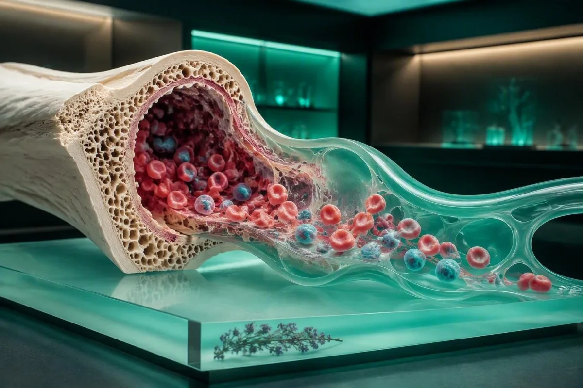
Кандының буялган препаратында полихромазия яшь кызыл кан тәнчекләренең гадәтидән тыш күренүен аңлата. Бу кан югалту яки дәвалаудан соң сөяк мие җыелуының ышанычлы билгесе булырга мөмкин, ләкин анемия плюс полихромазия ретикулоцитларны санауны һәм, күрсәтелгән очракта, гемолизны тикшерүне таләп итә.


- Полихромазия мәгънәсе: буялган слайдтагы күк-соры кызыл кан тәнчекләре, гадәттә, РНК калдыклары булган яңа чыгарылган ретикулоцитлар.
- Гадәти ретикулоцитлар: күпчелек олылар лабораторияләре якынча 0,5% - 2,5% ретикулоцит процентын хәбәр итә, анемиядә абсолют саннар ышанычлырак.
- Тергезү үрнәге: каты кан югалганнан, тимер белән дәвалаудан яки В12 витамины җитмәүне төзәткәннән соң 3-7 көн эчендә полихромазия күренергә мөмкин.
- Гемолиз билгесе: полихромазия, югары LDH, лаборатория диапазоныннан югарырак билгеле биллирубин һәм түбән һәпттоглобин белән анемия кызыл кан тәнчекләренең тиз җимерелүен хуплый.
- Төзәтелгән ретикулоцитлар: ретикулоцитларның 21% тан артык булуы гемоглобин түбән булганда да җитәрлек булмаска мөмкин; клиницистлар аны пациентның гемоглобинына төзәтәләр.
- Термінова картина: сары авыру, кара сидек, ял иткәндә сулыш алу, күкрәк авыртуы, хәлсезлек, яки шистоцитлар турында хәбәр итүче мазок шул ук көнне клиник тикшерү таләп итә.
- Nav diagnoze: “кечкенә” яки “уртача” полихромазия - лабораториянең күрү тасвирламасы, стандартлаштырылган авыру масштабы түгел.
- Киләсе тест: абсолют ретикулоцитлар санын, ретикулоцитлар җитештерү индексын, билирубин фракцияләрен, LDH, гаптоглобинны һәм туры антиглобулин тестын сезнең CBC үрнәгегезгә туры киләме, дип сорагыз.
Периферия препаратында полихромазия нәрсә аңлата
кан мазогында полихромазия яшь күзәнәкләр аркасында, алар кандагы РНК эчендәге яшь күзәнәкләр булу сәбәпле, гадәти алсу-роза төсенә караганда күк-соры төскә керә. Бу ачыш үзе гемолиз, рак яки тимер җитмәүне диагнозламый; ул безгә сөяк мие ретикулоцитларны циркуляциягә чыгарганын әйтә.
Гадәти CBC анализаторы ретикулоцитларны электрон рәвештә санарга мөмкин, шул ук вакытта кул белән мазок ясау лаборатория белгеченә аларның таралышын һәм форманың үзгәрүен күрергә мөмкинлек бирә. Минем клиник эшемдә, отчетта иң мөһиме “полихромазия” генә түгел, ә ул анемия, RDWның артуы яки фрагментланган күзәнәкләр янында торуы. КЗК нәрсә үз эченә ала бу нәтиҗәләр ни өчен бергә туры килүен аңлатырга ярдәм итә.
Гадәти олы кызыл күзәнәк якынча 120 көн яши, шул ук вакытта ретикулоцит гадәттә үзгәртеп кору алдыннан кан тамырларында 1-2 көн генә үткәрә. Шуңа күрә күренгән артыклык яисә сөяк миенең артуын яисә иртәрәк чыгарылуын күрсәтә. “Кечкенә”, “уртача”, һәм “зур” - лаборатория төшенчәләре; бу сүзләр өчен универсаль халыкара санлы масштаб юк.
Кантести - ЯИ анализы бу бер морфология флагны диагноз итеп караганчы, морфологияне гемоглобин, MCV, RDW, билирубин һәм алдагы нәтиҗәләр белән бергә интерпретацияли. 2026 елның 18 сентябренә кадәр, бу үрнәк-беренче подход, аеруча пациентлар клиницист караганчы морфология отчетын алганда файдалы.
Нигә күзәнәкләр зәңгәр төстә
Суправиталь буяулар ретикулар РНКны аеруча күренмәле итә, ләкин хәтта стандарт Райт-Гимза буялган слайд та полихромазияне булдыру өчен җитәрлек калдырылган РНКны күрсәтә ала. Төс - бу үзгәрешләрне аңлатучы күрсәткеч, лаборатория үзе “зәңгәр” яки пычранган дигән сүз түгел.
Лабораторияләр полихромазияне һәм аның чикләүләрен ничек ачыклый
Полихромазия буялган периферия күзәнәк үрнәге слайдын микроскоп аша карау аша билгеләнә, гадәттә автомат анализатор аномалияне ачканнан яки клиницист карауны сораганнан соң. Мазок күзәтүе - ярым-количественный, шуңа күрә ретикулоцитлар санын исәпләү - бу тәэсирне саннарга әйләндерә торган тест.
Лабораторияләр кул белән тикшерүне кайчан башлыйлар: кайберләре анализаторның таркалу үрнәкләрен, авыр анемия чикләрен, югары яшь ретикулоцит фракцияләрен яки алдагы аномаль морфологияне кулланалар. Шуңа күрә төрле лабораторияләрдән ике отчет бер үк дәрәҗәдәге полихромазияне төрлечә аңлатырга мөмкин. Нәтиҗә һәрвакыт лабораториянең үз CBC чикләре белән укылырга тиеш.
Бер абсолют ретикулоцитлар саны анемиядә проценттан күбрәк мәгълүмат бирә, чөнки процент кенә югары булырга мөмкин, чөнки бөтен кызыл күзәнәкләр саны азрак. Күпчелек олы лабораторияләр якынча 25-100 × 10⁹/л га кадәр чикне кулланганнар, ләкин җирле чикләр анализатор һәм халык буенча үзгәрә. Безнең аңлату югары ретикулоцит үрнәкләре клиник логиканы күбрәк аңлата.
Мазок шулай ук сфероцитларны, шистоцитларны, теш күзәнәкләрен, ядрәле кызыл күзәнәкләрне яки автоматик санаулар тулысынча классификацияли алмаган зурлыкны күрсәтә ала. Зини һ.б. (2012) шистоцитларны стандартлаштырылган идентификациясе мөһимлеген ассызыклады, чөнки фрагментация сөяк миенең гади кайтуыннан бик нык аерылып тора.
Мазоктагы тискәре нәтиҗә ретикулоцитозны инкар итми
Кечкенә ретикулоцитозны флуоресцентка нигезләнгән анализаторлар ярдәмендә визуаль яктан күренгәнче үк үлчәргә мөмкин. Киресенчә, кечкенә полихромазия турында хәбәр, ретикулоцитлар санын сорамаган очракта, раслауга лаек, шикләнүгә түгел.
Полихромазия кайчан сәламәт сөяк мие җыелуын күрсәтә
Полихромазия еш кына гемоглобинның күптән түгел кимегәнен һәм ретикулоцитлар саны берничә көн эчендә артканын күрсәткәндә, сөяк миенең тиешле кайтуын күрсәтә. Бу еш кына үз-үзеннән туктатылган кан югалту, уңышлы тимер өстәү яки вакытлыча сөяк мие репрессиясеннән кайткач көтелә.
After an acute loss of red cells, erythropoietin rises but the marrow response is not immediate; reticulocytosis usually becomes evident after about 3 to 5 days and can peak around 7 to 10 days. I often reassure patients who see polychromasia after treatment that this timing can be a sign the treatment is working, provided symptoms and hemoglobin are moving in the right direction.
A practical example is a patient with iron-deficiency anemia whose hemoglobin rises from 92 g/L to 103 g/L over 2 to 3 weeks of effective therapy. Their RDW may temporarily widen because older small cells and newer better-hemoglobinized cells coexist. The early RDW rise after iron therapy is easily mistaken for deterioration.
Кантести - AI кан анализы нәтиҗәләрен аңлату платформасы that compares CBC trends across visits, which can distinguish a one-time recovery response from persistent anemia with ongoing cell loss. For adults with iron deficiency, the cause still needs attention; replacement without asking why iron fell can miss menstrual, gastrointestinal, dietary, or absorption-related contributors.
Recovery after vitamin replacement
In vitamin B12 or folate deficiency, a brisk reticulocyte response can occur roughly 3 to 5 days after treatment begins. A falling potassium level is an uncommon but recognized concern during rapid hematologic response in severe deficiency, so clinicians may recheck electrolytes in frail or profoundly anemic patients.
Полихромазия кайчан гемолизны күрсәтә
Polychromasia suggests hemolysis when it appears with anemia and biochemical evidence of red-cell breakdown, especially raised LDH, increased unconjugated bilirubin, and reduced haptoglobin. No single one of these tests proves hemolysis; the combined pattern and the clinical history decide the likelihood.
Hemolysis can be inherited, immune-mediated, medication-related, mechanical, infection-associated, or linked to systemic illness. Barcellini and Fattizzo (2015) describe the classic laboratory constellation as reticulocytosis, increased LDH and unconjugated bilirubin, plus low haptoglobin. Haptoglobin can be low in liver disease without hemolysis, which is a nuance that prevents plenty of unnecessary alarm.
A direct antiglobulin test, also called DAT or Coombs testing, helps assess immune hemolysis when the history and chemistry fit. Spherocytes with a positive DAT may support warm autoimmune hemolytic anemia, while bite cells can suggest oxidative injury in a susceptible person. For related context, see our guide to mažas haptoglobinas be hemolizės.
I become more concerned when a person has fatigue, jaundice, dark tea-colored urine, back pain, or a rapid hemoglobin decline of more than about 20 g/L over days. These symptoms do not identify the cause, but they change the timeframe for evaluation from routine follow-up to urgent assessment.
Hemolysis outside the bloodstream
Extravascular hemolysis occurs mainly in the spleen and liver and may produce jaundice without obvious dark urine. Intravascular hemolysis more often lowers haptoglobin sharply and can produce hemoglobinuria, although hydration and timing affect what a urine sample shows.
Ачык яки яшерен кан югалганнан соң полихромазия
Polychromasia can follow blood loss because a healthy marrow responds by releasing more reticulocytes after roughly 3 to 5 days. The key next question is whether bleeding has stopped and whether the hemoglobin trend is stable, rising, or continuing to fall.
Heavy menstrual bleeding is a frequent cause of iron depletion and anemia in people who menstruate, particularly when pads or tampons need changing every 1 to 2 hours, clots are large, or bleeding lasts more than 7 days. A reticulocyte response does not make continued loss harmless. Our review of heavy menstrual bleeding warning signs outlines when prompt care is sensible.
Gastrointestinal loss may be invisible: black tarry stool, red rectal bleeding, persistent abdominal symptoms, regular NSAID use, and iron deficiency after menopause all deserve medical discussion. A normal reticulocyte response can coexist with low ferritin because the marrow may be trying to compensate while iron stores continue to drain.
One clinical trap is checking a hemoglobin level immediately after a sudden loss. In the first hours, it can underestimate severity because plasma and red cells have not equilibrated; repeat CBC testing after fluid shifts may tell a different story. Symptoms such as collapse, resting breathlessness, or persistent fast heartbeat trump any single early number.
Do not self-treat presumed loss
Oral iron is commonly used, but taking it before establishing the likely cause can obscure the diagnostic timeline and cause constipation or nausea. Ferritin, transferrin saturation, CRP, and a focused bleeding history usually give more useful direction than starting several supplements at once.
Кайсы ретикулоцит нәтиҗәләре препаратны ачыклый
A reticulocyte count clarifies polychromasia by measuring marrow output, and a corrected reticulocyte percentage or reticulocyte production index adjusts that output for the degree of anemia. In adults, a raw reticulocyte percentage of 0.5% to 2.5% is often reported as typical, but interpretation changes sharply when hematocrit is low.
.Әр сүзнең corrected reticulocyte percentage equals reticulocyte percentage multiplied by patient hematocrit divided by 45%. For example, 4% reticulocytes with a hematocrit of 27% corrects to 2.4%, not 4%. This matters because a percentage can look impressive while the actual response remains only modest for severe anemia.
.Әр сүзнең ретикулоцитлар җитештерү индексы (RPI) further divides the corrected percentage by a maturation factor, typically about 1.0 at hematocrit 45%, 1.5 at 35%, 2.0 at 25%, and 2.5 at 15%. An RPI above about 3 generally indicates an appropriate regenerative response; an RPI below 2 in significant anemia suggests underproduction, though cutoffs are not perfectly uniform.
Kantesti AI interprets reticulocyte data by pairing the absolute count with hematocrit and prior hemoglobin, not by flagging a percentage in isolation. The reticulocyte count and immature fraction guide explains why an immature reticulocyte fraction can rise before mature reticulocytes do.
Reticulocyte hemoglobin adds another layer
Reticulocyte hemoglobin content, often called Ret-He or CHr, estimates iron available to newly produced cells over the prior few days. Low values can support iron-restricted erythropoiesis, but device-specific cutoffs commonly range around 25 to 30 pg and should not be swapped between analyzers.
Полихромазия белән бергә укылырга тиешле CBC һәм химия нәтиҗәләре
Polychromasia is best interpreted with hemoglobin, MCV, RDW, bilirubin fractions, LDH, haptoglobin, and kidney function. A low hemoglobin plus a high reticulocyte response points toward loss or destruction, while low hemoglobin plus low reticulocytes points toward inadequate production.
Hemoglobin reference intervals differ by laboratory, altitude, sex, and pregnancy status, but many adult labs use approximately 120 to 160 g/L for nonpregnant women and 135 to 175 g/L for men. MCV below 80 fL often directs attention toward iron deficiency or thalassemia traits, whereas MCV above 100 fL raises questions about B12, folate, alcohol, liver disease, thyroid disease, medicines, or reticulocytosis itself. Review borderline MCV patterns before interpreting the index alone.
LDH is sensitive but nonspecific: skeletal muscle injury, liver conditions, sample hemolysis, and many other processes can elevate it. In suspected hemolysis, an LDH rise accompanied by indirect bilirubin elevation and a low haptoglobin is more persuasive than LDH alone. Our LDH test overview explains common non-red-cell causes.
A creatinine rise or reduced eGFR may blunt erythropoietin signaling and produce anemia with an inappropriately low reticulocyte response. That contrast is diagnostically useful: a person with hemoglobin 90 g/L and reticulocytes 0.6% needs a different work-up from someone with the same hemoglobin and reticulocytes 6.0%.
RDW is a timing marker, not a diagnosis
An RDW above roughly 14.5% is common in mixed cell populations, including early iron treatment and recent reticulocytosis. It can be normal in thalassemia trait and abnormal in dozens of unrelated settings, so it should never be used as a stand-alone screen for cause.
Мәгънәсен үзгәртә торган башка препаратлар
Polychromasia becomes more informative when another red-cell shape accompanies it. Spherocytes, schistocytes, bite cells, target cells, or nucleated red cells can narrow the differential diagnosis and sometimes change urgency immediately.
Шистоцитлар are fragmented red cells and can signal microangiopathic hemolysis, mechanical valve-related injury, severe hypertension, or disseminated coagulation depending on the clinical setting. The International Council for Standardization in Haematology uses a threshold of more than 1% schistocytes as suspicious for thrombotic microangiopathy when the morphology is otherwise appropriate, but interpretation belongs with an experienced laboratory and clinical team. Read about ашыгыч шистоцитлар табышлары if that exact word appears on your report.
Сфероцити are dense, round red cells lacking central pallor and may occur in hereditary spherocytosis or immune hemolysis. Polychromasia plus spherocytes often justifies a DAT, bilirubin, and family-history review; polychromasia alone does not. A high MCHC can strengthen suspicion but can also result from analytical artifacts.
Nucleated red cells in an adult peripheral sample are not simply “extra reticulocytes.” They can occur with severe marrow stress, profound hemolysis, hypoxia, marrow infiltration, or critical illness, and they deserve clinician review even when the reticulocyte count is high.
Shape reports need a real sample review
Automated analyzers are excellent screening tools but may generate morphology flags from cold agglutinins, marked leukocytosis, or sample handling issues. A manual review is particularly valuable when a result would lead to emergency referral or a major treatment decision.
Гемолиз һәм кан югалтудан тыш полихромазия сәбәпләре
Polychromasia can occur without active hemolysis or bleeding when the marrow is rebounding from suppression, responding to erythropoietin, or releasing cells early under physiologic stress. The reticulocyte count, medication list, and prior CBCs usually separate these possibilities.
Recovery after viral illness, chemotherapy, severe nutritional deficiency treatment, or transient marrow suppression can produce a temporary reticulocyte surge. In my experience, the trend is reassuring only when white cells and platelets are also recovering as expected and there are no blasts or unexplained persistent cytopenias. Aplastic anemia CBC warning patterns show the contrasting low-production picture.
Erythropoiesis-stimulating agents used in selected kidney disease or cancer-care settings can increase reticulocyte output. High altitude, chronic hypoxemia, and some congenital hemoglobin disorders may also raise baseline production. These are context-dependent effects, not reasons to dismiss new anemia.
Certain medicines can cause hemolysis in susceptible people, including oxidant drugs in G6PD deficiency; the timing after a new medicine, food exposure, or illness can be more revealing than a single test. If G6PD testing is performed during or soon after a hemolytic episode, young cells can make enzyme activity look falsely normal, so clinicians sometimes repeat it later.
Why a normal CBC can still carry a comment
Mild polychromasia with normal hemoglobin may be transient, especially after recent recovery. Persistent morphology comments across several draws merit discussion, but they do not automatically indicate a marrow disorder.
Йөклелек, яңа туганнар һәм яшькә бәйле аңлатма
Polychromasia is more common in newborn cell samples because neonatal red-cell turnover and marrow activity differ from adult physiology. During pregnancy, anemia and reticulocyte interpretation need trimester-specific ranges and a focused assessment for iron deficiency, hemolysis, and obstetric complications.
Newborns normally have higher reticulocyte counts than adults, often about 3% to 7% in the first days of life, then values fall over subsequent weeks. For that reason, a smear description that would be notable in a 40-year-old may be entirely expected in a newborn. Pediatric interpretation should use the reporting laboratory’s age-specific intervals.
Pregnancy expands plasma volume, which can lower hemoglobin concentration without equal loss of red-cell mass. The World Health Organization commonly defines anemia in pregnancy as hemoglobin below 110 g/L, although trimester-specific practices differ; a falling ferritin or low transferrin saturation adds useful evidence for iron-restricted production. See йөклелек ферритин диапазоны вакыт детальләре өчен.
Polychromasia with falling platelets, rising AST or ALT, elevated LDH, hypertension, upper abdominal pain, severe headache, or visual symptoms in pregnancy needs urgent obstetric evaluation. That combination can occur in HELLP syndrome or other serious conditions, and it should never be managed as an online lab question alone.
After delivery
Postpartum blood loss can trigger a reticulocyte response after several days, but symptoms matter more than the smear phrase. Heavy bleeding, faintness, chest symptoms, or breathlessness after delivery requires urgent in-person care, even if a prior CBC was acceptable.
Табу яки отчет артефакт булырга мөмкинме?
Polychromasia itself is usually a genuine cellular observation, but pre-analytical problems and automated flags can distort the surrounding CBC data. A repeat sample is reasonable when the result conflicts with how you feel, prior results, or other laboratory findings.
Delayed slide preparation can change cell appearance, and prolonged storage of an EDTA sample may cause red-cell crenation or other artifacts. This does not usually create true reticulocytosis, but it can complicate morphology interpretation. A fresh repeat sample is often preferable to overinterpreting an equivocal comment.
In-vitro hemolysis means cells broke down after collection, not necessarily inside the body. It can falsely raise potassium, LDH, AST, and phosphate, but it does not establish clinical hemolytic anemia. Our guide to potassium errors from sample hemolysis explains why the laboratory may request a redraw.
Cold agglutinins can make red cells clump in the tube, producing spuriously low red-cell counts and high MCV or MCHC until the sample is warmed and rerun. When indices look physically implausible, such as an unexpectedly high MCHC, I ask the laboratory whether analytical interference was assessed before assigning a disease label.
Trend before certainty
A repeat CBC after 1 to 2 weeks can be informative in a stable person with mild unexplained polychromasia, but faster retesting is appropriate when hemoglobin is falling or symptoms are progressing. Exact timing should be individualized by the clinician who knows the underlying history.
Полихромазия кайчан ашыгыч медицина ярдәмен таләп итә
Polychromasia needs urgent assessment when it accompanies symptoms or results suggesting rapid anemia, major hemolysis, or microangiopathy. Seek same-day medical care for chest pain, fainting, shortness of breath at rest, confusion, jaundice with dark urine, heavy ongoing bleeding, or a smear reporting schistocytes.
A hemoglobin below 70 g/L is commonly a threshold where transfusion is considered in stable hospitalized adults, though symptoms, active loss, heart disease, and the specific setting alter decisions. It is not a home-management cutoff and does not mean every person at 71 g/L is safe. A rapid decline is often more alarming than a chronic stable value.
Polychromasia with platelets below 150 × 10⁹/L, creatinine rising, neurologic symptoms, and schistocytes raises concern for thrombotic microangiopathy and requires emergency evaluation. The reason this combination matters is that it can reflect simultaneous red-cell fragmentation, platelet consumption, and organ injury—not simple iron deficiency.
Call emergency services rather than driving yourself if severe breathlessness, collapse, new weakness, or chest pressure develops. The blood-test guide for shortness of breath explains some laboratory patterns, but no online interpretation can safely rule out urgent cardiopulmonary causes.
Symptoms can outweigh a “mild” smear phrase
A report saying “slight polychromasia” does not make severe symptoms mild. Conversely, marked polychromasia in a person recovering comfortably with a rising hemoglobin can be less urgent than the wording sounds.
Практик адымнар һәм клиницист өчен сораулар
The most useful next step after polychromasia is to confirm whether anemia is present and whether reticulocyte production is appropriate for the hemoglobin level. Bring the full CBC, prior results, medication list, and a short timeline of bleeding, infection, new drugs, diet changes, and symptoms.
Useful questions include: “What is my absolute reticulocyte count?”, “Is my response corrected for hematocrit?”, “Do bilirubin, LDH, and haptoglobin suggest hemolysis?”, and “Should we check ferritin, transferrin saturation, B12, folate, kidney function, or a DAT?” Those questions are more productive than asking whether polychromasia is simply good or bad.
Dr. Thomas Klein, our Chief Medical Officer, advises recording recent events with dates: infection, donation, surgery, a heavy period, medication start, endurance event, or iron treatment. The marrow response has a clock, and a result taken 4 days after an event means something different from one taken 4 months later. Iron-study interpretation can help you prepare for that discussion.
Do not stop prescribed medicines or start high-dose iron, folate, or B12 solely because of a smear comment. Folic acid can improve the anemia of B12 deficiency while neurologic injury progresses, which is why B12 status should be checked first when macrocytosis or neurologic symptoms are present.
What to bring to a repeat draw
Bring the laboratory name, specimen date, whether you were recently ill, and a list of supplements including biotin. Hydrate normally unless instructed otherwise, and ask whether reticulocyte testing requires a separate order; in many systems it is not automatically included in a CBC.
Препарат комментары пәйда булганда, тенденцияләрне куркынычсыз куллану
Trend review is useful when polychromasia appears because the direction of hemoglobin, reticulocytes, RDW, bilirubin, and platelets often matters more than one isolated result. It cannot replace a clinician’s examination, a repeat smear, or urgent care when red flags are present.
Кантести - AI нигезендәге кан анализы тикшерү коралы that organizes uploaded reports into longitudinal patterns and flags combinations that deserve clinician follow-up. In practice, a hemoglobin rise of 10 to 20 g/L over a few weeks after identified iron deficiency is quite different from a fall of 20 g/L with increasing bilirubin and reticulocytes.
Our system can explain terminology in 75+ languages, but it deliberately treats a morphology comment as context rather than a verdict. Dr. Thomas Klein’s medical-review approach is to look for discordance: for example, high reticulocytes with no anemia may be transient, while low reticulocytes with marked anemia may point toward an underproduction problem. You can also review low reticulocyte symptoms for that opposite pattern.
For transparency around methodology and clinical boundaries, consult our медицина валидация стандартлары. Kantesti does not diagnose the cause of polychromasia from an image alone; our role is to help you recognize the relevant pattern, preserve trends, and prepare sharper questions for a qualified clinician.
Privacy and clinical limits
A laboratory PDF may contain identifiers and unrelated clinical information, so remove or protect details according to your preferences before sharing it. Digital interpretation is most valuable when it improves the next medical conversation rather than delaying needed care.
Төп фикер: төскне генә түгел, реакцияне аңлатыгыз
Polychromasia usually means the marrow is releasing young red cells, and the next step is to determine whether that response is appropriate recovery or a clue to ongoing loss or destruction. An absolute reticulocyte count plus hemoglobin trend is the most efficient starting point for most stable adults.
Most patients with a single mild smear comment and stable hemoglobin do not need to panic. They do need the comment placed beside symptoms, the CBC, and a reticulocyte result. Persistent polychromasia without an obvious explanation is a reasonable reason to ask for a clinician-led review, especially after age 50 or with recurrent anemia.
Kantesti AI supports this kind of structured review, while our physicians and clinical advisors retain clear boundaries around diagnosis and urgent triage. Readers who want to understand how medical oversight informs our work can meet the медицина консультатив советы.
For adjacent laboratory context, our ретикулоцит һәм LDH тикшерү кулланмасы links the turnover markers discussed here. The evidence is rarely black-and-white: timing, symptoms, and the pattern of companion results are what turn polychromasia from a microscope observation into a clinically useful answer.
Тикшеренү басмалары
Kantesti’s related educational publications are listed below for readers who want transparent source trails. They are supplementary resources and do not replace hematology guidelines, laboratory quality standards, or individualized medical care.
Еш бирелә торган сораулар
Какыслылыклы кан буялмасында куркынычмы?
Кандагы полихромазия яшь кызыл кан тәнчекләренең кан әйләнешенә керүен аңлата; бу гадәттә кан югалтудан яки анемияне дәвалаудан соң нормаль булырга мөмкин, гадәттә башланганнан соң 3-5 көн эчендә. Гемоглобин кимегәндә, ретикулоцитларның абсолют саны югары булганда, яки LDH, индирект билирубин һәм симптомнар гемолизны күрсәткәндә ул борчу тудыра. Сары тире, карасу сидек, эчпошыру, күкрәк авыртуы яки тын бетү тиз медицина ярдәме таләп итә.
Полихромазия һәрвакыт гемолизны аңлатамы?
Юк, полихромазия һәрвакыт гемолизны аңлатмый. Ул якын арада кан югалтканда, тимер яки витамин B12 белән дәвалаганда, вакытлыча сөяк мие эшчәнлеге кимегәндә сәламәтләнү чорында, эритропоэтин белән дәвалануда, яки яшь балаларда кызыл кан тәнчекләренең табигый югары әйләнеше булганда барлыкка килергә мөмкин. Анемия, гаптоглобинның түбән булуы, ЛДГның артуы, конъюгацияләнмәгән билирубинның күбәюе һәм ретикулоцитларның тиешле җавабы белән бергә полихромазия булганда гемолиз ихтималы югарырак. Иммун гемолиз шик тудырганда, туры антиглобулин сынавы өстәлергә мөмкин.
Полихромазия булганда ретикулоцитларның күпме күрсәткече югары санала?
Күпчелек өлкәннәр лабораторияләрендә ретикулоцитларның якынча 0.5% - 2.5% проценты һәм абсолют санын 25 - 100 × 10⁹/л тирәсендә күрсәтәләр, гәрчә лаборатория буенча диапазоны төрле булырга мөмкин. 100 × 10⁹/л дан артык абсолют ретикулоцитлар саны, гадәттә, сөяк миенең артык эшләвен күрсәтә. Анемиядә клиницистлар гематокритны кулланып төзәтелгән ретикулоцит процентын исәпләп чыгаралар һәм ретикулоцит җитештерү индексын кулланырга мөмкин; 3 дан артык RPI гадәттә тиешле регенератив җавапны күрсәтә. Төзетелмәгән югары процент гематокрит түбән булганда ялгыштырырга мөмкин.
Тимер җитмәү полихромазияга китерергә мөмкинме?
Укмаган тимер җитмәүлек ретикулоцитларның адекват булмаган реакциясенә ешрак китерә, чөнки гемоглобин ясау өчен тимер кирәк, ләкин полихромазия эффектив тимер терапиясе башланганнан соң барлыкка килергә мөмкин. Ретикулоцитларның күтәрелеше еш кына уңышлы дәвалау башланганнан соң 3-7 көн эчендә була, гемоглобин шактый күтәрелгәнче. Ферритин, трансферрин туендыру, MCV, RDW һәм кан югалту тарихы тимер җитмәүлекнең барлыгын ачыкларга ярдәм итә. Даими менструаль яки гастроинтестиналь кан югалту, күренеп торган ми реакциясенә карамастан, анемияне саклап кала ала.
Полихромазиядән соң канны кабат тапшырырга кирәкме?
Полихромазия аңлашылмаган очракларда, анемия булганда яки анализда катнашу булырга мөмкин булганда, кабатланган CBC һәм ретикулоцитлар санын тикшерү еш кына кирәк. Минималь үзгәрешләр булган тотрыклы кешеләрне 1-2 атнадан соң кабат тикшерергә мөмкин, ләкин бу вакыт симптомнарга, гемоглобин дәрәҗәсенә һәм шикләнелгән сәбәпкә бәйле. Гемоглобин кими башласа, актив кан китү булса яки гемолиз шикләнелсә, тизрәк тикшерү кирәк. Клиницист шулай ук LDH, билирубин фракцияләре, гаптоглобин, ферритин, B12, фолат, креатинин һәм DAT таләп итә ала.
Йөклелек яки яңа туган балаларда полихромазия нормаль булырга мөмкинме?
Яңа туган балаларда полихромазия ешрак очрый, чөнки тормышның беренче көннәрендә ретикулоцитлар саны еш кына 3% - 7% тәшкил итә, бу олылардагы күрсәткечләрдән югарырак. Йөклелек плазма күләмен һәм кызыл кан күзәнәкләре таләпләрен үзгәртә, шуңа күрә гемоглобин һәм тимер нәтиҗәләрен йөклелеккә специфик рәвештә аңларга кирәк; Бөтендөнья Сәламәтлек Саклау Оешмасы йөкле хатын-кызларда анемияне ачыклау өчен еш кына 110 г/л гемоглобинны куллана. Тромбоцитлар аз булганда, бавыр ферментлары артканда, югары LDH, гипертония, көчле баш авыртуы яки корсакның өске өлеше авыртканда полихромазия тиз акушерлык бәясен таләп итә. Йөкле бала үрнәкләренә олылар өчен стандарт аралар кулланылмый.
Бүген үк AI белән эшләнгән кан анализы тикшерүе
Дөнья буенча 2 миллионнан артык кулланучы кушылыгыз: алар Kantestiны тиз һәм төгәл лаборатория анализы өчен ышана. Кан анализы нәтиҗәләрегезне йөкләгез һәм 15,000+ биомаркерларын секундлар эчендә тулы аңлатма белән алыгыз.
📚 Сылтама бирелгән тикшеренү басмалары
Klein, T., Mitchell, S., & Weber, H. (2026). C3 C4 комплемент кан анализы һәм ANA титры буенча кулланма. Kantesti AI медицина тикшеренүе.
Klein, T., Mitchell, S., & Weber, H. (2026). Нипах вирусын кан анализы: иртә ачыклау һәм диагностикалау буенча кулланма 2026. Kantesti AI медицина тикшеренүе.
📖 Тышкы медицина белешмәләре
Phillips J, Henderson AC (2018). Hemolytic anemia: Evaluation and differential diagnosis. American Family Physician.
⚕️ Медицина кисәтүе
Бу мәкалә бары тик белем бирү максатларында гына һәм медицина киңәше булып тормый. Диагноз һәм дәвалау карарлары өчен һәрвакыт квалификацияле сәламәтлек саклау белгеченә мөрәҗәгать итегез.
E-E-A-T ышаныч сигналлары
Тәҗрибә
Табиб җитәкчелегендә лаборатория нәтиҗәләрен аңлату эш процессларын клиник тикшерү.
Белгечлек
Лаборатория медицинасы: биомаркерларның клиник контекстта үз-үзләрен тотышын аңлау.
Авторититет
Доктор Томас Кляйн тарафыннан язылган, доктор Сара Митчелл һәм профессор доктор Ханс Вебер тарафыннан тикшерелгән.
Ышанычлылык
Ачык күзәтү юллары белән куркуны киметү өчен дәлилләргә нигезләнгән аңлату.