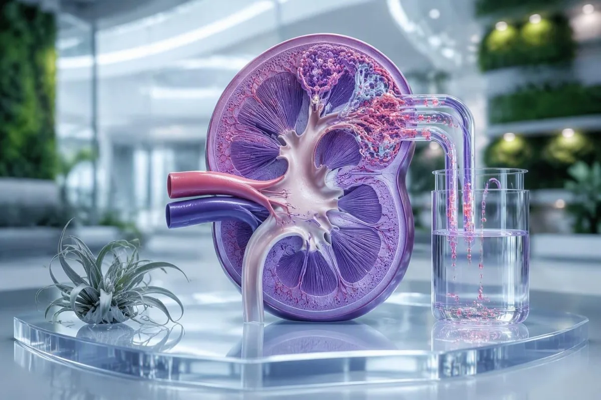
A spot urine sodium result is a snapshot of how the kidneys are handling salt at that moment. Its real value comes from reading it alongside serum sodium, urine osmolality, medicines and volume status.


- Spot urine sodium below 20 mmol/L usually means the kidneys are conserving sodium because effective circulating volume is low.
- Urine sodium above 30 mmol/L during true hypotonic hyponatremia suggests SIADH, diuretic exposure, adrenal insufficiency, or renal salt loss rather than simple dehydration.
- ออสโมลาลิตีในปัสสาวะต่ำกว่า 100 mOsm/kg indicates appropriately suppressed antidiuretic hormone and points toward excess water intake or low-solute intake.
- Urine osmolality above 100 mOsm/kg means antidiuretic hormone is active; the next discriminator is usually spot urine sodium.
- ยาขับปัสสาวะ can raise urine sodium for several hours and make a single result look like kidney salt wasting.
- Serum sodium below 125 mmol/L with confusion, seizure, severe headache, repeated vomiting, or marked drowsiness needs urgent emergency assessment.
- Fractional excretion of sodium below 1% can support reduced kidney perfusion, but it is less reliable with chronic kidney disease, diuretics, pigment injury, or sepsis.
- Do not increase salt intake solely because a urine sodium result is low; treatment depends on the cause, blood pressure, kidney function and serum sodium.
What a Spot Urine Sodium Result Actually Tells You
Spot urine sodium measures sodium excreted in one urine sample, usually reported in mmol/L, rather than the amount of sodium in your body. A low result often signals renal sodium conservation, while a high result means the kidneys are releasing sodium—but neither finding diagnoses dehydration or SIADH by itself.
In practice, I treat this as a kidney-behaviour test, not a dietary salt score. A person can eat a salty takeaway and still have urine sodium below 20 mmol/L if vomiting, diarrhoea, heart failure, cirrhosis or heavy sweating has triggered strong sodium retention.
The sample is most useful when collected near the blood draw. A urine sodium of 8 mmol/L collected after 2 litres of intravenous saline answers a different question than a value of 8 mmol/L collected before treatment, which is why hospital timestamps matter more than many patients realise.
Dr. Thomas Klein's clinical rule is simple: first confirm whether the serum sodium is genuinely low and whether serum osmolality is low; only then use urine tests to map the physiology. Our แนวทางตรวจอิเล็กโทรไลต์ explains why sodium values can be dangerous even when symptoms initially seem mild.
Urine Sodium Ranges: Useful Cutoffs, Not a Universal Normal
There is no single “normal” spot urine sodium range because sodium excretion changes with diet, fluid intake, timing and medication. In hypotonic hyponatremia, clinicians commonly use 20 to 30 mmol/L as a practical dividing line between sodium conservation and inappropriate renal sodium loss.
A random urine sodium below 20 mmol/L generally supports low effective arterial volume, especially when urine osmolality exceeds 100 mOsm/kg. A value above 30 mmol/L is more consistent with SIADH or renal sodium loss when the patient is not taking a diuretic and has normal kidney, thyroid and adrenal function.
These cutoffs are intentionally imperfect. The European hyponatremia guideline uses a urine sodium threshold of 30 mmol/L in hypotonic hyponatremia because the kidney should ordinarily conserve sodium below that level when circulation is underfilled (Spasovski et al., 2014).
คันเตสตีเป็น เครื่องวิเคราะห์ผลเลือด AI that reads serum sodium, creatinine, urea and glucose together; a urine sodium result should be added only when the clinical question is hyponatremia, acute kidney injury or unexplained fluid imbalance. For serum-side context, see our guide to อาการโซเดียมต่ำ.
A 24-hour urine sodium measurement, often roughly 40 to 220 mmol/day with ordinary dietary intake, answers a different nutritional or stone-risk question than a spot sample. Do not compare the two numbers as though they were interchangeable.
Low Urine Sodium Meaning: When the Kidney Is Saving Salt
Low urine sodium usually means the kidneys have switched into sodium-conservation mode because they sense reduced effective circulation. The most familiar causes are gastrointestinal fluid loss, sweating, poor intake, bleeding, heart failure and advanced liver disease.
With diarrhoea or vomiting, a urine sodium below 20 mmol/L plus concentrated urine is often physiologically appropriate. The key phrase is “effective” volume: someone with swollen ankles from heart failure may be fluid-overloaded overall while the kidneys still behave as if circulation is inadequate.
I see a common trap after endurance events. A runner who drinks plain water for hours, loses sodium in sweat and develops a serum sodium of 128 mmol/L may have low urine sodium from volume stress, but the immediate risk comes from excess retained water rather than simply “not enough salt.”
Low urine sodium can also follow a low-salt diet or very recent restriction, so it does not prove dehydration. Pair it with blood pressure, pulse, urea-to-creatinine pattern and weight change; our discussion of BUN เทียบกับครีเอตินีน shows why these clues often travel together.
Why oedema can coexist with sodium conservation
Heart failure and cirrhosis can lower the pressure sensed by the kidneys despite visible fluid accumulation. In those settings, a spot urine sodium below 20 mmol/L may reflect neurohormonal sodium retention, and self-treating with extra salt can worsen oedema or breathlessness.
High Urine Sodium Causes: SIADH, Medicines and Kidney Salt Loss
High urine sodium causes include diuretics, SIADH, adrenal insufficiency, renal tubular disorders, recovering acute tubular injury and chronic kidney disease. In a patient with low serum sodium and urine osmolality above 100 mOsm/kg, a urine sodium above 30 mmol/L makes simple volume depletion less likely.
SIADH produces a deceptively tidy pattern: low serum sodium, low serum osmolality, urine osmolality usually above 100 mOsm/kg and urine sodium commonly above 30 mmol/L. Patients often look clinically euvolemic, without obvious dehydration or marked oedema, although bedside volume assessment is far from perfect.
High urine sodium does not automatically mean dietary excess. A value of 74 mmol/L in a patient receiving furosemide 40 mg can reflect the intended drug effect, whereas the same value in a person taking no diuretic and with serum sodium 121 mmol/L raises a more focused question about SIADH, cortisol deficiency or tubular injury.
The American expert panel describes SIADH as a diagnosis of exclusion: hypotonicity, inappropriately concentrated urine, urine sodium generally above 30 mmol/L, and no better explanation such as adrenal, thyroid, renal or diuretic-related disease (Verbalis et al., 2013). Read our dedicated SIADH laboratory guide for the full pattern.
Why Blood Sodium Must Be Read Before Urine Sodium
A urine sodium result has its greatest diagnostic value when blood sodium is low, especially below 135 mmol/L, and measured serum osmolality confirms hypotonicity. A high urine sodium with normal serum sodium can simply reflect recent salt intake, a diuretic dose or normal daily variation.
Not all low serum sodium is hypotonic hyponatremia. Marked hyperglycaemia pulls water into the bloodstream and lowers measured sodium; serum sodium rises by about 1.6 to 2.4 mmol/L for every 100 mg/dL glucose above 100 mg/dL, depending on the clinical setting.
Normal or high serum osmolality with low sodium suggests a different branch of the diagnostic tree, such as hyperglycaemia or an exogenous osmole, rather than SIADH. This is one reason correcting sodium for glucose can prevent an incorrect fluid recommendation.
Kantesti AI interprets electrolyte patterns by placing sodium beside glucose, urea, creatinine and reported medications rather than presenting a urine result as a standalone verdict. In my experience, this reduces the especially risky mistake of calling every low sodium result “dehydration.”
How Urine Osmolality Changes the Interpretation
Urine osmolality below 100 mOsm/kg means the kidneys are making very dilute urine, while a value above 100 mOsm/kg indicates antidiuretic hormone activity. This distinction often matters more than the absolute urine sodium concentration at the beginning of a hyponatremia work-up.
Low serum sodium plus urine osmolality below 100 mOsm/kg points toward primary polydipsia, a low-protein and low-salt intake pattern, or rapid intake of hypotonic fluid. The kidney is doing its job; the problem is that water intake has exceeded its ability to clear free water.
Low serum sodium plus urine osmolality above 100 mOsm/kg means water excretion is constrained. Urine sodium then helps separate low effective volume, often below 20 mmol/L, from SIADH-like and renal-loss patterns, often above 30 mmol/L.
A urine osmolality of 650 mOsm/kg does not prove dehydration by itself—it can occur with nausea, pain, cortisol deficiency, lung disease or postoperative stress. Our detailed urine osmolality explanation covers these less obvious triggers.
Diuretics: The Most Common Reason a Spot Result Misleads
Loop and thiazide diuretics can raise urine sodium even when a patient is genuinely volume depleted. A spot sample collected within hours of a dose may therefore mimic SIADH or intrinsic renal salt wasting.
Loop diuretics such as furosemide block sodium reabsorption in the thick ascending limb, while thiazides act in the distal convoluted tubule. Both can produce urine sodium above 30 mmol/L, but thiazides are particularly associated with clinically significant hyponatremia in older adults.
When I review a confusing result, I ask for the drug name, dose and exact last-dose time before interpreting the number. A patient taking hydrochlorothiazide 25 mg each morning who gives urine at 10 a.m. has a very different pre-test probability from someone who stopped it 72 hours earlier.
Fractional excretion of uric acid above 12% may support SIADH even when diuretics cloud urine sodium interpretation, though it is not decisive and should be clinician-led. Medication lists also matter for kidney monitoring; see our แนวโน้มความปลอดภัยของยา.
The SIADH Pattern—and What Must Be Excluded First
SIADH is suggested by hypotonic hyponatremia, urine osmolality above 100 mOsm/kg and urine sodium above 30 mmol/L in a clinically euvolemic person. It cannot be diagnosed reliably until thyroid disease, cortisol deficiency, diuretics and significant kidney failure have been considered.
Common SIADH triggers include nausea, pain, pulmonary disease, central nervous system conditions and medicines such as selective serotonin reuptake inhibitors, carbamazepine and some cancer therapies. In hospital practice, postoperative nausea alone can raise antidiuretic hormone enough to distort urine studies for a day or two.
A morning cortisol test is usually preferable to a random afternoon value when adrenal insufficiency is on the table. Primary adrenal insufficiency can produce hyponatremia with high urine sodium because low aldosterone impairs sodium retention; potassium may be high, but not invariably.
คันเตสตีเป็น แพลตฟอร์มการแปลผลตรวจเลือดของ AI that highlights the combination of low sodium, low osmolality and non-suppressed urine concentration as a follow-up pattern—not as a diagnosis. The related clues in Addison disease testing are particularly relevant when fatigue, weight loss, low blood pressure or hyperpigmentation are present.
Kidney-Related Salt Loss: When the Tubules Cannot Hold Sodium
Kidney-related sodium loss occurs when damaged or hormonally impaired tubules cannot reabsorb sodium appropriately. Acute tubular injury, interstitial nephritis, post-obstructive diuresis, some inherited tubulopathies and adrenal insufficiency can all produce urine sodium above 30 mmol/L.
In classic prerenal physiology, fractional excretion of sodium, or FENa, is often below 1%; in intrinsic tubular injury it may exceed 2%. The calculation uses urine sodium, plasma sodium, urine creatinine and plasma creatinine, but it performs poorly after diuretics and in chronic kidney disease.
A rising creatinine plus granular casts, protein, blood or white cells on urinalysis shifts concern toward intrinsic renal disease rather than uncomplicated dehydration. A result of urine sodium 55 mmol/L is more meaningful if creatinine has risen from 0.8 to 2.1 mg/dL over 48 hours than if kidney function is stable.
Urine microscopy can add information a chemistry value cannot. Our guides to urine sediment findings และ granular casts explain when a kidney-focused review is appropriate.
FENa and FEUrea: Helpful Calculations With Real Limits
FENa estimates the percentage of filtered sodium that leaves in urine, and a value below 1% can support reduced kidney perfusion in selected acute kidney injury cases. It is an adjunct, not a replacement for history, examination, creatinine trend and urinalysis.
The formula is FENa = urine sodium × plasma creatinine divided by plasma sodium × urine creatinine, multiplied by 100. A FENa above 2% is classically associated with tubular injury, yet contrast exposure, chronic kidney disease, sepsis and diuretic treatment readily break that textbook rule.
Fractional excretion of urea, or FEUrea, below 35% is sometimes used when diuretics are present because urea handling is less directly altered by loop diuretics. The evidence is honestly mixed, particularly in sepsis and intensive care, so I would not use FEUrea to justify withholding fluids or delaying evaluation.
Kantesti AI can organize paired laboratory values and flag implausible timing gaps, but a clinician must decide whether a calculation fits the clinical episode. For a broader renal framework, review ระยะของโรคไตเรื้อรัง.
How Collection Timing, Diet and IV Fluids Alter the Result
Spot urine sodium can change substantially within hours after saline, a salty meal, exercise, vomiting or a diuretic dose. A technically accurate laboratory number may therefore be clinically misleading if the collection conditions are unknown.
Isotonic saline can increase urine sodium in a person with SIADH because the kidney excretes the sodium while retaining part of the water; serum sodium may not improve and can occasionally fall. Conversely, saline usually improves low-volume hyponatremia once the stimulus for antidiuretic hormone settles.
A morning first-void sample is not automatically “best” for this test. What matters is capturing urine before major treatment when possible, then documenting whether the person has eaten, received fluids, exercised intensely, vomited or taken a medication in the prior 6 to 12 hours.
Urine concentration also affects interpretation. A very dilute sample with urine creatinine of 12 mg/dL deserves more caution than a contemporaneous sample with 120 mg/dL; our guide to ครีเอตินินในปัสสาวะและการเจือจาง อธิบายว่าทำไม.
Four Clinical Patterns Doctors Use at the Bedside
The most useful interpretation comes from patterns: serum sodium, serum osmolality, urine osmolality, urine sodium, creatinine and medication history. The same urine sodium result can imply opposite things in dehydration, SIADH, diuretic use and kidney injury.
Pattern one: serum sodium 126 mmol/L, urine osmolality 540 mOsm/kg and urine sodium 9 mmol/L after three days of diarrhoea supports low effective volume. Treatment commonly targets the fluid loss and cause, not indiscriminate water restriction.
Pattern two: serum sodium 122 mmol/L, urine osmolality 420 mOsm/kg and urine sodium 62 mmol/L in a person without diuretics or renal failure supports an SIADH-like physiology. The next step is medication review and exclusion of cortisol and thyroid deficiency, not assuming excess salt intake.
Pattern three: serum sodium 129 mmol/L and urine sodium 68 mmol/L four hours after bumetanide may be drug effect. Pattern four: creatinine rapidly rising with urine sodium 58 mmol/L and abnormal sediment may require evaluation for tubular injury; acute eGFR changes after dehydration offer a useful contrast.
What Not to Do With a High or Low Urine Sodium Result
Do not change fluid intake, stop prescribed diuretics or take salt tablets solely on the basis of one spot urine sodium result. Those actions can be unsafe when the underlying issue is heart failure, cirrhosis, SIADH, adrenal insufficiency or acute kidney injury.
Salt tablets may have a role in carefully selected chronic SIADH plans, but they can aggravate swelling, hypertension and heart failure. Likewise, aggressive water drinking can worsen hypotonic hyponatremia when antidiuretic hormone is active, even if someone feels thirsty.
In my 15 years of clinical practice, the most preventable problem has been a well-intentioned patient taking extra salt after reading “low urine sodium.” Low urine sodium often means the body is already trying to retain sodium; the missing piece may be circulating volume, cortisol, medication adjustment or water balance.
คันเตสตีเป็น เครื่องมือวิเคราะห์ผลตรวจเลือดที่ขับเคลื่อนด้วย AI that can prepare a structured discussion of laboratory trends, but it does not replace urgent clinical assessment. If you are uncertain which results to bring, our รายการตรวจสอบสรุปผลเลือด สามารถช่วยคุณเตรียมตัวได้.
When a Sodium Pattern Needs Urgent Medical Care
Serum sodium below 125 mmol/L with confusion, seizure, reduced consciousness, severe headache, repeated vomiting or new unsteadiness requires urgent emergency assessment. Urine sodium helps identify cause, but it must never delay treatment of symptomatic hyponatremia.
A fall in serum sodium from 140 to 122 mmol/L over 24 to 48 hours is generally more dangerous than a stable value of 122 mmol/L present for months because brain adaptation takes time. Rapid correction is also hazardous: in many higher-risk patients, clinicians aim to limit correction to 8 mmol/L or less in 24 hours to reduce osmotic demyelination risk.
Seek same-day medical advice for sodium below 130 mmol/L accompanied by dizziness, falls, persistent nausea, significant weakness, new confusion, very low blood pressure or reduced urine output. People with kidney disease, heart failure, liver disease, pregnancy, cancer treatment or a new diuretic need a lower threshold for review.
As of September 29, 2026, our clinical team advises using trend data rather than isolated readings whenever possible. Kantesti's แนวทางการยืนยันทางการแพทย์ describes how our results are framed around follow-up triggers, and our คณะกรรมการที่ปรึกษาทางการแพทย์ provides clinical oversight for that safety-first approach.
คำถามที่พบบ่อย
ปัสสาวะโซเดียมต่ำหมายถึงอะไร
โซเดียมในปัสสาวะต่ำ โดยทั่วไปต่ำกว่า 20 mmol/L ในตัวอย่างที่ได้จากการเก็บปัสสาวะเพียงครั้งเดียว มักหมายถึงไตกำลังสงวนโซเดียมไว้เนื่องจากรับรู้ว่าปริมาตรเลือดไหลเวียนมีประสิทธิภาพลดลง สาเหตุทั่วไป ได้แก่ อาเจียน ท้องเสีย เหงื่อออก การรับประทานอาหารน้อย เลือดออก หัวใจล้มเหลว และตับแข็ง ค่านี้ไม่ได้พิสูจน์ภาวะขาดน้ำโดยอัตโนมัติ เนื่องจากอาหารโซเดียมต่ำและการรักษาล่าสุดก็สามารถทำให้ค่าลดลงได้เช่นกัน ผลลัพธ์จะมีประโยชน์มากที่สุดเมื่ออ่านควบคู่กับโซเดียมในเลือด ออสโมแลลิตีในปัสสาวะ ความดันโลหิต การทำงานของไต และเวลาการให้ยา.
สาเหตุของโซเดียมในปัสสาวะสูงคืออะไร?
สาเหตุของโซเดียมในปัสสาวะสูง ได้แก่ การใช้ยาขับปัสสาวะกลุ่ม loop หรือ thiazide เมื่อเร็วๆ นี้, SIADH, ภาวะต่อมหมวกไตทำงานบกพร่อง, ภาวะไตวายเฉียบพลัน, โรคไตเรื้อรัง และความผิดปกติของท่อไต ในภาวะโซเดียมในเลือดต่ำจากน้ำในร่างกายมากเกินไป (hypotonic hyponatremia) การมีโซเดียมในปัสสาวะสูงกว่า 30 mmol/L ร่วมกับความเข้มข้นของออสโมลาลิตีในปัสสาวะสูงกว่า 100 mOsm/kg บ่งชี้ว่าไตไม่สามารถสงวนโซเดียมไว้ได้อย่างเหมาะสม ผลที่สูงกว่า 80 mmol/L อาจเกิดขึ้นหลังการให้ยาขับปัสสาวะ หรือการให้สารน้ำเกลือ และไม่สามารถวินิจฉัยได้ด้วยตนเอง แพทย์ควรวินิจฉัยแยกโรคยา, ภาวะขาดคอร์ติซอล และความผิดปกติของไตออกก่อนที่จะวินิจฉัย SIADH.
ปัสสาวะโซเดียมสูงในภาวะขาดน้ำหรือไม่
โซเดียมในปัสสาวะมักจะต่ำในภาวะขาดน้ำที่ไม่ซับซ้อนเนื่องจากไตจะสงวนโซเดียมไว้ ทำให้มีค่าต่ำกว่า 20 mmol/L บ่อยครั้ง อย่างไรก็ตาม ภาวะขาดน้ำอาจเกิดขึ้นร่วมกับผลโซเดียมในปัสสาวะสูงได้หากบุคคลนั้นเพิ่งได้รับยาขับปัสสาวะ มีภาวะต่อมหมวกไตบกพร่อง มีการบาดเจ็บของท่อไต หรือได้รับน้ำเกลือทางหลอดเลือดดำ ออสโมลัลลิตีของปัสสาวะมักจะสูงกว่า 100 mOsm/kg ในภาวะขาดน้ำเนื่องจากฮอร์โมนต้านการขับปัสสาวะจะทำให้ปัสสาวะเข้มข้น ดังนั้น โซเดียมในปัสสาวะสูงจึงไม่สามารถตัดภาวะขาดน้ำออกได้หากไม่ทราบบริบททางคลินิก.
ระดับโซเดียมในปัสสาวะที่บ่งชี้ถึง SIADH คือเท่าใด
ค่าโซเดียมในปัสสาวะจากการเก็บตัวอย่างครั้งเดียว (spot urine sodium) ที่สูงกว่า 30 mmol/L สามารถสนับสนุนภาวะ SIADH ได้เมื่อระดับโซเดียมในเลือดต่ำ ระดับออสโมแลลิตีในเลือดต่ำ และระดับออสโมแลลิตีในปัสสาวะสูงกว่า 100 mOsm/kg ภาวะ SIADH ยังคงเป็นการวินิจฉัยที่ต้องแยกสาเหตุอื่นออกไป ดังนั้นแพทย์ต้องพิจารณาสาเหตุจากยาขับปัสสาวะ ภาวะต่อมหมวกไตทำงานบกพร่อง ภาวะพร่องไทรอยด์ และโรคไต ก่อน ผู้ป่วย SIADH หลายรายมีค่าโซเดียมในปัสสาวะระหว่าง 40 ถึง 80 mmol/L แต่ไม่มีค่าใดค่าหนึ่งที่สามารถวินิจฉัยได้อย่างชัดเจน การตรวจสภาพของเหลวที่ดูปกติไม่ได้เป็นการยืนยันภาวะ SIADH เนื่องจากการประเมินปริมาตรของเหลวเป็นสิ่งที่ไม่สมบูรณ์ในตัวเอง.
การดื่มน้ำช่วยลดโซเดียมในปัสสาวะได้หรือไม่
การดื่มน้ำปริมาณมากอาจทำให้ความเข้มข้นของโซเดียมในปัสสาวะเจือจางลง โดยเฉพาะอย่างยิ่งหากความออสโมลาลิตีของปัสสาวะต่ำ แต่ก็ไม่ได้ทำให้การขับโซเดียมรวมลดลงเสมอไป ในภาวะ polydipsia ขั้นต้น ความออสโมลาลิตีของปัสสาวะมักต่ำกว่า 100 mOsm/kg เนื่องจากฮอร์โมน ADH ถูกกดอย่างเหมาะสม ในภาวะ SIADH การดื่มน้ำมากขึ้นอาจทำให้โซเดียมในซีรั่มต่ำแย่ลง เนื่องจากปัสสาวะยังคงมีความเข้มข้นค่อนข้างสูงแม้จะได้รับน้ำเข้าไป นี่คือเหตุผลว่าทำไมการตรวจโซเดียมในปัสสาวะแบบสุ่ม (spot urine sodium) จึงต้องแปลผลควบคู่ไปกับความออสโมลาลิตีของปัสสาวะ แทนที่จะดูเพียงอย่างเดียว.
Should I eat more salt if my urine sodium is low?
You should not increase salt intake solely because a spot urine sodium is low. A value below 20 mmol/L often reflects an appropriate kidney response to reduced circulation, and extra salt may worsen fluid retention in heart failure, kidney disease or liver disease. If low urine sodium accompanies vomiting, diarrhoea or heavy sweating, oral rehydration may be appropriate, but the correct fluid and sodium plan depends on symptoms, blood pressure and serum sodium. Seek clinician advice if serum sodium is below 130 mmol/L or symptoms are present.
รับการวิเคราะห์ผลตรวจเลือดด้วย AI วันนี้
เข้าร่วมผู้ใช้งานมากกว่า 2 ล้านคนทั่วโลกที่ไว้วางใจ Kantesti สำหรับการวิเคราะห์ผลตรวจทางห้องแล็บแบบทันทีและแม่นยำ อัปโหลดผลตรวจเลือดของคุณ แล้วรับการอ่านผลตรวจเลือดอย่างครอบคลุมของไบโอมาร์กเกอร์ 15,000+ ภายในไม่กี่วินาที.
📚 งานวิจัยที่อ้างอิง
Klein, T., Mitchell, S., & Weber, H. (2026). คู่มือกรุ๊ปเลือดบีลบ การตรวจเลือด LDH และการนับเม็ดเลือดแดงตัวอ่อน.
Klein, T., Mitchell, S., & Weber, H. (2026). อาการท้องเสียหลังอดอาหาร, จุดดำในอุจจาระ และคู่มือระบบทางเดินอาหาร ปี 2026.
📖 อ้างอิงทางการแพทย์ภายนอก
⚕️ ข้อสงวนสิทธิ์ทางการแพทย์
บทความนี้จัดทำเพื่อวัตถุประสงค์ด้านการศึกษาเท่านั้น และไม่ถือเป็นคำแนะนำทางการแพทย์ โปรดปรึกษาผู้ให้บริการด้านสุขภาพที่มีคุณสมบัติเหมาะสมเสมอสำหรับการตัดสินใจด้านการวินิจฉัยและการรักษา.
สัญญาณความน่าเชื่อถือ E-E-A-T
ประสบการณ์
การทบทวนเชิงคลินิกโดยแพทย์ที่นำโดยกระบวนการตีความผลตรวจในห้องแล็บ.
ความเชี่ยวชาญ
โฟกัสด้านเวชศาสตร์ห้องปฏิบัติการเกี่ยวกับพฤติกรรมของไบโอมาร์กเกอร์ในบริบททางคลินิก.
อำนาจ
เขียนโดย ดร. โธมัส ไคลน์ (Dr. Thomas Klein) พร้อมทบทวนโดย ดร. ซาราห์ มิตเชลล์ (Dr. Sarah Mitchell) และ ศ.ดร. ฮันส์ เวเบอร์ (Prof. Dr. Hans Weber).
ความน่าเชื่อถือ
การตีความที่อิงหลักฐาน พร้อมเส้นทางการติดตามที่ชัดเจนเพื่อลดความตื่นตระหนก.