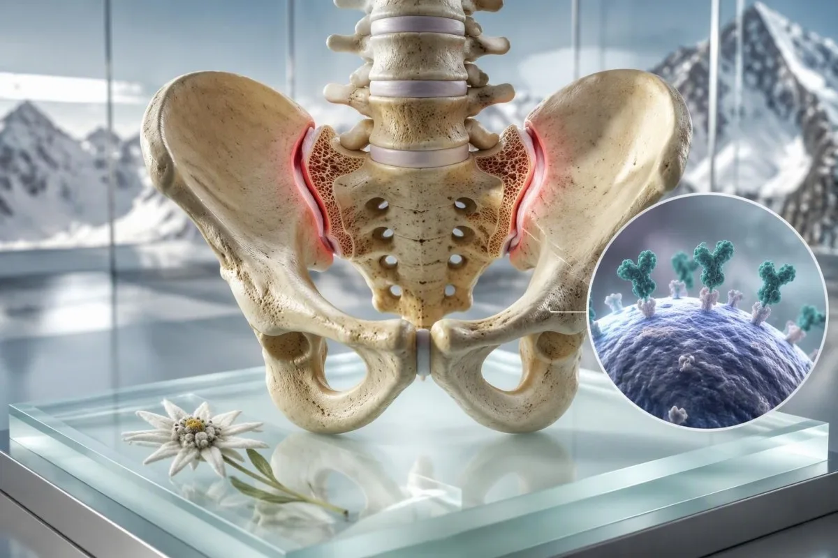
An HLA-B27 positive result identifies an inherited immune-system marker, not a diagnosis. It matters most when it accompanies back pain beginning before age 45, recurrent sudden-onset eye inflammation, psoriasis, bowel inflammation, or a close family history of spondyloarthritis.


- HLA-B27 positive means you carry a genetic marker; many healthy carriers never develop inflammatory disease.
- Inflammatory back pain usually starts before age 45, lasts longer than 3 months, improves with movement, and often disturbs sleep in the second half of the night.
- Acute anterior uveitis causes a red, painful, light-sensitive eye with blurred vision and needs same-day ophthalmology assessment.
- සාමාන්ය CRP does not exclude axial spondyloarthritis; CRP is raised in only a proportion of people with active disease.
- MRI sacroiliac joints can show active tissue response before plain X-rays show structural change.
- පවුලේ සෞඛ්ය ඉතිහාසය of ankylosing spondylitis, uveitis, psoriasis, or inflammatory bowel disease raises the value of a targeted rheumatology referral.
- HLA-B27 testing should answer a clinical question; it is not a useful screening test for uncomplicated mechanical low-back pain.
- Urgent eye symptoms outweigh laboratory results: do not wait for an HLA-B27 test before seeking care for eye pain or vision change.
What a positive HLA-B27 result actually tells you
HLA-B27 positive means that a person carries a particular HLA class I genetic variant; it does not mean they have ankylosing spondylitis or will develop it. The test becomes clinically useful only when symptoms and examination findings point toward a spondyloarthritis-spectrum condition. In my practice, the most damaging misunderstanding is treating a positive result as a forecast rather than one piece of probability-based evidence.
HLA-B27 is present in roughly 6.1% of adults in the United States, although prevalence varies sharply by ancestry and geography. It is more common in some northern European and Indigenous populations and uncommon in others, so a result has different predictive value in different communities. A negative result lowers the likelihood of HLA-B27-associated disease but cannot rule out axial spondyloarthritis.
The HLA-B27 test meaning is binary: detected or not detected, not high or low. Laboratories usually use molecular typing from a laboratory sample, and the result does not fluctuate with sleep, diet, a flare, or medication. That differs fundamentally from inflammatory markers such as ESR, which can change over days to weeks.
කන්ටෙස්ටි යනු AI රුධිර පරීක්ෂණ විශ්ලේෂකය that places an imported HLA result beside relevant inflammatory markers, but it cannot diagnose spondyloarthritis from a genetic marker alone. Thomas Klein, MD, advises patients to ask the simpler question: what clinical problem was the test ordered to answer? Our අවශ්ය වේ, explains why a flagged or positive result always needs its laboratory method and clinical context.
Why a positive result can feel more alarming than it is
Most HLA-B27-positive people never develop ankylosing spondylitis. The marker participates in antigen presentation, yet the biological route from carrying it to developing disease remains incomplete; intestinal microbes, immune regulation, and other genes appear to contribute. That uncertainty is real, and clinicians should not convert a genetic association into a prediction.
HLA-B27 and ankylosing spondylitis: association versus diagnosis
About 80% to 95% of people with radiographic ankylosing spondylitis are HLA-B27 positive in many northern European cohorts, but the marker is neither necessary nor sufficient for diagnosis. Doctors diagnose axial spondyloarthritis from the whole pattern: symptoms, physical findings, imaging, laboratory results, and competing explanations.
A positive HLA-B27 result has its greatest diagnostic value in someone with chronic back pain that began before age 45. In a 55-year-old whose first back pain began after lifting furniture at 52, the same result often changes very little. This is why a test ordered without a clinical pre-test probability can create more anxiety than clarity.
The ASAS classification framework allows an HLA-B27-positive person with chronic back pain beginning before 45 years to meet an axial spondyloarthritis pathway when at least two other spondyloarthritis features are present, such as uveitis, psoriasis, inflammatory bowel disease, arthritis, enthesitis, or a family history (Rudwaleit et al., 2009). Classification criteria help research consistency; they are not a home diagnostic checklist.
Radiographic ankylosing spondylitis describes established structural sacroiliac-joint change on X-ray, whereas non-radiographic axial spondyloarthritis may have normal X-rays. MRI can identify active bone-marrow tissue response earlier, although athletic loading, pregnancy-related changes, and degenerative disease can also make MRI interpretation tricky. A focused unexplained pain work-up පුළුල් වෙනස්කම් සැකිල්ලක් සකස් කිරීමට උපකාරී වේ.
The risk number patients usually want
First-degree relatives of a person with HLA-B27-associated ankylosing spondylitis have a higher risk than the general population, but family risk is not destiny. Risk estimates differ substantially by sex, ancestry, and whether the affected relative has radiographic disease. I do not recommend testing healthy children or siblings merely to settle worry unless a specialist identifies a specific clinical reason.
How inflammatory back pain differs from ordinary back pain
Inflammatory back pain typically begins gradually before age 45, persists for more than 3 months, improves with exercise, and does not improve reliably with rest. Mechanical pain more often follows a strain, worsens predictably with loading, and settles when the provoking movement stops.
Waking in the second half of the night because of back or buttock pain is a useful clue when it occurs repeatedly, not once after a poor mattress. Alternating buttock pain, morning stiffness lasting longer than 30 minutes, and marked improvement after getting moving add weight to the inflammatory pattern. None is individually diagnostic; together, they are more persuasive.
I often see a 32-year-old runner who says, I loosen up after the first kilometre. That is not proof of inflammation—many musculoskeletal problems warm up—but pain that returns after sitting, wakes them near 4 a.m., and has continued for 9 months deserves more than generic stretching advice. Pain severity is less informative than the pattern over time.
NICE advises rheumatology referral for back pain beginning before 45 and lasting more than 3 months when several spondyloarthritis features coexist; HLA-B27 testing can support referral when exactly three features are present. New bowel or bladder dysfunction, saddle numbness, fever, unexplained weight loss, cancer history, or rapidly progressive weakness requires urgent assessment for causes beyond spondyloarthritis. Review the practical distinction between inflammation and injury in our psoriasis laboratory guide, particularly if skin disease is also present.
A useful two-week symptom record
Record wake time, stiffness duration, exercise response, buttock location, and anti-inflammatory medicine response for 14 days before an appointment. This produces more diagnostic value than a generic pain score from 0 to 10, because rheumatologists are looking for a reproducible inflammatory rhythm.
When an HLA-B27-associated red eye needs same-day care
A painful red eye with light sensitivity, blurred vision, or a smaller-looking pupil can be acute anterior uveitis and needs same-day assessment by an eye specialist or emergency eye service. HLA-B27-associated uveitis often starts suddenly in one eye and can recur, sometimes in the other eye at a later date.
Acute anterior uveitis is not the same as conjunctivitis. Conjunctivitis commonly causes discharge or gritty discomfort; uveitis more often causes deep aching pain, photophobia, reduced visual clarity, and redness concentrated around the iris. A normal-looking external eye does not reliably exclude it, especially early on.
Roughly half of acute anterior uveitis cases in many European-derived cohorts are HLA-B27 positive, but eye doctors still consider infection, sarcoidosis, Behçet disease, inflammatory bowel disease, trauma, and medication effects. Do not use leftover steroid eye drops without an examination, because steroid drops can worsen undiagnosed corneal infection and raise eye pressure. නැවත පරීක්ෂණවල සැබෑ ප්රවණතා හඳුනාගැනීමට අපගේ මාර්ගෝපදේශය Behçet eye symptoms covers another condition where oral ulcers and eye symptoms change the urgency.
Treatment commonly uses prescription topical corticosteroid drops plus pupil-dilating drops, with a taper directed by ophthalmology. Follow-up matters because pressure elevation and swelling at the back of the eye can occur even when pain improves. A recurrent episode should trigger communication between ophthalmology and rheumatology rather than parallel, disconnected care.
සාමාන්ය හමුවීමක් සඳහා බලා නොසිටිය යුතු රෝග ලක්ෂණ
Sudden vision loss, a curtain or shower of new floaters, severe headache with nausea, halos around lights, or severe eye pain needs emergency evaluation immediately. These features can signal diagnoses other than uveitis, including acute angle-closure glaucoma or retinal disease, where delay can threaten sight.
The other symptoms that make HLA-B27 more meaningful
HLA-B27 becomes more clinically meaningful when back or eye symptoms occur with psoriasis, inflammatory bowel disease, heel enthesitis, swollen joints, or a close relative with related disease. This cluster is called the spondyloarthritis spectrum because different tissues may be involved at different times.
Enthesitis is inflammation where a tendon or ligament attaches to bone, commonly causing focal pain at the Achilles insertion or bottom of the heel. It differs from diffuse foot ache because the tenderness is usually sharply localized and may be worst after rest. Ultrasound can help, but body weight, sports loading, and footwear can produce similar symptoms.
Psoriasis-related nail pitting, separation of the nail plate, or a whole swollen finger or toe can be particularly helpful diagnostic clues. A person does not need widespread psoriasis for this to count; a small scalp patch or a long history of flaky plaques behind the ears may matter. Photographing an intermittent rash before it fades can save a surprising amount of time at review.
Chronic diarrhoea, nocturnal stool urgency, rectal bleeding, or unintended weight loss should prompt bowel evaluation rather than being attributed to stress. Faecal calprotectin is useful for detecting intestinal tissue response but is not specific for Crohn disease or ulcerative colitis; our calprotectin comparison explains its limitations. The connection matters because bowel inflammation can steer both imaging choices and treatment selection.
Symptoms that do not fit neatly
Fatigue is common in active inflammatory disease but is too non-specific to establish a diagnosis. Iron deficiency, sleep disruption, thyroid dysfunction, mood disorders, medications, and prolonged pain can all contribute, so I look for a coherent pattern rather than assigning fatigue to HLA-B27.
How family history changes the next step
A first-degree relative with ankylosing spondylitis, recurrent uveitis, psoriasis, inflammatory bowel disease, or reactive arthritis increases suspicion when compatible symptoms are present. Family history supports a referral decision; it does not make testing or imaging mandatory in a person with no symptoms.
Ask relatives about diagnoses and major symptoms, not only whether they carry HLA-B27. People may remember being told they had iritis, a stiff spine, Crohn disease, or psoriasis but have never heard the term spondyloarthritis. The age at symptom onset is especially valuable: back pain that began at 22 tells a different story from osteoarthritis diagnosed at 72.
Kantesti AI යනු AI රුධිර පරීක්ෂණ ප්රතිඵල වේදිකාව that can organise family-shared laboratory histories with consent, but family-history patterns still require a clinician to verify diagnoses. A useful record includes affected relative, relationship, condition, age at onset, eye episodes, and relevant imaging. Our පවුල් ඉතිහාස නිරීක්ෂකය offers a practical template for recording that detail.
Routine HLA-B27 testing in symptom-free relatives usually does not improve outcomes. The result can create false reassurance when negative and unnecessary fear when positive, because no proven preventive treatment is prescribed simply for carrier status. A child with persistent limp, eye symptoms, or swollen joints needs paediatric assessment based on symptoms, whatever the family genotype.
What to bring to a specialist appointment
Bring prior X-ray or MRI reports, eye-clinic letters, medication dates, and a one-page family history. Imaging discs or digital access are often more useful than repeating scans, particularly because sacroiliac-joint MRI requires experienced interpretation.
Why normal inflammation tests do not rule this out
Normal CRP and ESR do not rule out axial spondyloarthritis or HLA-B27-associated uveitis. These tests measure systemic inflammatory activity indirectly, and localized disease can be clinically active while both results remain within the laboratory reference interval.
CRP is often reported in mg/L, with many laboratories using less than 5 mg/L as a reference threshold, while ESR reference limits vary with age and sex. A CRP of 2 mg/L can coexist with active sacroiliac inflammation on MRI. Conversely, a CRP of 28 mg/L is non-specific and may reflect infection, obesity, dental inflammation, or another inflammatory condition.
A complete blood count, liver profile, kidney profile, CRP, and ESR are baseline context tests rather than diagnostic proof. Anaemia, raised platelets, low albumin, or iron deficiency can suggest persistent inflammation or bowel involvement, but each has alternative explanations. This is one reason inflammatory conditions should not be inferred from a single marker.
කන්ටෙස්ටි යනු AI මගින් බලගැන්වූ රුධිර පරීක්ෂණ විශ්ලේෂණ මෙවලම that can compare CRP, platelets, haemoglobin, and albumin across dates, helping users identify a pattern to discuss with their clinician. It cannot see sacroiliitis on an MRI or assess a painful eye. For antibody results that can be over-interpreted in a similar way, see our explanation of ANA පරීක්ෂාව.
Medication and test interpretation
NSAIDs can reduce CRP and improve symptoms without proving or disproving the diagnosis. Do not stop prescribed anti-inflammatory medication before testing unless the clinician managing your care specifically asks you to; an artificial flare is not a safe diagnostic strategy.
Which scans help, and what each scan can miss
MRI of the sacroiliac joints is the preferred scan when axial spondyloarthritis is suspected but X-rays are normal or inconclusive. X-rays show structural change that often develops slowly, whereas MRI can show active bone-marrow tissue response and other early changes.
A normal lumbar-spine MRI does not replace a dedicated sacroiliac-joint MRI protocol. The relevant joints are lower and more lateral than the area commonly imaged for a slipped disc. I have seen patients reassured by an ordinary lumbar scan that was simply not designed to answer the inflammatory question.
MRI findings require clinical correlation because bone-marrow oedema can occur after intense exercise, childbirth, mechanical stress, and degenerative change. The radiology report should describe location, depth, distribution, erosions, fat change, and structural features; a vague statement of oedema alone is not enough. A rheumatologist may ask for image review when the report and symptom pattern disagree.
Pelvic X-rays remain useful for documenting chronic sacroiliac structural changes, but a normal X-ray does not close the case in a younger person. CT shows structure very clearly but uses ionising radiation and is not generally the first choice for early inflammatory disease. If persistent inflammation affects mobility, bone-health assessment may also be relevant; read our bone loss laboratory guide.
Why the scan report can sound uncertain
Radiologists appropriately use terms such as compatible with or non-specific when the imaging pattern is not definitive. That wording is not evasive; it reflects a real overlap between inflammatory and mechanical findings, and it prevents imaging from being treated as a diagnosis in isolation.
When to ask for a rheumatology referral
Ask for rheumatology evaluation when chronic back pain began before age 45 and is accompanied by inflammatory features, HLA-B27 positivity, recurrent uveitis, psoriasis, bowel symptoms, heel enthesitis, or a relevant family history. A positive test plus one convincing clinical feature is more useful than a positive test discovered during broad screening.
A referral is reasonable even when CRP is normal and X-rays are unremarkable if the clinical pattern is compelling. The 2019 ACR/SAA/SPARTAN guideline recommends treatment decisions based on disease activity, function, comorbidities, and patient preferences rather than a genetic result alone (Ward et al., 2019). This approach prevents both delayed diagnosis and reflexive overtreatment.
At the appointment, expect questions about age at onset, morning stiffness, night waking, response to NSAIDs, eye attacks, skin lesions, bowel symptoms, infection preceding arthritis, and family history. A rapid and clear response to a full-dose NSAID may support suspicion, but it is not a diagnostic test. NSAIDs also improve ordinary back pain.
Thomas Klein, MD, recommends making a dated timeline rather than trying to remember every symptom in the consulting room. Include medication dose and duration, especially if using ibuprofen, naproxen, or diclofenac, because kidney function, blood pressure, ulcer risk, and anticoagulant use alter the safety calculation. Our medication trend guide explains why repeat kidney and liver results may matter during longer courses.
When an orthopaedic or physiotherapy assessment may be better first
Back pain starting after age 45 following a specific injury, with clear load-related triggers and no inflammatory features, more often benefits initially from musculoskeletal care. That is a probability statement, not a dismissal; persistent uncertainty or new eye, skin, bowel, or joint symptoms warrants reassessment.
What treatment looks like if axial spondyloarthritis is confirmed
Confirmed axial spondyloarthritis is treated with education, regular movement, targeted physiotherapy, and anti-inflammatory medicines; biologic or targeted synthetic medicines are reserved for persistent active disease. HLA-B27 status does not determine treatment by itself.
Exercise is treatment, not a test of willpower. A programme that maintains spinal extension, hip mobility, chest expansion, and aerobic capacity is generally more sustainable than sporadic intense workouts. Most patients find short daily mobility work of 10 to 20 minutes easier to keep than a single ambitious weekend session.
NSAIDs are usually considered first-line when safe, but dose and duration must be individualised. Naproxen 500 mg twice daily is a common adult anti-inflammatory dose, yet it may be unsuitable with chronic kidney disease, prior ulcer bleeding, heart failure, uncontrolled hypertension, anticoagulants, or pregnancy. Gastroprotection may be appropriate for some people, but it does not remove kidney or cardiovascular risk.
Biologic medicines such as TNF inhibitors and IL-17 inhibitors require screening and specialist monitoring because infection risk and comorbidities affect selection. Recurrent uveitis, psoriasis, and inflammatory bowel disease influence which agent best fits the whole person. This is one reason a diagnosis based only on an HLA-B27 result can lead to the wrong treatment conversation.
Avoid the supplement trap
No supplement has been shown to prevent HLA-B27-associated spondyloarthritis in healthy carriers. Correcting a documented vitamin D, iron, or B12 deficiency can improve general health, but it is not a substitute for assessment of inflammatory back pain or a painful red eye.
How to track results without becoming captive to them
The most useful record combines symptoms, function, medications, eye episodes, imaging, and laboratory trends rather than repeatedly retesting HLA-B27. Once accurately typed, HLA-B27 status does not need serial monitoring because your inherited marker does not change.
Track functional measures that mean something to you, such as minutes until morning stiffness eases, uninterrupted sleep, walking tolerance, or ability to turn while driving. These details show treatment response more honestly than a single good or bad day. Note whether an eye flare preceded a back-pain change by days or weeks.
Kantesti can organise imported laboratory reports across time and flag changes for discussion, but it should not be used to self-start or stop prescription treatment. Our clinical framework is described in the AI තාක්ෂණික මාර්ගෝපදේශය: results are interpreted as context for professional care, not as a replacement for examination or imaging.
Repeating CRP during a flare can be useful when a clinician is also following symptoms and treatment response, but routine weekly testing is rarely helpful. In my experience, a symptom record every day and blood work at clinically meaningful intervals yields less noise and fewer false alarms. Laboratory trends only become informative when collection dates, infections, exercise, and medicines are documented.
A note about privacy and family data
Genetic information can affect relatives as well as the person tested, so share reports deliberately. Keep the original laboratory report, remove unrelated identifiers when appropriate, and obtain consent before adding another adult's results to a shared family record.
Common misconceptions about being HLA-B27 positive
HLA-B27 positivity does not mean your immune system is attacking your spine, that you are contagious, or that you need treatment while symptom-free. It is a common inherited allele whose significance depends on the clinical setting.
Myth: a positive HLA-B27 test explains every ache. Fact: mechanical back pain remains common among HLA-B27 carriers. Strain, disc disease, hip disorders, hypermobility, fibromyalgia, kidney stones, and pelvic conditions still deserve a normal differential diagnosis. The marker should sharpen clinical reasoning, not end it.
Myth: a negative HLA-B27 test rules out ankylosing spondylitis. Fact: HLA-B27-negative axial spondyloarthritis occurs, especially across populations with lower background carrier frequency. When imaging and clinical features are persuasive, a negative result should not be used to deny specialist review.
Myth: genes are the same as destiny. Fact: genetics influence risk, but no preventive biologic therapy is indicated for a well HLA-B27 carrier. This is conceptually similar to the limits of MTHFR result interpretation: genetic information is medically useful only when the result changes an evidence-based decision.
Can stress cause a flare?
Stress can worsen pain, sleep, fatigue, and coping, but it does not explain away objective uveitis or inflammatory arthritis. Treating sleep and stress is worthwhile alongside medical care; it should never delay eye assessment or replace a work-up for persistent inflammatory symptoms.
Questions that make a first appointment more useful
The best first appointment questions focus on probability, alternative diagnoses, and the next decision—not on whether HLA-B27 is good or bad. A clinician should be able to explain what finding would increase or decrease their suspicion and what test would change management.
Ask whether your symptoms meet an inflammatory-back-pain pattern and which features argue against it. Then ask whether dedicated sacroiliac MRI is appropriate, whether an ophthalmology referral is needed, and whether psoriasis or bowel symptoms warrant coordinated review. This approach respects uncertainty without letting it become inaction.
Ask how long to try a treatment, what outcome defines success, and which adverse effects need a call. For NSAIDs, the specific safety discussion should include kidney function, blood pressure, gastrointestinal history, pregnancy plans, and interacting drugs. For biologics, ask about screening, vaccines, infection precautions, and how uveitis or bowel disease changes the choice.
Kantesti's clinical content is medically reviewed with structured quality controls; readers can examine our වෛද්ය සත්යාපන (medical validation) ප්රවේශය සහ අපගේ වෛද්ය උපදේශක මණ්ඩලය. A laboratory interpretation is most valuable when it helps you ask a better question of the clinician who can examine you.
A concise appointment script
Say: My back pain started at age 29, has lasted 14 months, wakes me at 4 a.m., improves after 30 minutes of movement, and I had a painful light-sensitive eye last spring. That one sentence gives a rheumatologist more usable information than stating only that an HLA-B27 result was positive.
Urgent symptoms and a safe next-step plan
Seek same-day urgent eye care for eye pain, light sensitivity, red eye with blurred vision, or any sudden visual change; seek emergency care for new weakness, bladder or bowel dysfunction, saddle numbness, high fever, or severe unremitting back pain. Do not wait for repeat blood work, an MRI booking, or a rheumatology appointment when these red flags occur.
For non-urgent symptoms, book a primary-care review within weeks if back pain has lasted more than 3 months and began before age 45, especially with HLA-B27 positivity or family history. Bring your HLA report, symptom timeline, medication list, eye-clinic letters, and prior images. A targeted referral is generally faster and more useful than repeating broad autoimmune panels.
As of September 27, 2026, the central clinical principle remains unchanged: HLA-B27 is a risk marker, not a diagnosis. Thomas Klein, MD, has found that patients do best when they treat a positive result as a prompt to notice patterns—then obtain the right eye, primary-care, or rheumatology assessment rather than catastrophising or ignoring symptoms.
කන්ටෙස්ටි යනු AI රසායනාගාර පරීක්ෂණ අර්ථකථන සේවාව built to explain laboratory reports in plain language while preserving the boundary between information and diagnosis. Learn how our organisation approaches this work on our About Us පිටුවේ and find further patient education in the කන්ටෙස්ටි බ්ලොග් අඩවිය.
පර්යේෂණ ප්රකාශන කොටස
Kantesti LTD. (2026). B Negative රුධිර වර්ගය, LDH රුධිර පරීක්ෂණය සහ රෙටිකියුලොසයිට් ගණන් කිරීමේ මාර්ගෝපදේශය. Figshare. https://doi.org/10.6084/m9.figshare.31333819. Related author records may be located through පර්යේෂණ ද්වාරය සහ ඇකඩමියා.එඩු.
Kantesti LTD. (2026). නිරාහාරව සිටීමෙන් පසු පාචනය, මළපහ වල කළු ලප සහ ආමාශ ආන්ත්රික රෝග මාර්ගෝපදේශය 2026. Figshare. https://doi.org/10.6084/m9.figshare.31438111. Related author records may be located through පර්යේෂණ ද්වාරය සහ ඇකඩමියා.එඩු.
These publications are not evidence for diagnosing HLA-B27-associated disease and should not replace the rheumatology and ophthalmology references cited above. They are listed as Kantesti research records for transparency.
නිතර අසන ප්රශ්න
HLA-B27 ධනාත්මක වීම යනු ඔබට ඇන්කලෝසින් ස්පොන්ඩිලයිටිස් ඇති බවද?
නැත. HLA-B27 ධනාත්මක ප්රතිඵලයක් යනු ඔබ ankylosing spondylitis සහ ඒ ආශ්රිත තත්වයන් සමඟ සම්බන්ධ ජානමය සලකුණක් දරන බවයි, නමුත් බොහෝ වාහකයෝ ජීවිත කාලය පුරාම නිරෝගීව සිටිති. රූපීය ankylosing spondylitis සහිත කණ්ඩායම් වල, ආසන්න වශයෙන් 80% ත් 95% ත් HLA-B27 ධනාත්මක වන නමුත්, අනෙක් අතට එය සත්ය නොවේ: ධනාත්මක පුද්ගලයන්ගෙන් වැඩි දෙනෙකුට මෙම තත්වය නොමැත. රෝග විනිශ්චය සඳහා අනුකූල රෝග ලක්ෂණ, පරීක්ෂාව, රූපගත කිරීම සහ වෙනත් හේතු බැහැර කිරීම අවශ්ය වේ.
HLA-B27-සම්බන්ධිත දැවිල්ලක් යෝජනා කරන කොන්දේ වේදනාවේ රෝග ලක්ෂණ මොනවාද?
වයස 45ට අඩු, ක්රමයෙන් ආරම්භ වන, මාස 3කට වඩා පවතින, චලනයෙන් සමනය වන, සහ විනාඩි 30කට වඩා උදෑසන තද ගතියක් ඇති කරන කොන්දේ වේදනාව අක්ෂීය ස්පොන්ඩිලෝආතරයිටිස් (axial spondyloarthritis) සඳහා වැඩි වශයෙන් යෝජනා කරයි. රාත්රියේ දෙවන භාගයේදී නැවත නැවත අවදි වීම සහ මාරුවෙන් මාරුවට පපුවේ වේදනාව (buttock pain) එකතු කිරීමෙන් ප්රයෝජනවත් ඉඟි ලැබේ. HLA-B27 ධනාත්මක ප්රතිඵලය මෙම රටාවේ අගය වැඩි කරන නමුත්, යාන්ත්රික හේතු (mechanical causes) සහ රතු කොඩි (red flags) තක්සේරු කිරීමට වෛද්යවරයෙකුට තවමත් අවශ්ය වේ.
HLA-B27 නිසා යුවියිටිස් ඇති විය හැකිද?
HLA-B27 සෑම අක්ෂි ගැටලුවකටම සෘජුවම හේතු නොවේ, නමුත් එය උග්ර ඉදිරිපස යුවේටිස් (acute anterior uveitis) සමඟ ශක්තිමත්ව සම්බන්ධ වේ. මෙම යුවේටිස් වර්ගය සාමාන්යයෙන් එක් ඇසක හදිසි රතු පැහැය, ගැඹුරු වේදනාව, ආලෝකයට සංවේදීතාවය සහ පෙනීම බොඳවීම ඇති කරයි; එය අක්ෂි විශේෂඥයෙකු විසින් එදිනම ඇගයීමට ලක් කළ යුතුය. බොහෝ යුරෝපීය සම්භවයක් ඇති පුද්ගලයන් අතර උග්ර ඉදිරිපස යුවේටිස් (acute anterior uveitis) ආබාධවලින් අඩක් පමණ HLA-B27 ධනාත්මක වන අතර, ජනගහනය අනුව ව්යාප්තිය වෙනස් වන අතර, විකල්ප හේතු ද සලකා බැලිය යුතුය.
Should I see a rheumatologist if I am HLA-B27 positive but have no symptoms?
Most symptom-free HLA-B27-positive people do not need routine rheumatology follow-up or preventive treatment. A review becomes sensible if chronic back pain began before age 45, recurrent uveitis, psoriasis, persistent bowel symptoms, heel pain from enthesitis, swollen joints, or a strong family history develops. Repeating HLA-B27 testing is not useful because inherited HLA status does not change over time.
Can CRP and ESR be normal with ankylosing spondylitis?
Yes. CRP is commonly reported as normal below 5 mg/L in many laboratories, but a normal CRP and ESR do not exclude axial spondyloarthritis or active uveitis. These markers reflect systemic inflammatory activity and can stay normal when inflammation is localized to the sacroiliac joints or eye. MRI, symptom pattern, physical examination, and specialist assessment may be more informative than a single normal inflammation result.
Is HLA-B27 testing done from blood?
HLA-B27 typing is commonly performed from a laboratory sample using molecular or flow-cytometry-based methods, and many laboratories use a blood sample. The test is qualitative, reported as detected or not detected, rather than as a concentration with a normal range. Diet, exercise, and disease activity do not change the result, because HLA-B27 is inherited rather than an inflammatory marker.
අදම AI බලයෙන් රුධිර පරීක්ෂණ විශ්ලේෂණය ලබාගන්න
තත්පර කිහිපයකින් ක්ෂණික හා නිවැරදි රසායනාගාර පරීක්ෂණ විශ්ලේෂණය සඳහා Kantesti විශ්වාස කරන ලොව පුරා මිලියන 2කට වැඩි පරිශීලකයන්ට එක්වන්න. ඔබගේ රුධිර පරීක්ෂණ ප්රතිඵල උඩුගත කර, තත්පර කිහිපයකින් 15,000+ ජෛව සලකුණු පිළිබඳ සවිස්තර අර්ථකථනය ලබාගන්න.
📚 යොමු කර ඇති පර්යේෂණ ප්රකාශන
ක්ලයින්, ටී., මිචෙල්, එස්., සහ වෙබර්, එච්. (2026). B Negative රුධිර වර්ගය, LDH රුධිර පරීක්ෂණය සහ රෙටිකියුලොසයිට් ගණන මාර්ගෝපදේශය. Kantesti AI වෛද්ය පර්යේෂණ.
ක්ලයින්, ටී., මිචෙල්, එස්., සහ වෙබර්, එච්. (2026). නිරාහාරව සිටීමෙන් පසු පාචනය, මළපහ වල කළු ලප සහ ආමාශ ආන්ත්රික රෝග මාර්ගෝපදේශය 2026. Kantesti AI වෛද්ය පර්යේෂණ.
📖 බාහිර වෛද්ය යොමු
⚕️ වෛද්ය වියාචනය
මෙම ලිපිය අධ්යාපනික අරමුණු සඳහා පමණක් වන අතර වෛද්ය උපදෙස් ලෙස නොසැලකේ. රෝග නිદાન සහ ප්රතිකාර තීරණ සඳහා සෑම විටම සුදුසුකම් ලත් සෞඛ්ය සේවා සපයන්නෙකුගෙන් උපදෙස් ලබාගන්න.
E-E-A-T විශ්වාස සංඥා
අත්දැකීම්
වෛද්යවරයා විසින් මෙහෙයවන ලද රසායනාගාර අර්ථකථන ක්රියාවලි පිළිබඳ සමාලෝචනය.
ප්රවීණතාව
සායනික සන්දර්භය තුළ ජෛව සලකුණු (biomarkers) හැසිරෙන ආකාරය පිළිබඳ රසායනාගාර වෛද්ය විද්යා අවධානය.
අධිකාරීත්වය
ආචාර්ය තෝමස් ක්ලයින් විසින් ලියන ලද අතර ආචාර්ය සාරා මිචෙල් සහ මහාචාර්ය ආචාර්ය හෑන්ස් වෙබර් විසින් සමාලෝචනය කරන ලදී.
විශ්වසනීයත්වය
අනතුරු ඇඟවීම් අඩු කිරීමට පැහැදිලි පසුකැඳවීම් මාර්ග සහිත සාක්ෂි-පාදක අර්ථකථනය.